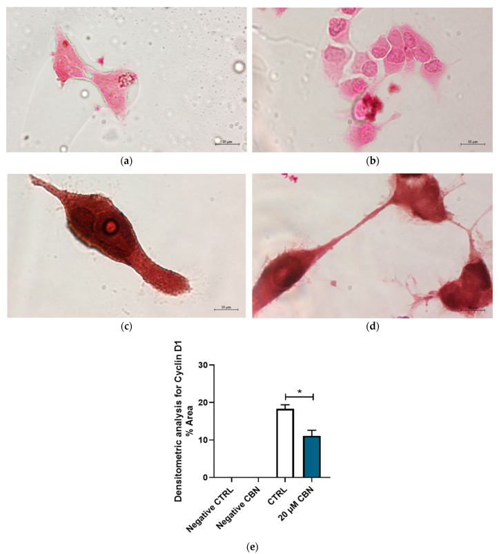
Figure 11.
Immunocytochemistry analysis of differentiated NSC-34 cells. Untreated differentiated NSC-34 cells without primary antibody (Negative CTRL) (a), 20 µM CBN-treated differentiated NSC-34 cells without primary antibody (20 µM CBN Negative CTRL) (b), untreated differentiated NSC-34 cells immunoprofiled for Cyclin D1 (c), and 20 µM CBN-treated differentiated NSC-34 cells immunoprofiled for Cyclin D1 (d). Objective: 100×. Densitometric analysis for Cyclin D1 (e). The data represent the percentage of staining resulting from the immunohistochemistry analysis. We report the mean ± SEM of the samples. Statistical significance was evaluated by t test. * indicates a significant difference (p < 0.05) between Cyclin D1 expression level in 20 µM CBN compared to CTRL.

