Case presentation
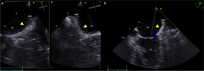
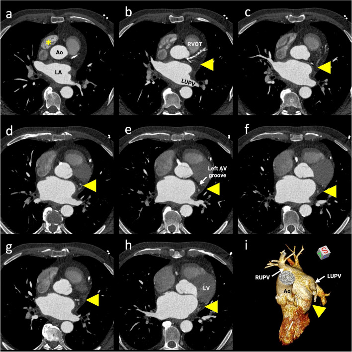
A 65-year-old man with persistent atrial fibrillation was referred to our institute for catheter ablation treatment. Initial three-dimensional transesophageal echocardiogram using multiplane imaging failed to detect a patent left atrial appendage (LAA), and there was no Doppler flow detected from the usual LAA location (Fig. 1). Cardiac contrast-enhanced computed tomography (CCT) confirmed the congenital absence of LAA (Fig. 2). Pulmonary vein isolation was successfully achieved without complications, and anticoagulation was managed based on the CHA2DS2-VASc score.
Fig. 1.
Initial three-dimensional (3D) transesophageal echocardiogram (TOE). A Magnified multiplane 3D TOE did not detect a patent left atrial appendage in its normal position (arrowheads). B Color Doppler unable to detect flow from the usual left atrial appendage location (arrowhead). LA, left atrium; LOM, ligament of Marshall; LV, left ventricle
Fig. 2.
Cardiac contrast-enhanced computed tomography (CCT) images. A–H CCT axial-plane images cranial to caudal. Arrowheads show absence of the left atrial appendage in its normal position (arising from the anterolateral wall of the left atrium [LA] at the level of the left upper pulmonary vein [LUPV] and typically extending anteriorly, overlapping the left border of the pulmonary trunk and the circumflex artery). Asterisks represent the normally developed right atrial appendage. I Three-dimensional volume-rendered reconstruction of a left chamber comprehensively demonstrates the absence of the left atrial appendage (arrowhead). Ao, aorta; RVOT, right ventricular outflow tract; AV, atrioventricular; LV, left ventricle; RUPV, right upper pulmonary vein
Discussion
While transesophageal echocardiogram remains the gold standard for identifying cardiac embolic sources, CCT can provide highly accurate imaging of the LAA if the appropriate scan algorithm, including a late pass scan, is employed [1]. Absence of the LAA is a rare finding, often incidentally discovered during preprocedural thrombus evaluation. In patients without a history of open-heart surgery, advanced imaging with CCT is crucial to distinguish between LAA absence and total thrombotic occlusion of the LAA. Decisions regarding anticoagulation in patients with LAA absence can be challenging due to limited evidence and the use of heterogeneous strategies [2].
Abbreviations
- CCT
Contrast-enhanced computed tomography
- LAA
Left atrial appendage
Authors’ contributions
FM, CV, and VHP contributed to the manuscript conception. The first draft of the manuscript was written by FM, and all authors reviewed and commented on previous versions of the manuscript. All authors read and approved the final manuscript.
Funding
None.
Availability of data and materials
Not applicable.
Declarations
Ethics approval and consent to participate
Not applicable.
Consent for publication
Not applicable.
Competing interests
The authors declare that they have no competing interests.
Footnotes
Publisher’s Note
Springer Nature remains neutral with regard to jurisdictional claims in published maps and institutional affiliations.
References
- 1.Donal E, Lip GY, Galderisi M, Goette A, Shah D, Marwan M, et al. EACVI/EHRA Expert Consensus Document on the role of multi-modality imaging for the evaluation of patients with atrial fibrillation. Eur Heart J Cardiovasc Imaging. 2016;17:355–383. doi: 10.1093/ehjci/jev354. [DOI] [PubMed] [Google Scholar]
- 2.Arguelles E, Mihalatos D, Leung A, Colangelo RG, Jayam V, Fujikura K. Congenital absence of the left atrial appendage: role of multimodality imaging. CASE (Phila) 2023;7:220–225. doi: 10.1016/j.case.2023.01.003. [DOI] [PMC free article] [PubMed] [Google Scholar]
Associated Data
This section collects any data citations, data availability statements, or supplementary materials included in this article.
Data Availability Statement
Not applicable.


