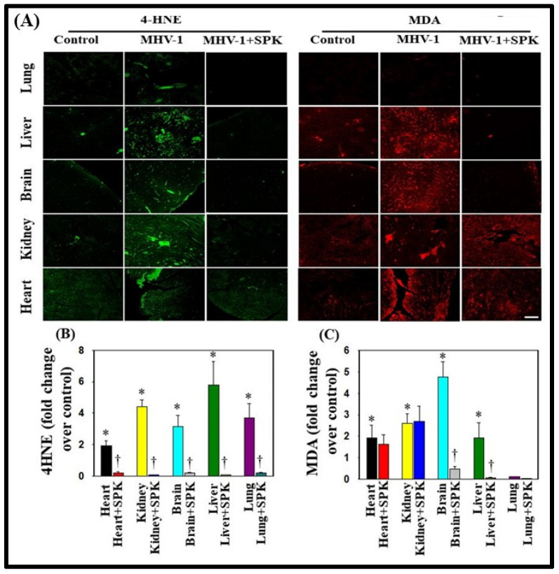
Figure 9.
Oxidative stress post-MHV-1 infection in mice. (A) Representative immunofluorescence images from four individual animals show increased levels of 4-hydroxynonenol (4-HNE) and malondialdehyde (MDA) in the lung, liver, kidney, brain, and heart. Treatment of MHV-1-inoculated mice with SPK (5 mg/kg) prevented such an increase. (B,C) Quantitation of 4-HNE and MDA levels with and without SPK post-MHV-1 infection. ANOVA, n = 4. * = p < 0.05 versus control; † = p < 0.05 verses MHV-1-infected mice. Scale bar = 25 μm. Error bars represent mean ± SEM [13].

