Figure 4.
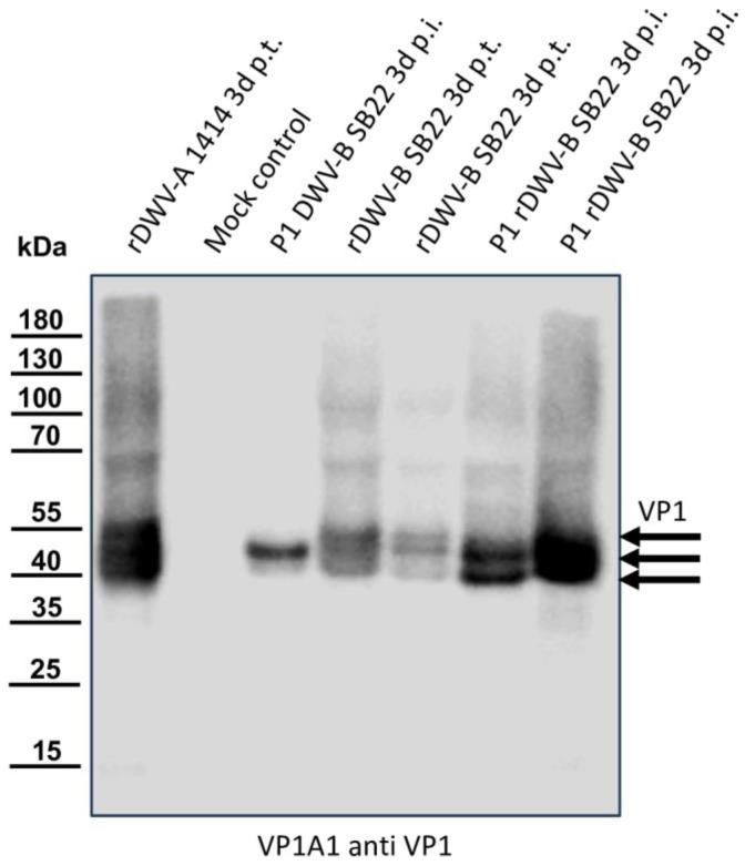
Western blot analyses of bee pupae transfected or infected with the DWV-B clone. Honey bee pupae were either transfected with the in vitro-transcribed RNA of DWV strain 1414 (rDWV-A 1414 3d p.t.) or mock-transfected (Mock control). One pupa was injected with 1 µL of a DWV-B-positive bee sample (P1 DWV-B SB22 3d p.i.), and two pupae were transfected with the in vitro-transcribed RNA of rDWV-B Austria-SB22 (rDWV-B SB22 3d p.t.). Subsequently, two naïve pupae were injected with 1 µL of lysate from rDWV-B-transfected pupae (P1 rDWV-B SB22 3d p.i.). Total bee protein was resolved via SDS-PAGE, blotted, and analyzed using a VP1-specific antibody. In the rDWV-A-transfected pupa, a typical VP1 pattern with bands at 47, 42, and 39 kDa was observed, whereas the mock-transfected pupa showed no signals. The pupa inoculated with the original DWV-B SB22 material displayed a strong protein band at 42 kDa and a faint band at 39 kDa. The two bee pupae transfected with in vitro-transcribed RNA displayed protein bands at 47, 42, and 39 kDa, similar to rDWV-A but less intense. However, pupae infected with lysates from these transfected animals exhibited very strong bands at 43 and 39 kDa, indicating further spread and growth of rDWV-B in the first passage. Arrows indicate the protein bands of the characterized target proteins of the antibody used.

