Abstract
The SD0 mutant of influenza virus A/WSN/33 (WSN), characterized by a 24-amino-acid deletion in the neuraminidase (NA) stalk, does not grow in embryonated chicken eggs because of defective NA function. Continuous passage of SD0 in eggs yielded 10 independent clones that replicated efficiently. Characterization of these egg-adapted viruses showed that five of the viruses contained insertions in the NA gene from the PB1, PB2, or NP gene, in the region linking the transmembrane and catalytic head domains, demonstrating that recombination of influenza viral RNA segments occurs relatively frequently. The other five viruses did not contain insertions in this region but displayed decreased binding affinity toward sialylglycoconjugates, compared with the binding properties of the parental virus. Sequence analysis of one of the latter viruses revealed mutations in the hemagglutinin (HA) gene, at sites in close proximity to the sialic acid receptor-binding pocket. These mutations appear to compensate for reduced NA function due to stalk deletions. Thus, balanced HA-NA functions are necessary for efficient influenza virus replication.
Influenza A viruses contain eight segments of negative-sense, single-stranded RNA (reviewed in reference 16). Each RNA segment encodes at least one protein, and two of these proteins, hemagglutinin (HA) and neuraminidase (NA), project through the viral envelope and are available for interactions with cellular molecules. The abundance of each protein varies among virus subtypes, with the HA-NA ratio of influenza virus A/WSN/33 (H1N1) being approximately 10 to 1 (21). Since HA and NA recognize the same molecule (sialic acid) with conflicting activities, it can be assumed that drastic changes in either activity would affect viral replication.
The HA, a type I integral membrane glycoprotein, is cleaved into two disulfide-linked chains, HA1 and HA2, by host proteases. Such cleavage is critical for viral infectivity, because it exposes the membrane fusion peptide located at the amino terminus of the HA2 subunit (reviewed in reference 14). The HA functions as a homotrimer of noncovalently linked monomers and plays two major roles during the replication of influenza A virus in host cells. First, it attaches the virus to the cell surface by binding to sialic-acid-containing receptors and promotes viral penetration by mediating fusion of the endosomal and viral membranes. The conserved sialic acid receptor-binding pocket, located on the HA1 subunit at the distal end of the molecule, binds to monovalent sialic acid receptor analogs with relatively low affinity (dissociation constant, approximately 0.1 to 1 mM [11]); however, the high abundance of HA molecules on the virion surface permits a sufficient number of low-affinity interactions to allow virus attachment and entry into host cells.
The NA molecule, a type II integral membrane glycoprotein (7, 28), consists of a box-like catalytic head, a centrally attached stalk with a hydrophobic transmembrane-spanning region that attaches the molecule to the plasma and viral membranes, and a cytoplasmic tail of six amino acids (1). The NA functions as a homotetramer, facilitating the mobility of virions by removing sialic acid residues from viral glycoproteins and infected cells during both entry and release from cells (1, 15, 25). Many studies have documented that influenza virus particles with low NA enzymatic activity cannot be efficiently released from infected cells, resulting in the accumulation of large aggregates of progeny virions on the cell surface (17, 21, 25). Since the formation of aggregates results directly from HA binding to sialic acid receptors on cellular and viral surfaces, a balance of competent HA and NA activities appears critical. In brief, there should be enough HA activity to ensure virus binding and enough NA activity to ensure the release of progeny virus.
Observing how viruses adapt to restricted conditions in the host can provide important information on the requirements for efficient viral replication. In most instances, influenza viruses overcome barriers to replication by one of three mechanisms: (i) genetic drift (mutations due to errors introduced by viral polymerase), (ii) genetic shift (reassortment of RNA segments between two different viruses), and (iii) RNA-RNA recombination (exchange of genetic information between RNA segments). Although rarely observed in nature, examples of RNA-RNA recombination of influenza A viruses have been documented in the laboratory. Khatchikian et al. (12) discovered 54 nucleotides of the cellular 28S rRNA inserted into the HA1/HA2 cleavage site, while Orlich et al. (24) found 60 nucleotides of the nucleoprotein (NP) gene within the HA1/HA2 cleavage site. In each case, the resultant virus had acquired the necessary alteration for efficient replication in the restrictive host. Moreover, mutant viruses have been generated by introducing foreign nucleotides into the NA gene during ribonucleoprotein transfection experiments (2).
Castrucci et al. (6) and Luo et al. (18) investigated the biologic importance of the NA stalk by generating WSN viruses with a complete deletion of the NA stalk region. The virus made by Castrucci et al. (6), designated SD0, grew to the same titer as wild-type virus on cultured Madin-Darby canine kidney (MDCK) cells; however, it did not grow in embryonated chicken eggs. In these studies, the length of the NA stalk correlated with growth of the virus in eggs; also, the longer the NA stalk, the more active NA was in eluting virions from chicken erythrocytes.
To understand the molecular mechanism by which an influenza virus compensates for a defect in NA function necessary for growth, we passaged SD0 virus until it efficiently grew in chicken eggs and identified the molecular events that occurred during its adaptation.
MATERIALS AND METHODS
Viruses and cells.
Influenza virus A/WSN/33 (H1N1) (WSN) was obtained from the repository at St. Jude Children's Research Hospital, Memphis, Tenn. SD0 virus, which contains a 24-amino-acid deletion of the NA stalk region, was generated and characterized previously (6). Madin-Darby bovine kidney (MDBK) cells were maintained in Eagle's minimal essential medium in the presence of 10% fetal calf serum. MDCK cells were maintained in Eagle's minimal essential medium in the presence of 5% newborn calf serum.
Passage of SD0 virus in embryonated chicken eggs.
Approximately 107.3 PFU of MDCK cell-grown SD0 virus (1 ml) was injected into 10-day-old embryonated chicken eggs (done in duplicate). After incubation at 35°C for 2 days, the egg allantoic fluid was harvested, clarified, and tested for viral growth by hemagglutination assays at room temperature, by using 0.5% turkey red blood cells (tRBCs). Virus was serially passaged by inoculating eggs with undiluted allantoic fluid until hemagglutination was observed, at which time the allantoic fluid (100 μl) was injected into eggs in limiting dilutions (usually 10−1 to 10−5). To biologically clone the egg-adapted viruses, we plaque assayed the fluid sample that produced hemagglutination and then picked five separate plaques. Each plaque was injected into eggs, and hemagglutination assays were performed to confirm viral growth in eggs. The HA and NA genes of the egg-adapted viruses (plaques that resulted in hemagglutination) were sequenced and then grown in MDCK cells to make stock viruses for further characterization.
NA and HA sequence analysis.
Viral genes from egg- and MDCK cell-grown viruses were sequenced after the isolation of viral RNA and cDNA, with 20 U of avian myeloblastosis virus reverse transcriptase (Life Sciences, Inc.) and 1 μg of Uni12 primer (5′AGCGAAAGCAGG3′, corresponding to viral noncoding nucleotides 1 to 12 [15]). The HA and NA genes were then amplified by PCR by using 2.5 U of cloned Pfu polymerase (Stratagene) and were sequenced with specific HA and NA primers. The viral genes were sequenced by TaqFS Dye Terminator Chemistry.
PCR verification of NP insertion.
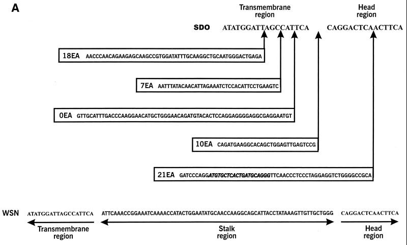
To determine the egg passage at which NP sequences were inserted into the NA stalk region, we designed a specific NP reverse primer (NPinsert) corresponding to NP nucleotides 532 to 551 (5′TACACGAGTGACTACGTCCC3′), representing the NP (antisense) sequences inserted into the NA stalk region (Fig. 1A, italic sequences). A specific NA forward primer (N1W26), corresponding to the coding nucleotides 26 to 46 (5′CCATTGGGTCAATCTGTATGG3′), and NPinsert were then used in PCRs (Pfu polymerase; Stratagene). Viral RNA was isolated from aliquots of each egg passage and was used as a template for cDNA synthesis. To confirm RNA isolation and cDNA synthesis, we used a specific NA reverse primer (N1R836), corresponding to nucleotides 836 to 817 (5′TCACTTTGCCGGTATCAGGG3′), instead of NPinsert, as a positive control.
FIG. 1.
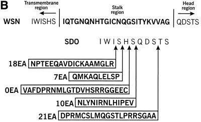
(A) Nucleic acid sequences and the locations of inserts in the NAs of five SD0 egg-adapted strains. The proposed transmembrane and catalytic head domains of the NA are indicated. Inserted sequences are identical to the PB1, PB2, and NP gene segments of WSN virus. The 21EA strain contains NP nucleotides 523 to 586; 18EA contains PB2 nucleotides 928 to 981; 10EA contains PB1 nucleotides 1814 to 1851; 7EA contains PB1 nucleotides 2063 to 2092; and 0EA contains PB2 nucleotides 720 to 784. Bold italic sequences in 21EA indicate the NP-specific primer (primer NPinsert) synthesized for PCR detection (see Table 2). (B) Deduced amino acid sequence and locations of inserted amino acids in five SD0 egg-adapted strains.
Glycoprotein incorporation into virions.
MDBK cells were infected with wild-type or egg-adapted virus, and 4 h later, were starved of glucose for 30 min and were labeled with 0.2 mCi of [3H]mannose (Amersham) for 18 h. Virus in the culture supernatant was purified by centrifugation (1 h) at 130,000 × g through 30% sucrose. The virus pellet was then disrupted with lysis buffer (50 mM Tris-HCl [pH 7.5], 600 mM KCl, and 0.5% Triton X-100). Viral proteins were analyzed by sodium dodecyl sulfate–10% polyacrylamide gel electrophoresis (13).
Hemagglutination test.
Hemagglutination activity was determined in microtiter plates by using 0.5% tRBCs. The reactions were performed in phosphate-buffered saline (PBS), either on ice or at room temperature (approximately 20°C). To avoid possible destruction of the sialyloligosaccharide receptors, a 2 μM concentration of the NA inhibitor zanamivir (2,3-didehydro-2,4-dideoxy-4-guanidino-N-acetyl-d-neuraminic acid, GG167 [29]), kindly provided by R. Bethell (Research and Development, Glaxo Wellcome), was included in the reaction mixture.
Assay of virus binding of sialylglycopolymers.
For the receptor-binding assay, virus in culture fluids was partially purified by removing cellular debris by low-speed centrifugation prior to pelleting by high-speed centrifugation. The pelleted viruses were suspended in 50% glycerol–0.1 M Tris buffer, pH 7.3, and were stored at −20°C. The general procedure of this solid-phase receptor-binding assay was described previously (9). The modifications applied in this study included the use of synthetic sialylglycopolymers and a biotin-streptavidin detection system. Monospecific biotinylated sialylglycopolymers bearing pendant Neu5Ac(α2-3)Gal(β1-4)Glc residues (3′SL-PAA) or Neu5Ac(α2-6)Gal(β1-4)GlcNAc residues (6′SLN-PAA) were synthesized as previously described (4). Polyvinyl chloride EIA microplates (Costar) were coated with 5 μg of bovine fetuin per ml in PBS (50 μl/well) overnight and were washed with distilled water. The viruses, diluted in PBS to an HA titer of 1/32 to 1/64, were incubated in the wells of fetuin-coated plates (40 μl/well) at 4°C overnight; then the wells were washed with an ice-cold 0.2× PBS–0.01% Tween 80 solution washing buffer. Serial twofold dilutions of sialylglycopolymer in the reaction buffer (0.02% bovine serum albumin, 0.01% Tween 80, 1 μM zanamivir in PBS) were added to the wells (20 μl/well), followed by 2 h of incubation at 4°C. After five washings with washing buffer, 25 μl of streptavidin-peroxidase conjugates (ICN Biomedicals, Inc.), diluted 1/2,000 in reaction buffer, was added to each well, followed by 1 h of incubation at 4°C.
The plates were washed, and the peroxidase activities in the wells were determined with o-phenylenediamine, which was used as a chromogenic substrate. The absorbency data were converted to Scatchard plots graphing A492 per degree centigrade versus A492. Affinity values (Kaff), formally equivalent to dissociation constants of the virus-sialylglycopolymer complexes, were obtained by regression analysis of these plots (9). The absolute Kaff varied in replicate experiments performed on different days, but the relative affinities of the variants were highly reproducible, permitting the use of averaged data for this variable.
RESULTS
Adaptation of SD0 to embryonated chicken eggs.
To determine the molecular requirements for efficient growth of SD0 in eggs, we serially passaged the virus in eggs (10 independent lines), as outlined in Materials and Methods. tRBCs were used because they are more sensitive than chicken red blood cells to hemagglutination by virus (unpublished data). Ten viruses capable of hemagglutination activity were generated after 8 to 12 passages (Table 1), suggesting that the SD0 virus requires several mutations to grow efficiently in eggs. The hemagglutination titers varied among the adapted viruses: some had a hemagglutination titer of only 1:2, while others had titers as high as 1:64. There was no apparent relationship between the hemagglutination titer and passage number (i.e., viruses that became hemagglutination positive at later passages did not always produce higher titers than viruses that became hemagglutination positive earlier).
TABLE 1.
Properties of egg-adapted SD0 strainsa
| Virus | Egg passage at which positive hemagglutination was detected | NA stalk insertion
|
|
|---|---|---|---|
| No. of amino acids inserted | Origin of gene insertion | ||
| 0EA | 12 | 22 | PB2 |
| 7EA | 12 | 10 | PB2 |
| 10EA | 10 | 13 | PB1 |
| 18EA | 8 | 18 | PB2 |
| 21EA | 10 | 20 | NP |
| 2EA | 12 | 0 | NAb |
| 5EA | 8 | 0 | NA |
| 14EA | 12 | 0 | NA |
| 19EA | 12 | 0 | NA |
| 25EA | 10 | 0 | NA |
Approximately 107.3 PFU of SD0 virus (1 ml) were injected into 10-day-old embryonated chicken eggs. After 2 days of incubation at 35°C, egg allantoic fluid was harvested and tested for viral growth by hemagglutination assays. Viruses were serially passaged by inoculating the eggs with undiluted allantoic fluid until hemagglutination was observed. The passage at which hemagglutination was first observed is shown. Only strains that yielded positive hemagglutination are indicated. The NA stalk insertion was detected by sequencing the NA gene.
NA, not applicable.
Sequence analysis of NA genes.
Castrucci et al. (6) showed that the length of the SD0 NA stalk correlates with improved viral growth in eggs, prompting us to investigate the NA gene for molecular changes. Sequence analysis of cDNAs by reverse transcriptase PCR revealed two types of egg-adapted viruses, one containing nucleotide insertions in the NA gene and the other containing no insertions in this gene (Table 1).
Five viruses (0EA, 7EA, 10EA, 18EA, and 21EA) had insertions between the transmembrane and catalytic head regions of the NA gene (Fig. 1). The insertions originated from three viral gene segments: PB1, PB2, and NP. One virus, 0EA, contained 22 amino acids inserted from the PB2 gene (Fig. 1, nucleotides 720 to 784). Egg-adapted virus 10EA contained sequences from the PB1 gene (Fig. 1A, 13 amino acids, nucleotides 1814 to 1851). Two other PB2 sequences were inserted into the NA gene (Fig. 1): 10 amino acids for 7EA (nucleotides 2063 to 2092) and 18 amino acids for 18EA (nucleotides 928 to 981). In addition, 21EA acquired sequences from the NP gene, resulting in an addition of 21 amino acids (nucleotides 523 to 586) between the NA transmembrane and catalytic head regions.
By computer analysis, the inserted nucleotide and amino acid sequences lacked homology with the wild-type NA stalk sequences (Fig. 1) and with each other. Of the five insertions, three were in the same open reading frames of the respective gene segments (10EA, 18EA, and 21EA), while the remaining two (0EA and 7EA) were not.
PCR detection of NP insertion.
Additional studies focused on 0EA and 21EA, which acquired new NA stalks, and 2EA, which still lacked an NA stalk. To determine the passage number at which the insertion occurred, we tested each passage of 21EA for incorporation of NP sequences by using PCR with a specific primer that corresponded to the inserted NP sequences (Fig. 1A, 21EA, italic boldface sequences) and an NA sequence-specific primer. With this combination, we could identify cDNAs containing the NP insertion by the detection of a 0.8-kb PCR product; the absence of this product indicated that RNA recombination had not yet occurred. Two NA sequence-specific primers were used as a positive control. Viruses from all passages produced a 0.8-kb band in the positive control reaction (Table 2), but only those from the fourth and subsequent passages yielded this product when the alternative primer was used, suggesting that the NP insertion had occurred between passages 3 and 4. The lack of appreciable improvement in viral replication in eggs with the acquisition of new sequences (recombination event), as judged by hemagglutination of virus in allantoic fluid, indicated that other mutations were likely needed to establish productive growth in embryonated eggs.
TABLE 2.
Egg passage during which a new sequence was inserted into the NA stalka
| Assay | Egg passage number
|
|||||||||
|---|---|---|---|---|---|---|---|---|---|---|
| 1 | 2 | 3 | 4 | 5 | 6 | 7 | 8 | 9 | 10 | |
| PCR with: | ||||||||||
| NP primer | − | − | − | + | + | + | + | + | + | + |
| NA primer (positive control) | + | + | + | + | + | + | + | + | + | + |
| Hemagglutination of allantoic fluid | − | − | − | − | − | − | − | − | − | + |
Viral RNA was isolated from each egg passage of the 21EA strain and was used as a template for cDNA synthesis. The NP-specific reverse primer (primer NPinsert), which corresponded to the inserted NP nucleotide sequences (5′TACACGAGTGACTACGTCCC3′ [see Fig. 1A, italic boldface sequences]), was combined with a specific NA forward primer (N1W26) (5′CCATTGGGTCAATCTGTATGG3′) in PCRs with the Pfu polymerase. A specific NA reverse primer (N1R836), corresponding to the NA-coding nucleotides 836 to 817 (5′TCACTTTGCCGGTATCAGGG3′), was included instead of primer NPinsert as a positive control. After 20 cycles, PCR products were separated on 1% agarose gels. Viral hemagglutination activity for each passage was determined by using tRBCs. All passages produced a 0.8-kb band in the positive control reaction.
Incorporation of recombinant NA molecules into virions.
An increased number of NA molecules in virions may compensate for the functional defect in the SD0 NA. To assess the level of incorporation of NA molecules containing newly acquired foreign sequences (new NA stalks), we grew egg-adapted viruses on MDBK cells in the presence of [3H]mannose, followed by purification and analysis by sodium dodecyl sulfate–10% polyacrylamide gel electrophoresis. Each recombinant NA molecule was incorporated to the same extent as the NA of the parental (SD0) virus (data not shown), indicating that insertion of foreign sequences into the NA stalk region does not affect NA incorporation.
Sequence analysis of HA genes.
Influenza A viruses cultivated in the presence of NA inhibitors contain mutations in their HA genes (10, 19). Thus, since some egg-adapted viruses did not contain insertions in the NA, we sequenced and compared the HA genes of recombinant and nonrecombinant viruses. In addition to an insertion in the NA gene, 0EA had two mutations in the HA, one in the HA1 region (asparagine 95 to aspartic acid)(H3 numbering) and one in the HA2 region (asparagine 72 to lysine), while 21EA contained a valine-135-to-isoleucine change in HA1 (Table 3). 2EA lacked an insertion in the NA gene but contained three HA mutations, serine 146 to glycine and arginine 262 to lysine in HA1 and arginine 106 to lysine in HA2.
TABLE 3.
Mutations in the HA gene of egg-adapted virusesa
| Virus | NA insertion | Mutation(s) in:
|
|
|---|---|---|---|
| HA1 | HA2 | ||
| 0EA | YES | 95Asn→Asp | 72Asn→Lys |
| 21EA | YES | 135Val→Ile | |
| 2EA | NO | 146Ser→Gly | 106Arg→Lys |
| 262Arg→Lys | |||
For each adapted strain, HA cDNA was synthesized by reverse transcriptase PCR followed by cDNA sequencing as described in Materials and Methods.
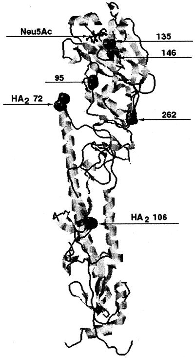
Figure 2 is a schematic diagram of the three-dimensional structure of the HA monomer (30), indicating the location of each mutation in relation to its sialic acid substrate. As shown, the mutations at residues 135HA1 and 146HA1 are in close proximity to the receptor binding site, while the mutation at residue 95HA1 is somewhat farther away, though still in the globular portion of the HA molecule. Potentially, each mutation could alter the characteristics of HA receptor binding and thus the replicative properties of the virus. Mutations at residues 262HA1, 72HA2, and 106HA2 are more distant from the receptor-binding site; hence, a contribution of these changes to receptor-binding activity is unlikely.
FIG. 2.
Three-dimensional structure of the HA monomer (30), indicating the location of each HA mutation discovered in three SD0 egg-adapted strains, relative to sialic acid (Neu5Ac) binding.
HA receptor-binding properties.
One explanation for the above results is that HA mutations can compensate for the loss of NA activity (3, 20). We therefore evaluated the receptor-binding properties of our egg-adapted viruses with two distinct viral binding assays: an HA assay at two different temperatures and a sialylglycopolymer binding assay (Table 4). In both assays, an NA inhibitor, zanamivir, was included in the incubation mixtures to preclude any contribution from viral NA enzymatic activity during the assays. The patterns of hemagglutination at two temperatures indicated that 2EA (which did not acquire an NA stalk insertion) possesses substantially lower receptor-binding activity than do the other adapted viruses (Table 4). Lowering the incubation temperature to 4°C decreased the dissociation rate of 2EA and increased its relative HA titer to more than 30 times that observed at 20°C, consistent with the large reduction in the virus binding affinity to sialylglycopolymers (Table 4). The relative HA titer was the same among the other viruses tested. In contrast to results of the hemagglutination test, we observed several distinctions in the receptor-binding properties of the adapted viruses in assays with specific polymers. In particular, 0EA displayed a slight decrease in affinity for both 3′SL-PAA and 6′SLN-PAA, while 21EA bound less avidly only to 6′SLN-PAA.
TABLE 4.
Comparison of the receptor-binding activities of WSN, SD0, and egg-adapted strains of SD0
| Virus | NA stalk | Relative HA titera | Binding to sialylglycopolymersb
|
|
|---|---|---|---|---|
| 3′SL-PAA | 6′SLN-PAA | |||
| WSN | YES | 1 | 1 | 1 |
| SD0 | NO | 1 | 1.4 | 1.6 |
| 2EA | NO | >32 | 40 | 20 |
| 0EA | YES | 1 | 3.4 | 2.7 |
| 21EA | YES | 1 | 1 | 2.5 |
Ratio of hemagglutinating titers determined at 4 and 20°C (ratio = HA at 4°C/HA at 20°C). A higher ratio indicates a lower affinity for sialic acid substrates.
The relative binding affinities for each sialylglycopolymer were calculated by the formula Krel = Kaff(x)/Kaff(WSN). Higher values of Krel correspond to lower affinities. For each polymer, Kaff of WSN = 1.
In principle, the decrease in affinity shown by 2EA could be explained by incomplete removal of sialic acid from the oligosaccharides on the viral HA and NA (23) due to a deficit in the NA activity of this strain (deletion in the stalk). To pursue this possibility, we treated the viruses with exogenous NA (Vibrio cholerae) and compared the abilities of treated and nontreated viruses to bind sialylglycopolymers. Neither strain showed an increase in binding affinity after NA treatment (data not shown), suggesting that incomplete desialylation of the virus does not account for the low binding affinity of 2EA. The most plausible explanation is that acquired mutations in the HA (S146G and R262K in HA1 and, to a lesser extent, R106K in HA2) specifically contribute to the observed decreased in 2EA binding affinity for sialic-acid-containing receptors.
DISCUSSION
We demonstrate that an influenza A virus with restricted growth in embryonated chicken eggs can acquire replicative competence by either of two distinct mechanisms: restoration of the NA stalk by RNA-RNA recombination or a decrease in viral (HA) binding affinity to sialylglycoconjugates. Both mechanisms of adaptation involve interplay between the NA and HA, two virion glycoproteins needed for viral attachment to and release from host cells. Of 10 adapted strains of SD0 (an A/WSN/33 mutant with a truncated NA stalk) examined, five underwent RNA-RNA recombination involving three separate viral RNA segments (PB1, PB2, and NP). Since the parental virus does not grow efficiently in eggs, we reasoned that insertions into the NA stalk through recombination of RNA segments increased accessibility of the NA enzymatic pocket, and hence the enzyme's receptor-destroying activities. One of the five noninserted adapted strains (2EA) showed a decrease in HA binding affinity for sialic acid substrates, presumably compensating for a defect in functional sialidase activity due to a deletion in the NA stalk, thereby preventing virion aggregation. These findings emphasize the importance of HA-NA interplay in the replicative capacity of influenza A viruses.
Others have reported RNA-RNA recombination during the adaptation of influenza A viruses (2, 12, 24). Our results are consistent with theirs (2, 12, 24) in that the mechanism appears to operate through copy choice, nonhomologous RNA-RNA recombination due to polymerase jumping during viral RNA transcription. Since no consensus motifs have emerged from the inserted sequences, the mechanism does not appear to be specific. It appears unlikely that the RNA-RNA recombination frequency is inherently greater in the NA stalk region; rather, the insertions seem to be selected by the growth restriction (low viral NA function) imposed on the virus in embryonated eggs. Indeed, we suggest that any gene segment could participate in recombination with any sequence inserted into the NA stalk; thus, RNA-RNA recombination during influenza A virus replication may occur more often than previously thought. In this study, we only examined the NA and HA genes because the initial virus used for egg adaptation was defective in NA activity. Whether mutations (such as mutator mutations [26, 27]) occurred in the polymerase genes of these strains, which might have enhanced the recombination frequency, is unknown. Also, involvement of other viral gene products in the adaptation process is unknown, since sequence analysis was not performed for all gene segments.
Castrucci et al. (6) have shown that the longer the NA, the better the virus replicates in eggs. Our findings substantiate this observation and additionally show that although the NA stalk presumably contains 24 amino acids, a minimum of 10 in this region is sufficient for growth adaptation in eggs. Although new sequences were acquired early in the adaptation process, at passage 4 the resulting mutant did not begin to replicate efficiently until several passages later. This suggests that insertions in the NA gene are not by themselves sufficient for adaptation and that additional mutations are needed for full conversion, as discussed below.
All egg-adapted viruses, and 2EA in particular, displayed changes in their receptor-binding properties, regardless of the type of sialic acid linkage displayed by the sialylglycopolymers. Among the three HA mutations we identified (Table 3), the Ser-to-Gly substitution at residue 146 is most likely responsible for the decrease in affinity. 146Ser is conserved among most of the 15 known HA subtypes (22), suggesting that a mutation at this position could alter HA function. In the three-dimensional model of X31 HA, residue 146 lies immediately proximal to residue 136, which participates in van der Waals contacts and hydrogen bond formation with the carboxylic group of sialic acids (30). All of these interactions can contribute significantly to HA binding affinity. Thus, a mutation at residue 146 could decrease the affinity for sialic acid, thereby inhibiting aggregation of viral particles during infection and budding of progeny particles. Of course, any acquired HA mutations must reduce virus affinity for sialic acid without completely abrogating sialic acid binding, which is required for virus entry into the host cell.
It is interesting that HA mutations are also important for replication of zanamivir-resistant variants (10, 19). Resistant mutants selected in the presence of zanamivir contained substitutions in the HA that decreased the molecule's affinity for sialylglycopolymers. These variants emerge during the first stages of adaptation, after which mutations in the NA that decrease enzyme sensitivity to zanamivir emerge and outgrow the parental virus. These results are compatible with ours and further demonstrate the functional relationship between HA and NA activities.
Elucidating the mechanisms of influenza virus adaptation to restricted hosts is essential to understanding the molecular basis for the emergence of new strains, especially those implicated in global outbreaks. If we could understand how viruses adapt to otherwise replication-incompetent hosts, we might be in a position to target antiviral therapies to the adapted strains. Results of the present study promote this aim by demonstrating the critical role of balanced HA-NA activities in efficient replication of influenza A viruses and by identifying the mechanisms operating to achieve this balance of protein activity.
ACKNOWLEDGMENTS
We thank Robert Webster for providing the monoclonal antibodies to the HA and NA of the WSN virus.
This work was supported in part by Public Health Service research grants from the National Institute of Allergy and Infectious Diseases, Cancer Center Support (CORE) grant CA-21765, and the American Lebanese Syrian Associated Charities (ALSAC). This work was also supported by an American Lung Association (Memphis, Tenn., chapter) research grant to L.J.M. M.N.M. was supported by a Karnofsky fellowship from St. Jude's Children's Research Hospital.
REFERENCES
- 1.Air G M, Laver W G. The neuraminidase of influenza virus. Proteins Struct Funct Genet. 1989;6:341–356. doi: 10.1002/prot.340060402. [DOI] [PubMed] [Google Scholar]
- 2.Bergmann M, Garcia-Sastre A, Palese P. Transfection-mediated recombination of influenza A virus. J Virol. 1992;66:7576–7580. doi: 10.1128/jvi.66.12.7576-7580.1992. [DOI] [PMC free article] [PubMed] [Google Scholar]
- 3.Blick T J, Sahasrabudhe A, McDonald M, Owens I J, Morley P J, Fenton R J, McKimm-Breschkin J L. The interaction of neuraminidase and hemagglutinin mutations in influenza virus resistance to 4-guanidino-Neu5Ac2en. Virology. 1998;246:95–103. doi: 10.1006/viro.1998.9194. [DOI] [PubMed] [Google Scholar]
- 4.Bovin N V, Korchagina E Y, Zemlyanukhina T V, Byramova N E, Galanina O E, Zemlyakov A E, Ivanov A E, Zubov V P, Mochalova L V. Synthesis of polymeric neoglycoconjugates based on N-substituted polyacrylamides. Glycoconj J. 1993;10:142–151. doi: 10.1007/BF00737711. [DOI] [PubMed] [Google Scholar]
- 5.Castrucci M R, Bilsel P, Kawaoka Y. Attenuation of influenza A virus by insertion of a foreign epitope into the neuraminidase. J Virol. 1992;66:4647–4653. doi: 10.1128/jvi.66.8.4647-4653.1992. [DOI] [PMC free article] [PubMed] [Google Scholar]
- 6.Castrucci M R, Kawaoka Y. Biologic importance of neuraminidase stalk length in influenza A virus. J Virol. 1993;67:759–764. doi: 10.1128/jvi.67.2.759-764.1993. [DOI] [PMC free article] [PubMed] [Google Scholar]
- 7.Colman P M, Laver W G, Varghese J N, Baker A T, Tulloch P A, Air G M, Webster R G. The three-dimensional structure of a complex of influenza virus neuraminidase and an antibody. Nature (London) 1987;326:358–363. doi: 10.1038/326358a0. [DOI] [PubMed] [Google Scholar]
- 8.Crecelius D M, Deom C M, Schulze I T. Biological properties of a hemagglutinin mutant of influenza virus selected by host cells. Virology. 1984;139:164–177. doi: 10.1016/0042-6822(84)90337-4. [DOI] [PubMed] [Google Scholar]
- 9.Gambaryan A S, Matrosovich M N. A solid-phase enzyme-linked assay for influenza virus receptor-binding activity. J Virol Methods. 1992;39:111–123. doi: 10.1016/0166-0934(92)90130-6. [DOI] [PubMed] [Google Scholar]
- 10.Gubareva L V, Bethell R C, Hart G J, Murti K G, Webster R G. Characterization of mutants of influenza A virus selected with the neuraminidase inhibitor 4-guanidino-Neu5Ac2en. J Virol. 1996;70:1818–1827. doi: 10.1128/jvi.70.3.1818-1827.1996. [DOI] [PMC free article] [PubMed] [Google Scholar]
- 11.Hanson J E, Sauter N K, Skehel J J, Wiley D C. Proton nuclear magnetic resonance studies of the binding of sialosides to intact influenza virus. Virology. 1992;189:525–533. doi: 10.1016/0042-6822(92)90576-b. [DOI] [PubMed] [Google Scholar]
- 12.Khatchikian D, Orlich M, Rott R. Increased viral pathogenicity after insertion of a 28S ribosomal RNA sequence into the haemagglutinin gene of an influenza virus. Nature (London) 1989;340:156–157. doi: 10.1038/340156a0. [DOI] [PubMed] [Google Scholar]
- 13.Laemmli U K. Cleavage of structural proteins during the assembly of the head of bacteriophage T4. Nature (London) 1970;227:680–685. doi: 10.1038/227680a0. [DOI] [PubMed] [Google Scholar]
- 14.Lamb R. The influenza virus RNA segments and their encoded proteins. In: Palese P, Kingsbury D W, editors. Genetics of influenza viruses. Vienna, Austria: Springer-Verlag; 1983. pp. 26–69. [Google Scholar]
- 15.Lamb R A, Choppin P W. The gene structure and replication of influenza virus. Annu Rev Biochem. 1983;52:467–506. doi: 10.1146/annurev.bi.52.070183.002343. [DOI] [PubMed] [Google Scholar]
- 16.Lamb R A, Krug R M. Orthomyxoviridae: the viruses and their replication. In: Fields B N, Knipe D M, Howley P M, editors. Fields virology. 3rd ed. Philadelphia, Pa: Lippincott-Raven Publishers; 1996. pp. 1353–1445. [Google Scholar]
- 17.Liu C, Eichelberger M C, Compans R W, Air G M. Influenza type A virus neuraminidase does not play a role in virus entry, replication, assembly, or budding. J Virol. 1995;69:1099–1106. doi: 10.1128/jvi.69.2.1099-1106.1995. [DOI] [PMC free article] [PubMed] [Google Scholar]
- 18.Luo G, Chung J, Palese P. Alterations of the stalk of the influenza virus neuraminidase: deletions and insertions. Virus Res. 1993;29:141–153. doi: 10.1016/0168-1702(93)90055-r. [DOI] [PubMed] [Google Scholar]
- 19.McKimm-Breschkin J L, Blick T J, Sahasrabudhe A, Tiong T, Marshall D, Hart G J, Bethell R C, Penn C R. Generation and characterization of variants of NWS/G70C influenza virus after in vitro passage in 4-amino-Neu5Ac2en and 4-guanidino-Neu5Ac2en. Antimicrob Agents Chemother. 1996;40:40–46. doi: 10.1128/aac.40.1.40. [DOI] [PMC free article] [PubMed] [Google Scholar]
- 20.McKimm-Breschkin J L, Sahasrabudhe A, Blick T J, McDonlad M, Colman P M, Grahm J H, Bethell R C, Varghese J N. Mutations in a conserved residue in the influenza virus neuraminidase active site decreases sensitivity to Neu5Ac2en-derived inhibitors. J Virol. 1998;72:2456–2462. doi: 10.1128/jvi.72.3.2456-2462.1998. [DOI] [PMC free article] [PubMed] [Google Scholar]
- 21.Mitnaul L, Castrucci M R, Murti K G, Kawaoka Y. The cytoplasmic tail of influenza neuraminidase (NA) affects NA incorporation into virions, virion morphology, and virulence in mice but is not essential for virus replication. J Virol. 1996;70:873–879. doi: 10.1128/jvi.70.2.873-879.1996. [DOI] [PMC free article] [PubMed] [Google Scholar]
- 22.Nobusawa E, Aoyama T, Kato H, Suzuki Y, Tateno Y, Nakajima K. Comparison of complete amino acid sequences and receptor-binding properties among 13 serotypes of hemagglutinins of influenza A viruses. Virology. 1991;182:475–485. doi: 10.1016/0042-6822(91)90588-3. [DOI] [PubMed] [Google Scholar]
- 23.Ohuchi M, Feldmann A, Ohuchi R, Klenk H D. Neuraminidase is essential for fowl plague virus hemagglutinin to show hemagglutinating activity. Virology. 1995;212:77–83. doi: 10.1006/viro.1995.1455. [DOI] [PubMed] [Google Scholar]
- 24.Orlich M, Gottwald H, Rott R. Nonhomologous recombination between the hemagglutinin gene and the nucleoprotein gene of an influenza virus. Virology. 1994;204:462–465. doi: 10.1006/viro.1994.1555. [DOI] [PubMed] [Google Scholar]
- 25.Palese P, Tobita K, Ueda M, Compans R W. Characterization of temperature sensitive influenza virus mutants defective in neuraminidase. Virology. 1974;61:397–410. doi: 10.1016/0042-6822(74)90276-1. [DOI] [PubMed] [Google Scholar]
- 26.Scholtissek C, Ludwig S, Fitch W M. Analysis of influenza A virus nucleoproteins for the assessment of molecular genetic mechanisms leading to new phylogenetic virus lineages. Arch Virol. 1993;131:237–250. doi: 10.1007/BF01378629. [DOI] [PubMed] [Google Scholar]
- 27.Suárez P, Valcárcel J, Ortín J. Heterogeneity of the mutation rates of influenza A viruses: isolation of mutator mutants. J Virol. 1992;66:2491–2494. doi: 10.1128/jvi.66.4.2491-2494.1992. [DOI] [PMC free article] [PubMed] [Google Scholar]
- 28.Varghese J N, Laver W G, Colman P M. Structure of the influenza virus glycoprotein antigen neuraminidase at 2.9Å resolution. Nature. 1983;303:35–40. doi: 10.1038/303035a0. [DOI] [PubMed] [Google Scholar]
- 29.von Itzstein M, Wu W-Y, Kok G B, Pegg M S, Dyason J C, Jin B, Van Phan T, Smythe M L, White H F, Oliver S W, Colman P M, Varghese J N, Ryan D M, Woods J M, Bethell R C, Hotham V J, Cameron J M, Penn C R. Rational design of potent sialidase-based inhibitors of influenza virus replication. Nature. 1993;363:418–423. doi: 10.1038/363418a0. [DOI] [PubMed] [Google Scholar]
- 30.Weis W, Brown J H, Cusack S, Paulson J C, Skehel J J, Wiley D C. Structure of the influenza virus hemagglutinin complexed with its receptor, sialic acid. Nature. 1988;333:426–431. doi: 10.1038/333426a0. [DOI] [PubMed] [Google Scholar]


