Abstract
Unique to pestiviruses, the N-terminal protein encoded by the bovine viral diarrhea virus (BVDV) genome is a cysteine protease (Npro) responsible for a self-cleavage that releases the N terminus of the core protein (C). This unique protease is dispensable for viral replication, and its coding region can be replaced by a ubiquitin gene directly fused in frame to the core. To develop an antiviral assay that allows the assessment of anti-hepatitis C virus (HCV) NS3 protease inhibitors, a chimeric BVDV in which the coding region of Npro was replaced by that of an NS4A cofactor-tethered HCV NS3 protease domain was generated. This cofactor-tethered HCV protease domain was linked in frame to the core protein of BVDV through an HCV NS5A-NS5B junction site and mimicked the proteolytic function of Npro in the release of BVDV core for capsid assembly. A similar chimeric construct was built with an inactive HCV NS3 protease to serve as a control. Genomic RNA transcripts derived from both chimeric clones, PH/B (wild-type HCV NS3 protease) and PH/B(S139A) (mutant HCV NS3 protease) were then transfected into bovine cells (MDBK). Only the RNA transcripts from the PH/B clone yielded viable viruses, whereas the mutant clone, PH/B(S139A), failed to produce any signs of infection, suggesting that the unprocessed fusion protein rendered the BVDV core protein defective in capsid assembly. Like the wild-type BVDV (NADL), the chimeric virus was cytopathic and formed plaques on the cell monolayer. Sequence and biochemical analyses confirmed the identity of the chimeric virus and further revealed variant viruses due to growth adaptation. Growth analysis revealed comparable replication kinetics between the wild-type and the chimeric BVDVs. Finally, to assess the genetic stability of the chimeric virus, an Npro-null BVDV (BVDV−Npro in which the entire Npro coding region was deleted) was produced. Although cytopathic, BVDV−Npro was highly defective in viral replication and growth, a finding consistent with the observed stability of the chimeric virus after serial passages.
The Flaviviridae family currently comprises three genera of single-stranded positive-sense RNA viruses: flaviviruses, pestiviruses, and hepaciviruses (36). Bovine viral diarrhea virus (BVDV) is a prototype virus in the genus Pestivirus, which also includes Classical swine fever virus (CSFV) and Border disease virus. The RNA genome of BVDV is one of the largest (12.5 kb) among members of the Flaviviridae family (8). Similar to hepatitis C virus (HCV), it consists of a long 5′ untranslated region (UTR) which contains an internal ribosomal entry site (IRES) for the translation of viral proteins (6, 15, 35). The single large open reading frame encodes a polyprotein of approximately 3,900 amino acids (8, 29) that is processed into at least 12 functional proteins (Npro-C-Erns-E1-E2/p7-NS2-NS3-NS4A-NS4B-NS5A-NS5B) by both host and viral proteases (10, 36, 49). The first virally encoded protein is a unique protease (Npro for N-terminal protease), responsible for the cleavage between Npro and the core protein (C) (38, 41). A study by Rümenapf et al. showed that Npro is a novel type of cysteine proteinase which required cysteine69 for proteolytic activity (38). Interestingly, partial and complete replacement of the Npro protein by a ubiquitin or fusion with a chloramphenicol acetyltransferase in pestivirus genomes had been shown to produce viable viruses (32, 45). The resulting chimeric viruses were demonstrated to have growth properties similar to the wild-type viruses.
As one of the most characterized members of the Flaviviridae family, BVDV provides a good model system for HCV, a major etiologic agent for non-A non-B hepatitis (1, 7). It shares many important features with HCV. Both viruses utilize an IRES within the 5′ UTR, for the translation of the viral polyprotein (6, 15, 35). Furthermore, the viral NS3 proteases of both viruses require NS4A as a cofactor for polyprotein processing (11, 25, 42). The cytopathic and plaque-forming properties of BVDV in cell cultures allow rapid and quantitative analysis of viral replication and growth. The availability of infectious clones (28, 46, 49) provides opportunities for genetic manipulation to alter viral functions and to construct chimeric viruses. Indeed, a recent report by Frolov et al. found that the entire BVDV IRES could be replaced by HCV IRES. The resulting chimeric viruses relied on the HCV IRES for growth (15), which should allow the in vitro efficacy evaluation of HCV IRES inhibitors.
HCV infection is prevalent and a major global health issue. A recently completed population-based survey revealed that in the United States alone the overall prevalence of anti-HCV was 1.8%, corresponding to an estimated 3.9 million individuals infected by HCV nationwide. A total of 74% of these seropositive individuals tested positive for HCV RNA, indicating that an estimated 2.7 million persons were chronically infected (2). Currently, the combination of alpha 2b interferon and ribavirin (Rebetron; Schering Plough, Kenilworth, N.J.) has been shown to have clinical efficacy in only a proportion (<50%) of patients with chronic HCV infection (9, 26). Vaccine development has been hampered by the high immune evasion rate with poor or no protection against reinfection with a heterologous or homologous inoculum in chimpanzees (12, 40, 48). Development of small molecule inhibitors directed against specific viral targets has thus become the major focus of anti-HCV drug development.
Extensive characterization of the HCV NS3 serine protease (3, 11, 16, 19, 21, 24) has shed light in developing assays and identifying inhibitors of HCV. Major advances in the determination of crystal structures for NS3 protease have begun to delineate important features for the development of potent and specific anti-HCV inhibitors (23, 50, 51). Many high-throughput enzyme-based screening assays, targeting HCV NS3 serine protease, have been developed. Further development of potential inhibitors has to rely on a convenient and reliable cell-based assay system to demonstrate their antiviral efficacy. The lack of a bona fide cell culture system that permits HCV infection makes it a daunting task to evaluate the antiviral efficacy of candidate inhibitors prior to in vivo studies in animals and humans.
Several HCV NS3 protease-dependent chimeric viruses using the genetic backbones of Sindbis virus and poliovirus have been created, providing potential cell-based antiviral assays to evaluate the efficacy of candidate inhibitors against HCV protease (4, 13, 14, 18). Similar schemes were adopted to create these chimeric viruses in which HCV NS3 protease-containing genes were inserted and fused in frame to an essential viral protein through an HCV junction site cleavable by HCV NS3 protease. Failure to cleave the junction by a mutant protease or in the presence of any potent HCV NS3 protease inhibitors would render the unprocessed viral proteins unable to perform their designated functions for viral growth (4, 13, 18). However, genetic stability of such chimeric viruses with foreign gene inserts was a major issue since RNA viruses recombined at a high frequency (29, 47). Indeed, the HCV NS3 genes inserted in the Sindbis viral genome were quickly deleted during initial viral passages and the revertant viruses appeared rapidly and exhibited similar advantageous growth properties as the wild-type viruses (13). Although a second generation of chimeric Sindbis viruses was generated in which a second HCV NS3 cleavage site was created, these viruses were rather defective and unable to replicate at the normal physiological temperature (13). This would limit the development of animal models for in vivo testing of the protease inhibitors.
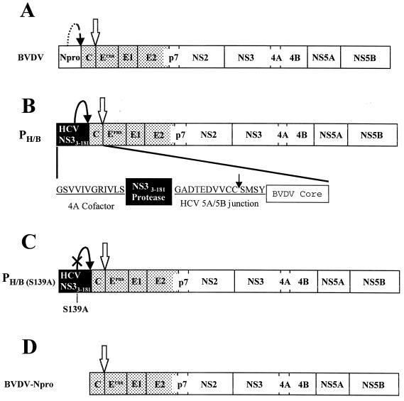
We describe here the generation of a chimeric BVDV in which the Npro coding region is replaced by that of an NS4A cofactor-tethered HCV NS3 protease. This tethered HCV protease domain is fused in frame with the BVDV core protein via an HCV NS5A-NS5B junction site. In this chimeric design, the normal proteolytic function of the Npro is substituted by that of the HCV NS3 serine protease. We demonstrated that viable and cytopathic chimeric viruses were produced. They had growth kinetics comparable to that of the wild-type BVDV and were stable during subsequent serial passages. Our results suggest that the development of a cell-based antiviral assay is feasible using the HCV NS3 protease-dependent BVDV chimeric virus for in vitro testing of potential HCV NS3 protease inhibitors.
MATERIALS AND METHODS
Bacterial strains, oligonucleotides, and plasmids.
Bacterial strains JM109(DE3) and XL1-Blue were purchased from Promega (Madison, Wis.) and Stratagene (La Jolla, Calif.), respectively. DNA oligonucleotides were purchased from Life Technologies (Gaithersburg, Md.). Expression vector, pET-28a, was purchased from Novagen, Inc. (Madison, Wis.). The full-length molecular clone (pVVNADL) of the cytopathic BVDV (NADL isolate) was described previously (46). The entire BVDV genome was subcloned into a medium-copy-number p15A vector, resulting in a molecular clone, NADLp15a cl.4, with improved stability.
Construction of plasmids for in vitro expression of HCV NS3 and BVDV core fusion proteins.
A single-chain HCV NS3 protease domain (H77 isolate), in which the NS4A cofactor peptide (GSVVIVGRIVLS) was fused in frame to the N terminus of the protease domain (amino acids 3 to 181) through a linker tetrapeptide (GSGS), was engineered and described previously (43). The mutation at amino acid 139 (from serine to alanine) of HCV NS3 protease was generated by using the QuickChange mutagenesis kit (Stratagene). A DNA fragment encoding the NS4A-tethered HCV NS3 protease and BVDV core was generated by using the standard PCR method as follows. A 5′ PCR primer containing an NdeI site and coding region of NS4A cofactor (amino acids 21 to 25) was designed as the forward primer. A 3′ PCR primer covering the coding region of NS3 (amino acids 175 to 181) and bearing a BamHI site was engineered as the reverse primer. The resulting PCR amplification of an NS4A-tethered HCV NS3 protease cDNA fragment consisted of NdeI and BamHI sites on either termini. The BVDV core cDNA was isolated similarly by using a long 5′ primer encompassing a BamHI site, the NS5A-NS5B junction site (GADTEDVVCC-SMSY) and the N-terminal 1 to 7 amino acids of the core, and a 3′ primer covering the C-terminal 97 to 102 amino acids of the core and an EcoRI site. The resulting PCR fragments were digested with the appropriate restriction enzymes and cloned in between the NdeI and EcoRI sites of pET-28a vector via a three-way ligation. Plasmid pNS3-C encodes a fusion protein consisting of an N-terminal NS4A cofactor-tethered single-chain HCV NS3 protease domain, a C-terminal BVDV core and an HCV NS5A-NS5B junction site as a linker in the middle. The plasmid pNS3mt-C was constructed similarly with a mutant HCV NS3 protease (S139A). Sequences of all clones were confirmed by dideoxynucleotide sequencing using an Automated Sequencer (ABI377; Perkin-Elmer, Foster City, Calif.). The amino acid sequences corresponding to the junctions of the fusion proteins are shown (see Fig. 1 and 5).
FIG. 1.
Schematic design of chimeric HCV NS3 protease-dependent BVDV and Npro-null BVDV. (A) Genome organization of BVDV. (B and C) Genome structures of the HCV NS3 protease-dependent BVDV (PH/B) and the mutant chimeric BVDV with an inactive HCV NS3 protease (PH/B(S139A), respectively. (D) Genome organization of BVDV−Npro. The shaded and open boxes represent BVDV structural and nonstructural polyproteins, respectively. The black boxes represent the HCV NS3 protease domain (residues 3 to 181). The open arrow indicates the cleavage site between the capsid (C) and Erns of BVDV by host signal peptidase. The arrows with dotted and solid lines show the cis-cleavages of Npro of BVDV and HCV NS3 protease, respectively. Inactive HCV NS3 protease was generated by alanine substitution of serine 139 (vertical line with S139A). In panel B the amino acid sequences of the HCV NS4A, the NS5A-NS5B junction between HCV NS3 protease, and BVDV C are indicated by single-letter amino acid codes. The cleavage site of HCV NS3 protease is marked by a solid arrow.
FIG. 5.
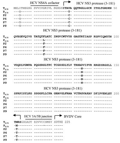
Amino acid sequence analysis of the HCV NS3 protease coding region in the chimeric viruses at different serial passages. cDNA fragments (1.4 kb) encompassing part of the BVDV 5′ UTR, NS4A-tethered HCV NS3 protease, the HCV NS5A-NS5B junction site, the BVDV core, and the Erns were generated by RT-PCR from viral genomic RNAs and cloned into pCR2.1-TOPO vector by using TOPO TA Cloning Kit (Invitrogen). All clones were verified by dideoxynucleotide sequencing. The amino acid sequences encoding the N terminus of Npro, the HCV NS4A cofactor (underlined), the HCV NS3 protease domain (boldface type), the HCV 5A-5B junction site (underlined), and the N terminus of BVDV core are compared. TH/B represents the template sequences for all fusion components derived from their respective parent clones (43, 46). PH/B represents the starting chimeric BVDV clone used to generate infectious RNA transcripts and transfect MDBK cells. The sequences from chimeric BVDV at different passages (PH/B, passage 0; P2, passage 2; P4, passage 4; P7, passage 7; P9, passage 9) were compared and aligned with the lead sequence of TH/B. –, Identical amino acid compared to the corresponding one in TH/B.
In vitro transcription and translation.
HCV NS3 protease and BVDV core fusion proteins were expressed from the plasmids pNS3-C and pNS3mt-C by using the in vitro transcription and translation system (Promega) and labeled with [35S]methionine (Amersham-Pharmacia Biotech, Arlington Heights, Ill.). The transcription-translation reactions were terminated by mixing with 2× sample buffer, and the protein products were separated on a 10 to 20% polyacrylamide gradient gel and analyzed by autoradiography.
Construction of chimeric BVDV plasmid.
The chimeric clone (PH/B) was constructed by the overlapping-extension PCR method (27) and standard molecular cloning techniques. Briefly, the 5′ UTR was amplified by PCR with a ClaI/T7 promoter attached to the 5′ end and a 3′ end at the seventh codon of BVDV Npro. The cDNA fragment encoding the NS4A-tethered HCV NS3-BVDV core fusion protein was amplified from pNS3-C or pNS3mt-C. A third fragment covering the first amino acid of BVDV Erns and amino acid 347 of E2 was also isolated by PCR. These three PCR fragments were joined together by using the TaqPlus long PCR system from Stratagene. The resulting PCR fragment (3,684 bp) was purified and digested with ClaI and RsrII and cloned into the BVDV pVVNADL clone between ClaI and RsrII. Both wild-type and mutant HCV NS3 protease chimeric BVDV plasmids PH/B and PH/B(S139A) were constructed (Fig. 1).
Construction of Npro-null BVDV.
The Npro-null deletion mutant clone was constructed by using a PCR method known as gene splicing via overlapping extension (22, 39). Precise deletion of the entire open reading frame of Npro was accomplished by this PCR method so that the N terminus of core was fused directly in frame with the start codon of the BVDV genome.
Cell culture and virus stock.
Madin-Darby bovine kidney (MDBK) cells were obtained from the American Type Culture Collection (CCL-22). MDBK cells were propagated in Eagle modified minimal essential medium (EMEM) supplemented with 2 mM l-glutamine, 0.1 mM nonessential amino acids, 1.0 mM sodium pyruvate, 1.5 g of sodium biocarbonate (Bio-Whitaker) per liter, and 10% heat-inactivated horse serum (HS; Sigma, St. Louis, Mo.). Cell cultures were maintained at 37°C with 5% CO2. The wild-type BVDV was derived from the molecular clone, and the high-titer viral stock was amplified in MDBK cells (46).
Large-scale production of full-length genomic RNA by in vitro transcription.
The chimeric HCV NS3 protease-dependent BVDV plasmid (PH/B) was used as the template for PCR amplification to generate the linearized cDNA of the entire genome with a 5′ T7 promoter. The resulting long PCR fragment contains the entire BVDV genome under the T7 promoter and ends with the authentic 3′ terminus. Two micrograms of the cDNA was transcribed into RNA by using the T7-MEGAscript Kit from Ambion (Austin, Tex.), according to the manufacturer's protocol. The RNA transcripts were extracted by phenol-chloroform and ethanol precipitated. The integrity of RNA transcripts was determined by 0.8% agarose gel electrophoresis, and the RNAs were stored at −80°C.
Transfection of MDBK cells with chimeric RNA transcripts.
The in vitro-transcribed RNAs from PH/B were transfected into MDBK cells by electroporation as described (17, 28). Briefly, 5 μg of RNA transcripts was mixed with 0.1 ml of the cell suspension (2 × 10⁷ cells/ml) and pulsed twice with a Gene Pulser (set at 0.4 kV and 25 μF with infinite resistance) from Bio-Rad (Hercules, Calif.). The electroporated cell suspension was mixed with EMEM supplemented with 10% HS and plated on a T-75 (75-cm2) tissue culture flask (Becton Dickinson, Franklin Lakes, N.J.). The virally induced cytopathic effect was carefully examined. The culture media containing the progeny viruses were collected at 3 to 4 days posttransfection and used to infect fresh MDBK cells. The mutant RNA transcripts from PH/B(S139A) were produced and transfected into MDBK cells in a similar manner.
Serial infections and generation of high-titer viral stocks.
The chimeric viruses (VH/B) produced by RNA transfection of MDBK cells were used to infect naive MDBK cells (5 × 10⁶ cells plated in a T-75 tissue culture flask). After incubation at 37°C for 1 h, the inoculum was removed and replaced with 20 ml of fresh EMEM with 10% HS. The cells were incubated at 37°C for 2 days or until the cytopathic effect (CPE) was observed. The infection was repeated 10 times in MDBK cells using one-tenth of the previously infected culture media to infect the next plate of fresh MDBK cells. At each passage, extra culture media were either saved by storing at −80°C or used to infect more cells to amplify the virus stock.
Plaque assay and viral isolation.
The plaque assay was described previously by Mendez et al. (28). The chimeric virus VH/B stocks were serially diluted in EMEM. The MDBK cells (0.5 × 10⁶ cells seeded in each well of the six-well culture dish) were infected at 37°C with 0.5 ml of each dilution inoculum. After 1 h of adsorption, the inoculum was removed. The cell monolayer was overlaid with 1% low-melting-point agarose dissolved in EMEM containing 10% HS. The dishes were incubated for 3 days at 37°C. The monolayer of MDBK cells was fixed and stained with crystal violet staining solution containing 2.5% formaldehyde and 25% ethanol (37). Four well-separated plaques generated by the chimeric viruses were carefully removed with a pipette tip before fixation, and viruses in the agarose plugs were recovered in phosphate-buffered saline (PBS) at room temperature. The recovered viruses from each plaque were amplified in MDBK cells and used as the initial inoculum for serial passage of viral infection. A total of 10 passages were performed (P1 to P10), and viral stocks from each passage were collected and stored at −80°C.
One-step single cycle growth kinetic analysis.
To determine the viral replication efficiency, MDBK cells (2 × 10⁴ cells) in each well of a 24-well dish were infected with virus at a multiplicity of infection (MOI) of 5. After 1 h of incubation at 4°C, the inoculum was removed and the cell monolayer was washed with EMEM thoroughly to remove any unabsorbed viruses. Fresh EMEM with 10% HS was added, and the dish was incubated at 37°C. Media in individual wells were harvested at various time points as described in Results. The virus titers were determined by plaque assay and plotted against time to generate the one-step growth curves.
Isolation of viral RNA and RT-PCR analysis.
Virus-containing culture medium (20 ml) was centrifuged at a low speed (10,000 rpm; Beckman SS34 Rotor) for 10 min at 4°C and then loaded onto a 10-ml sucrose cushion (30% in PBS). The viruses were pelleted by centrifugation at 25,000 rpm at 4°C for 10 h (Beckman SW28 Rotor). The virus pellet was resuspended in PBS and treated with RNase A (0.5 μg/μl) from Boehringer Mannheim (Indianapolis, Ind.) and RQ1-DNase I (1 U/μl) from Promega at 37°C for 3 h. Viral RNA was extracted by using phenol-chloroform and precipitated by ethanol in the presence of 0.2 M sodium chloride. Reverse transcription-PCR (RT-PCR) was performed on the viral RNA samples by using Thermoscript RT-PCR System from Life Technologies. The RT-PCR primers were as follows: 5′-GAGTACAGGACAGTCGTCAG-3′ (forward primer corresponding to nucleotides 210 to 229 of the BVDV 5′ UTR) and 5′-ACCAGTTGCACCAACCATG-3′ (reverse primer complementary to nucleotides 1620 to 1635 of the BVDV Erns). Potential DNA contamination was assessed by PCR with omission of the RT step. Medium from mutant chimeric RNA (S139A) transfection was processed similarly, and RT-PCR was performed as described above. The RT-PCR yielded expected products of 1.4 kb. The PCR products were purified and cloned into the pCR2.1-TOPO vector (Invitrogen, Carlsbad, Calif.) for direct sequencing and into the pET-28a vector for in vitro expression and cis-cleavage activity analysis of HCV NS3 protease-BVDV core fusion protein. All clones were verified by dideoxynucleotide sequencing by using an ABI377 Automated Sequencer.
Northern blotting analysis.
Total intracellular RNA was prepared by using the NorthernMax Kit from Ambion (Austin, Tex.). Psoralen-biotinylated DNA probes derived from either the HCV NS3 protease or the BVDV NS5B were produced by using the BrightStar Psoralen-Biotin Labeling Kit (Ambion). Viral RNA was denatured by using glyoxal-dimethyl sulfoxide at 50°C for 30 min, separated by 1% agarose gel electrophoresis, and transferred onto the BrightStar Plus Membrane (Ambion). The membrane was incubated in hybridization solution at 42°C for 1 h, followed by overnight incubation in fresh hybridization solution supplemented with a 0.1 nM concentration of biotinylated DNA probes. The membrane was then washed, and the viral RNA was detected by using the BrightStar BioDetect Kit (Ambion).
Western blotting analysis.
Cell lysates from infected cells were denatured and subjected to SDS-PAGE (on a 10 to 20% gradient gel) analysis. After electrophoresis, the proteins were electrotransferred onto a nitrocellulose membrane (Novex, San Diego, Calif.). The rabbit polyclonal antibodies raised against HCV NS3 protease and the monoclonal antibody raised against BVDV NS3 were used as the primary antibodies. Horseradish peroxidase-conjugated anti-mouse immunoglobulin G (IgG) and anti-rabbit IgG antibodies (Promega) were used as the secondary antibodies. The immunoreactive protein bands were detected by ECL Western Blot Detection Kit (Amersham-Pharmacia Biotech) and recorded on an X-ray film.
Nucleotide sequence accession number.
The chimeric viral genome sequence was deposited in the GenBank database (accession no. AF268278).
RESULTS
Chimeric concept and experimental designs.
HCV and BVDV are closely related (33). It is conceivable that the two viruses share similar replication strategies and regulatory interactions inside the host cells (36). Similar intracellular viral replication and assembly pathways may render BVDV a better choice to provide the genetic background for making HCV-dependent chimeric virus that may be superior to those viruses that are distantly related to HCV. Another advantage of using BVDV is its genomic plasticity (29), which allows recombinant manipulations with fewer concerns about genome compatibility. BVDV also consists of one of the largest genomes among members of the Flaviviridae family and encodes two unique viral proteins: Npro and Erns. While the biological functions of these “extra” viral proteins are unclear, it has been shown that Npro can be deleted or replaced without significant effect on the viability and infectivity of BVDV and CSFV (30, 31, 44, 45). Taking advantage of the fact that the BVDV Npro protease is dispensable for viral replication and growth, its coding region was replaced by an NS4A cofactor-tethered HCV NS3 protease domain linked in frame to the BVDV core protein through an HCV NS5A-NS5B junction site (Fig. 1B, PH/B). In this chimeric construct, as guided by the crystal structure of NS3-NS4A complexes (23, 50), the NS4A cofactor peptide (GSVVIVGRIVLS) was covalently linked to the N terminus of the HCV NS3 protease catalytic domain (amino acids 3 to 181) through a flexible “GSGS” spacer (43). It has been shown that this N-terminal tethered “single-chain” NS3 represents the activated form of the HCV protease with improved stability and solubility (34, 43). The proximity of the NS4A peptide to the N terminus of the protease allows tighter intercalation and proper folding of the protease domain mimicking that in the “two-chain” NS3-NS4A complexes (23, 50, 51). Due to this economic coupling of the NS3 protease and NS4A cofactor, a much smaller gene was used to replace the Npro coding region, which may have greatly enhanced the genome stability of the resulting chimeric viruses compared to those containing the full-length NS3-NS4A genes (14, 18). Among other functions, Npro is believed to free the BVDV core protein for genome encapsidation and capsid assembly by autoproteolysis of its C terminus. To mimic this function, an NS5A-NS5B junction site (P10-P4′) was engineered to link the NS4A-tethered NS3 protease to the BVDV core. This linkage, once cleaved by the N-terminal HCV NS3 protease, would release the BVDV core with an additional tetrapeptide “SMSY” at its N terminus (Fig. 1B). As a control, a similar construct, PH/B(S139A), was created with an inactive HCV NS3 protease in which the catalytically essential serine 139 was mutated to alanine (Fig. 1C). This mutation should render the BVDV core unable to be released from the C terminus of the HCV protease domain, resulting in a fusion protein that may not be able to perform its related functions in capsid assembly and virion production. In order to further assess the stability of the chimeric viruses and to address whether the NS4A-tethered HCV NS3 protease can be deleted from the viral genome, an Npro-null BVDV construct (BVDV−Npro) was created (Fig. 1D). In this construct, the entire Npro coding region was removed, and the core protein was directly fused to the start codon (methionine) of the polyprotein.
Feasibility analysis of the chimeric constructs by in vitro translation cleavage assay.
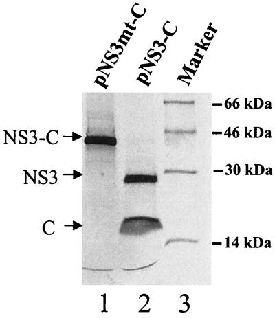
To demonstrate whether the newly created chimeric fusion protein would cleave itself as designed to release the BVDV core protein, we cloned the HCV-NS3/BVDV core cDNA (containing the coding regions of the NS4A-tethered HCV NS3 protease plus the core of BVDV) into the pET expression plasmid. The fusion proteins were produced and labeled in rabbit reticulocyte lysate using the in vitro transcription and translation system. The labeled protein products were separated on a sodium dodecyl sulfate-polyacrylamide gel electrophoresis (SDS-PAGE) gel and analyzed as described in Materials and Methods. As shown in Fig. 2, the fusion protein that contains the active HCV NS3 protease domain (pNS3-C) cleaved itself rapidly and yielded two smaller products corresponding to the predicted sizes for HCV NS4A-tethered NS3 and BVDV core (Fig. 2, lane 2). In contrast, the construct with the mutant HCV NS3 protease (pNS3mt-C) failed to be processed and remained as a single and larger fusion processor (Fig. 2, lane 1). These results confirmed that the designed chimeric fusion protein was functional and processed the NS5A-NS5B cleavage site correctly during expression of the polyprotein, indicating that the N-terminal HCV NS3 protease would substitute the proteolytic function of Npro in the polyprotein processing during BVDV infection.
FIG. 2.
In vitro expression of the HCV NS3-BVDV core fusion proteins using an in vitro transcription and translation system. The proteins were labeled with [35S]methionine. Lane 1, production of the fusion protein with a catalytically inactive HCV NS3 protease from plasmid pNS3mt-C; lane 2, expression of the fusion protein with the wild-type HCV NS3 protease from plasmid pNS3-C plasmid. The protein products were separated by SDS-PAGE on a 10 to 20% gradient gel and visualized by autoradiography. The fusion protein with the active HCV NS3 protease self-cleaved at the NS5A-NS5B junction between HCV NS3 and BVDV core protein. Lane 3, 14C-labeled molecular mass markers (kilodaltons).
Generation of an HCV NS3 protease-dependent BVDV chimeric virus.
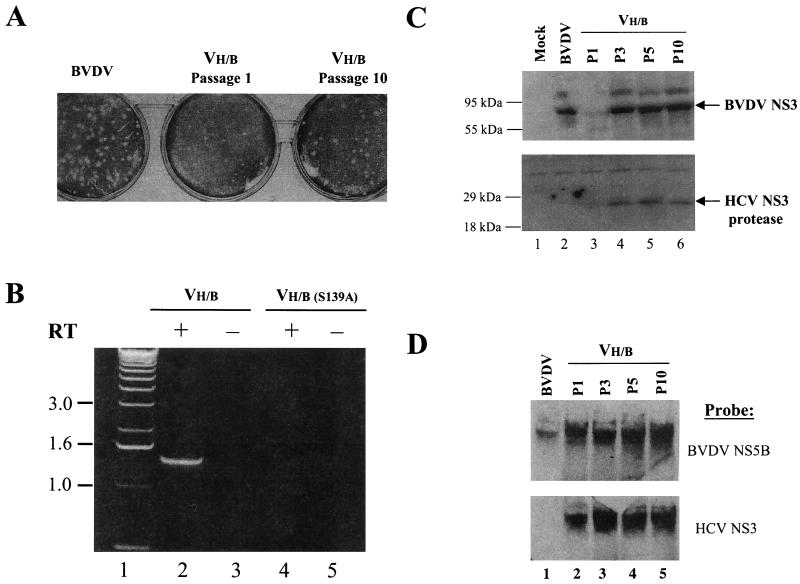
Having shown that the HCV NS3 protease was processed correctly in the context of the chimeric setting, we built two full-length chimeric clones, PH/B and PH/B(S139A), from the infectious clone of BVDV (isolate NADL) as described previously (46) (also refer to Fig. 1). Full-length RNA transcripts were produced by T7 RNA polymerase in vitro and transfected into MDBK cells by electroporation. Production of viable viruses was examined microscopically for virus-induced CPE and visualized by standard BVDV plaque assay. As shown in Fig. 3A (middle panel), the RNA transcript from the PH/B clone produced viable chimeric viruses (VH/B) that formed somewhat smaller plaques (compared to the plaques from the wild-type BVDV in the left panel) on an MDBK cell monolayer 2 to 3 days posttransfection. As expected, the RNA transcript from mutant chimeric clone, PH/B(S139A), failed to produce any signs of infection. Extracellular supernatant or medium from each cell culture was collected and used to infect naive MDBK cells. After a series of reinfection or passages in fresh MDBK cells, the chimeric viruses from the PH/B clone exhibited larger plaque phenotype (Fig. 3A, right panel), suggesting that viral adaptation or revertant mutation might have occurred. No CPE or plaque was observed in cells inoculated with supernatants derived from the mutant chimeric RNA transfection (data not shown). This was most likely due to the fact that the mutant HCV NS3 protease failed to cleave the NS5A-NS5B junction, preventing the BVDV core from being released from the fusion protein to support virus growth. These data also confirmed that the BVDV NS3 could not substitute HCV NS3 to cleave the HCV NS5A-NS5B junction. Taken together, these results support the idea that the chimeric BVDV depends on HCV NS3 protease activity for replication and growth.
FIG. 3.
Characterization of chimeric viruses VH/B obtained from serial passages. (A) Plaque phenotypes. The wild-type BVDV and the chimeric viruses (VH/B) from different passages were compared. MDBK cells were infected with BVDV and VH/B from the first passage (P1) and the tenth passage (P10) for 3 days at 37°C. The plaque formation were revealed by crystal violet staining of the MDBK cell monolayer. Plaques formed by the wild-type BVDV (left panel) appeared to be larger in size compared with that formed by the P1 VH/B viruses (central panel). After nine serial passages, P10 VH/B viruses generated larger plaques (right panel). (B) RT-PCR analysis. Viral RNAs were isolated from the third passages of both the chimeric viruses (VH/B, lanes 2 and 3) with active HCV NS3 protease and the mutant chimeric viruses (VH/B(S139A), lanes 3 and 4). The HCV NS3-BVDV core coding region in the viral RNAs was amplified in the presence (lanes 2 and 4) or absence (lanes 3 and 5) of reverse transcriptase (RT). DNA molecular size standards (1-kb ladder) is shown in lane 1. (C) Western blot analysis. Cell lysates from mock-infected MDBK cells (lane 1) or from MDBK cells infected by the wild-type BVDV (lane 2) and the chimeric viruses VH/B of various passages (P1, lane 3; P3, lane 4; P5, lane 5; and P10, lane 6). The cell lysates were fractionated by SDS-PAGE on a 10 to 20% gel, transferred to nitrocellulose membrane, and analyzed by immunoblotting with a monoclonal antibody against BVDV NS3 (upper panel) and the polyclonal antibody against HCV NS3 (lower panel). The molecular mass standards (in kilodaltons) are indicated at the left side of each panel. (D) Northern blot analysis. Total RNA was isolated from cells infected by the wild-type BVDV (lane 1) or by the chimeric viruses VH/B of various passages (P1, lane 3; P3, lane 4; P5, lane 5; and P10, lane 6). Glyoxal-denatured viral RNAs were separated by 1% agarose gel electrophoresis and transferred to a nylon membrane. The blot was incubated in the hybridization solution with psoralen-biotinylated DNA probes derived from the BVDV NS5B (upper panel) and the HCV NS3 protease (lower panel). Bands on the blot were visualized by using the BrightStar BioDetect detection kit from Ambion.
The chimeric viruses from the third-passage inoculum (P3 VH/B) were further purified on a sucrose gradient. Viral RNA was extracted, and RT-PCR was performed to isolate the cDNA fragment that encompassed the HCV protease and BVDV core fusion (∼1.4 kb). The results in Fig. 3B demonstrated that the expected RT-PCR product was only detected in the supernatant from VH/B (Fig. 3B, lane 2) and not from the supernatant of the S139A mutant chimeric RNA (Fig. 3B, lane 4). As a control, PCR alone (without RT) did not produce any products, confirming that the PCR product originated from viral RNA and not from any contaminating DNA sources. The presence of the NS4A-tethered NS3 protease and the HCV NS5A-NS5B junction in the genome of the chimeric viruses was further authenticated by direct sequencing of the RT-PCR products (see Fig. 5). In addition, no BVDV Npro sequence was detected in any of the viral RNA samples (data not shown). This further suggests that the viruses from the chimeric RNA transfection were not due to contaminating cytopathic BVDV (strain NADL).
Genome stability of the chimeric virus.
The plaque-purified chimeric viruses (passage 1) were amplified through 10 serial passages up to 10 times by reinfection of fresh MDBK cells. The infectivity of the chimeric viruses was analyzed by microscopic evaluation of CPE or plaque assay. The size of the plaques was larger at later passages than that at passage 1 (Fig. 3A, compare middle and right panels), a size comparable to that of the wild-type BVDV (isolate NADL). To assess the genome stability and to determine whether the HCV NS3 protease was deleted during serial passages of the chimeric viruses, the infected cells at different passages were subjected to Western blot (Fig. 3C) and Northern blot (Fig. 3D) analyses. The results from the Western blot analysis demonstrated the presence of HCV NS3 protease (recognized by the rabbit polyclonal antibodies raised against a purified HCV NS3 protease) throughout all passages of the chimeric viruses (Fig. 3C, lower panel, lanes 3 to 6). The cells infected by the wild-type BVDV did not express the HCV NS3 protease (lane 2). Meanwhile, all infected cells produced the BVDV NS3 and NS2-3 proteins detected by antibodies reactive to BVDV NS3 (Fig. 3C, upper panel). The presence of abundant BVDV NS3 compared to NS2-3 correlated with the observed CPE of the cells infected with the chimeric viruses (Fig. 3C, upper panel, lanes 3 to 6). The Northern blot analysis (Fig. 3D) revealed similar results in that the probe derived from BVDV NS5B hybridized with intracellular viral RNAs from both the wild-type BVDV and the chimeric viruses (upper panel), whereas the probe derived from the HCV NS3 only detected viral RNAs from cells infected by the chimeric viruses (lower panel). The detection of HCV NS3 protease in infected cells as well as its sequence in viral genome RNA following multiple serial passages supports the hypothesis that the chimeric virus are stable, further confirming the dependence of the chimeric viruses on the HCV NS3 protease activity.
One-step viral growth kinetic analysis.
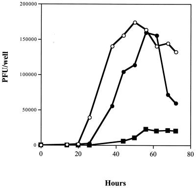
Because Npro is dispensable for BVDV replication, it seemed likely that the chimeric virus could lose the tethered HCV NS3 protease, resulting in a Npro-null BVDV. This deletion may occur rapidly if the Npro-null BVDV has certain growth advantages. To better understand the observed stability of the chimeric viruses, an Npro-null virus, BVDV−Npro, was constructed (Fig. 1D). Surprisingly, BVDV−Npro retained the cytopathogenic phenotype of BVDV, although to a lesser extent, and formed smaller plaques on the MDBK cell monolayer. One-step growth kinetic analysis was performed to compare the viral growth rates, eclipse phases of replication, and the maximum viral yields among wild-type BVDV, chimeric BVDV, and Npro-null BVDV. The results presented in Fig. 4 demonstrated that despite a slight delay of ca. 5 h, the chimeric viruses reached a similar replication efficiency, as reflected by the similar maximum viral yields compared to the wild-type BVDV. In contrast, the Npro-null BVDV had a prolonged eclipse phase and achieved a viral yield at a greatly reduced level, one at least 10 times lower than those of the chimeric and wild-type BVDVs. This suggests that although cytopathic, the propagation of the Npro-null BVDV is greatly compromised, a finding consistent with the hypothesis that the stability of the chimeric viruses is a consequence of the reduced fitness of the potential deletion mutants. Thus far, we have not been able to isolate any Npro-null BVDV-like viruses from chimeric virus-infected cells under optimal viral infection conditions.
FIG. 4.
Single cycle growth kinetic analysis. A total of 20,000 MDBK cells in each well of a 24-well dish were infected by 100,000 PFU of wild-type BVDV (○), chimeric BVDV (●), and BVDV−Npro (■). After 1 h of incubation at 4°C, the inocula were removed, and the cell monolayers were washed thoroughly to remove any residual viruses. Then, 0.5 ml of fresh EMEM with 10% HS was added, and the dish was incubated at 37°C. Newly secreted BVDVs in the media were harvested at various time points postinfection. The virus titers were determined and plotted against time to generate the growth curves.
Isolation of variant viruses and evidence of viral adaptation.
Genomic RNAs were isolated from chimeric viruses after different numbers of passages. The region corresponding NS4A-tethered HCV NS3 protease plus the NS5A-NS5B junction was amplified by RT-PCR and sequenced directly. The sequences from different passages were compared with the template sequence (TH/B) shown in the multiple alignment in Fig. 5. The template sequence was derived from the parent HCV and BVDV clones (43, 46) used to build the chimeric plasmid (PH/B). As shown in Fig. 5, three PCR mutations were generated during the construction of the chimeric clone (PH/B) (compare the sequence of PH/B with that of TH/B). Two mutations occurred in the coding region of HCV NS3 protease, F43 (phenylalanine at amino acid position 43 numbered according to the sequence of HCV NS3) to L (leucine) and D112 (aspartic acid at amino acid position 112) to N (asparagine), and one occurred in the N-terminal coding region for Npro, I4 (isoleucine at position 4) to N (asparagine). After two serial passages, one of the mutations (F43L) quickly reverted to the wild-type at P2, suggesting that this mutation is detrimental to the chimeric viruses. Interestingly, two additional point mutations were identified in the HCV NS3 coding region from the chimeric viruses of later passages: Y6 (tyrosine at amino acid position 6) to C (cysteine) and M179 (methionine at position 179) to T (threonine). Both mutations were near the chimeric junctions, which might allow better folding of the fusion protein.
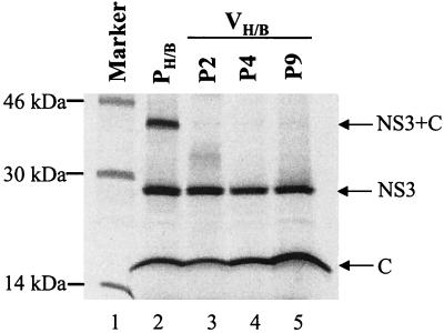
To correlate these mutations or variations with the protease activity, the cDNA fragments encompassing the HCV NS3 protease and BVDV core fusion protein were cloned into the pET expression plasmid as described for Fig. 2. Results from the in vitro transcription and translation cleavage assay demonstrated that the starting chimeric clone (PH/B) harboring the F43L mutation exhibited reduced protease activity (Fig. 6, lane 2), as reflected by the incomplete cleavage of the fusion precursor. The fusion proteins from the later passages (P2 to P9) had normal levels of the protease activity comparable to that of the wild-type construct (pNS3-C, equivalent of TH/B, in Fig. 2, lane 2). These data suggest that there is a correlation between viral adaptation and enhancement of HCV protease activity. Studies by others using the Sindbis chimeric viruses also reported a similar point mutation (C16Y) without a clear correlation with the in vitro protease activity (14). This is consistent with our observations that some of the mutations or variations (Y6C and M179T) do not correlate with any detectable changes in the protease activity (compare Fig. 2 and 6). Other mutations (I4N in Npro and D112N in HCV NS3) can be tolerated and sustained throughout serial passages. We have not observed any deletions in the HCV NS3 region, as reported by others who used the full-length NS3-NS4A for the construction of chimeric viruses (14), possibly due to the economic packing of the minimum NS4A cofactor and the small catalytic domain of HCV NS3 protease in our construct.
FIG. 6.
Correlation of the HCV NS3 protease activity with viral adaptation. The coding regions for the HCV NS3 protease and BVDV core were isolated either from plasmid PH/B by PCR or from the chimeric viruses VH/B at various passages (P2, P4, and P9) by RT-PCR. These coding regions were subcloned into the pET-28a expression vector (see Materials and Methods). The fusion proteins were expressed and labeled with [35S]methionine by using the in vitro transcription and translation system. The labeled fusion precursor and the cleavage products were separated by SDS-PAGE and detected by autoradiography. Lane 1 indicates the 14C-labeled molecular mass marker (in kilodaltons).
DISCUSSION
The lack of a cell culture system that permits HCV infection imposes a major obstacle for anti-HCV drug development. Although substantial efforts have been devoted to the characterization of HCV NS3 protease (23, 50), the inhibitor development against HCV NS3 protease is limited without a good cell-based system to evaluate its cellular potency. Several cell-based trans-cleavage assays have been developed relying on the coexpression of an NS3 protease-containing plasmid and a substrate-containing plasmid in the same cells (5, 20). However, these systems often yield less-reproducible results and, more importantly, fail to reflect the polyprotein processing which occurs within the subcellular microenvironment of viral replication. Such intrapolyprotein processing may resemble the kinetics of a cis-cleavage which is insensitive to dilution.
Several HCV NS3 protease-dependent chimeric viruses have been created (13, 14, 18) using two plus-stranded RNA viruses from unrelated virus families as the carriers. In these chimeric viruses, HCV NS3 protease-mediated protein processings (mostly cis-cleavages) are essential for virus growth. However, these chimeric viruses are less stable because the inserted HCV NS3-NS4A can be easily deleted, resulting in reversion to the wild-type viruses which have many growth advantages over the chimeric viruses (14). In this approach, we took advantage of the dispensability of Npro for BVDV replication and substituted its proteolytic function with that of HCV NS3 protease. The resulting chimeric BVDV was cytopathic and easy to quantify. Its growth properties were comparable to those of the wild-type BVDV. Most importantly, under normal cell culture conditions (with a low MOI and without any selection pressures against the chimeric viruses), this HCV protease-dependent BVDV was very stable, lacking any detectable deletions in the HCV coding regions.
One of the key benefits of developing stable chimeric viruses is the possibility to assess potential drug resistance to anti-HCV NS3 protease inhibitors. This will require that the chimeric viruses be very stable. Whether the chimeric viruses reported here were stable enough to allow the selection of drug-resistant variants remains to be addressed. Based on a prediction that the HCV NS3 protease might be deleted to yield a virus without Npro, we created a molecular clone lacking Npro (BVDV−Npro) in which the entire Npro coding region was removed (Fig. 1). To our surprise, the Npro-null BVDV was not only viable but also cytopathic and formed small plaques on a cell monolayer. Further growth analysis revealed that this Npro-null BVDV was highly defective in replication and achieved a virus production level at least 10 times lower than that of the chimeric viruses or the wild-type viruses. While this partially explained the observed stability of the chimeric BVDV, it also limited the chimeric viruses from being used to select potential mutations that conferred drug resistance. Under a strong selection against the chimeric viruses, such as a potent HCV NS3 protease inhibitor, the Npro-null BVDV would appear quickly and may overtake the virus production.
In a cell culture dish, the chimeric BVDV appeared to retain comparable growth properties and CPE as the wild-type virus. Whether the chimeric virus replicates in vivo in calves and duplicate the course of disease progression as the wild-type virus will be an interesting subject to address. A better understanding of the chimeric virus correlated with the Npro-null BVDV will help to elucidate the biological function of this unique Npro protein acquired by BVDV. The one-step growth analysis has revealed that this virus is defective in replication, suggesting that BVDV without the Npro is attenuated. If the in vivo studies demonstrate that the Npro-null BVDV, or even the chimeric virus, is attenuated with self-limiting infection which induces anti-BVDV responses, it may be an excellent candidate for vaccine development. Finally, the HCV NS3 protease-dependent BVDV may provide an alternative and much more affordable animal model for the in vivo testing of any HCV protease inhibitors.
ACKNOWLEDGMENTS
We thank Gregory R. Reyes for support and Bahige M. Baroudy, Michael Endres, Seung-Ki Chon, and Fred Lahser for helpful discussions. We also appreciate the excellent assistance of Jacquelyn Wright-Minogue and Barbara Kerr.
REFERENCES
- 1.Alter H J, Purcell R H, Shih J W, Melpolder J C, Houghton M, Choo Q-L, Kuo G. Detection of antibody to hepatitis C virus in prospectively followed transfusion recipients with acute and chronic non-A non-B hepatitis. N Engl J Med. 1989;321:1494–1500. doi: 10.1056/NEJM198911303212202. [DOI] [PubMed] [Google Scholar]
- 2.Alter M J, Kruszon-Moran D, Nainan O V, McQuillan G M, Gao F, Moyer L A, Kaslow R A, Margolis H S. The prevalence of hepatitis C virus infection in the United States, 1988 through 1994. N Engl J Med. 1999;341:556–562. doi: 10.1056/NEJM199908193410802. [DOI] [PubMed] [Google Scholar]
- 3.Bartenschlager R, Ahlborn-Laake L, Mous J, Jacobsen H. Nonstructural protein 3 of the hepatitis C virus encodes a serine-type proteinase required for cleavage at the NS3/4 and NS4/5 junctions. J Virol. 1993;67:3835–3844. doi: 10.1128/jvi.67.7.3835-3844.1993. [DOI] [PMC free article] [PubMed] [Google Scholar]
- 4.Cho Y G, Moon H S, Sung Y C. Construction of hepatitis C-SIN virus recombinants with replicative dependency on hepatitis C virus serine protease activity. J Virol Methods. 1997;65:201–207. doi: 10.1016/s0166-0934(97)02183-6. [DOI] [PubMed] [Google Scholar]
- 5.Cho Y G, Yang S H, Sung Y C. In vivo assay for hepatitis C viral serine protease activity using a secreted protein. J Virol Methods. 1998;72:109–115. doi: 10.1016/s0166-0934(98)00010-x. [DOI] [PubMed] [Google Scholar]
- 6.Chon S K, Perez D R, Donis R O. Genetic analysis of the internal ribosome entry segment of bovine viral diarrhea virus. Virology. 1998;251:370–381. doi: 10.1006/viro.1998.9425. [DOI] [PubMed] [Google Scholar]
- 7.Choo Q-L, Kuo G, Weiner A J, Overby L R, Bradley D W, Houghton M. Isolation of a cDNA clone derived from a blood-borne non-A, non-B viral hepatitis genome. Science. 1989;244:359–364. doi: 10.1126/science.2523562. [DOI] [PubMed] [Google Scholar]
- 8.Collett M S, Larson R, Gold C, Strick D, Anderson D K, Purchio A F. Molecular cloning and nucleotide sequence of the pestivirus bovine viral diarrhea virus. Virology. 1988;165:191–199. doi: 10.1016/0042-6822(88)90672-1. [DOI] [PubMed] [Google Scholar]
- 9.Davis G L, Esteban-Mur R, Rustgi V, Hoefs J, Gordon S C, Trepo C, Shiffman M L, Zeuzem S, Craxi A, Ling M H, Albrecht J. Interferon alpha-2b alone or in combination with ribavirin for the treatment of relapse of chronic hepatitis C. International Hepatitis Interventional Therapy Group. N Engl J Med. 1998;339:1493–1499. doi: 10.1056/NEJM199811193392102. [DOI] [PubMed] [Google Scholar]
- 10.Elbers K, Tautz N, Becher P, Stoll D, Rumenapf T, Thiel H-J. Processing in the pestivirus E2-NS2 region: identification of proteins p7 and E2p7. J Virol. 1996;70:4131–4135. doi: 10.1128/jvi.70.6.4131-4135.1996. [DOI] [PMC free article] [PubMed] [Google Scholar]
- 11.Failla C, Tomei L, De Francesco R. both NS3 and NS4A are required for proteolytic processing of hepatitis C virus nonstructural proteins. J Virol. 1994;68:3753–3760. doi: 10.1128/jvi.68.6.3753-3760.1994. [DOI] [PMC free article] [PubMed] [Google Scholar]
- 12.Farci P, Alter H J, Govindarajan S, Wong D C, Engle R, Lesniewski R R, Mushahwar I K, Desai S M, Miller R H, Ogata N, Purcell R H. Lack of protective immunity against reinfection with hepatitis C virus. Science. 1992;258:135–140. doi: 10.1126/science.1279801. [DOI] [PubMed] [Google Scholar]
- 13.Filocamo G, Pacini L, Migliaccio G. Chimeric Sindbis viruses dependent on the NS3 protease of hepatitis C virus. J Virol. 1997;71:1417–1427. doi: 10.1128/jvi.71.2.1417-1427.1997. [DOI] [PMC free article] [PubMed] [Google Scholar]
- 14.Filocamo G, Pacini L, Nardi C, Bartholomew L, Scaturro M, Delmastro P, Tramontano A, De Francesco R, Migliaccio G. Selection of functional variants of the NS3-NS4A protease of hepatitis C virus by using chimeric Sindbis viruses. J Virol. 1999;73:561–575. doi: 10.1128/jvi.73.1.561-575.1999. [DOI] [PMC free article] [PubMed] [Google Scholar]
- 15.Frolov I, McBride M S, Rice C M. cis-acting RNA elements required for replication of bovine viral diarrhea virus-hepatitis C virus 5′ nontranslated region chimeras. RNA. 1998;4:1418–1435. doi: 10.1017/s1355838298981031. [DOI] [PMC free article] [PubMed] [Google Scholar]
- 16.Grakoui A, McCourt D W, Wychowski C, Feinstone S M, Rice C M. Characterization of the hepatitis C virus-encoded serine proteinase: determination of the proteinase-dependent polyprotein cleavage sites. J Virol. 1993;67:2832–2843. doi: 10.1128/jvi.67.5.2832-2843.1993. [DOI] [PMC free article] [PubMed] [Google Scholar]
- 17.Grassmann C W, Isken O, Behrens S-E. Assignment of the multifunctional NS3 protein of bovine viral diarrhea virus during RNA replication: an in vitro and in vivo study. J Virol. 1999;73:9196–9205. doi: 10.1128/jvi.73.11.9196-9205.1999. [DOI] [PMC free article] [PubMed] [Google Scholar]
- 18.Hahm B, Back S H, Lee T G, Wimmer E, Jang S K. Generation of a novel poliovirus with a requirement of hepatitis C virus protease NS3 activity. Virology. 1996;226:318–326. doi: 10.1006/viro.1996.0659. [DOI] [PubMed] [Google Scholar]
- 19.Hijikata M, Mizushima H, Akagi T, Mori S, Kakiuchi N, Kato N, Tanaka T, Kimura K, Shimotohno K. Two distinct proteinase activities required for the processing of a putative nonstructural precursor protein of hepatitis C virus. J Virol. 1993;67:4665–4675. doi: 10.1128/jvi.67.8.4665-4675.1993. [DOI] [PMC free article] [PubMed] [Google Scholar]
- 20.Hirowatari Y, Hijikata M, Shimotohno K. A novel method for analysis of viral proteinase activity encoded by hepatitis C virus in cultured cells. Anal Biochem. 1995;225:113–120. doi: 10.1006/abio.1995.1116. [DOI] [PubMed] [Google Scholar]
- 21.Hong Z, Ferrari E, Wright-Minogue J, Chase R, Risano C, Seelig G, Lee C-G, Kwong A D. Enzymatic characterization of hepatitis C virus NS3/4A complexes expressed in mammalian cells using the herpes simplex virus amplicon system. J Virol. 1996;70:4261–4268. doi: 10.1128/jvi.70.7.4261-4268.1996. [DOI] [PMC free article] [PubMed] [Google Scholar]
- 22.Horton R M, Cai Z L, Ho S N, Pease L R. Gene splicing by overlap extension: tailor-made genes using the polymerase chain reaction. BioTechniques. 1990;8:528–535. [PubMed] [Google Scholar]
- 23.Kim J L, Morgenstern K A, Lin C, Fox T, Dwyer M D, Landro J A, Chambers S P, Markland W, Lepre C A, O'Malley E T, Harbeson S L, Rice C M, Murcko M A, Caron P R, Thomson J A. Crystal structure of the hepatitis C virus NS3 protease domain complexed with a synthetic NS4A cofactor peptide. Cell. 1996;87:343–355. doi: 10.1016/s0092-8674(00)81351-3. [DOI] [PubMed] [Google Scholar]
- 24.Kwong A D, Kim J L, Rao G, Lipovsek D, Raybuck S A. Hepatitis C virus NS3/4A protease. Antiviral Res. 1999;41:67–84. [PubMed] [Google Scholar]
- 25.Lin C, Thomson J A, Rice C M. A central region in the hepatitis C virus NS4A protein allows formation of an active NS3-NS4A serine proteinase complex in vivo and in vitro. J Virol. 1995;69:4373–4380. doi: 10.1128/jvi.69.7.4373-4380.1995. [DOI] [PMC free article] [PubMed] [Google Scholar]
- 26.McHutchison J G, Gordon S C, Schiff E R, Shiffman M L, Lee W M, Rustgi V K, Goodman Z D, Ling M H, Cort S, Albrecht J K. Interferon alpha-2b alone or in combination with ribavirin as initial treatment for chronic hepatitis C. Hepatitis Interventional Therapy Group. N Engl J Med. 1998;339:1485–1492. doi: 10.1056/NEJM199811193392101. [DOI] [PubMed] [Google Scholar]
- 27.Mehta R K, Singh J. Bridge-overlap-extension PCR method for constructing chimeric genes. BioTechniques. 1999;26:1082–1086. doi: 10.2144/99266bm17. [DOI] [PubMed] [Google Scholar]
- 28.Mendez E, Ruggli N, Collett M S, Rice C M. Infectious bovine viral diarrhea virus (strain NADL) RNA from stable cDNA clones: a cellular insert determines NS3 production and viral cytopathogenicity. J Virol. 1998;72:4737–4745. doi: 10.1128/jvi.72.6.4737-4745.1998. [DOI] [PMC free article] [PubMed] [Google Scholar]
- 29.Meyers G, Thiel H-J. Molecular characterization of pestiviruses. Adv Virus Res. 1996;47:53–118. doi: 10.1016/s0065-3527(08)60734-4. [DOI] [PubMed] [Google Scholar]
- 30.Mittelholzer C, Moser C, Tratschin J D, Hofmann M A. Generation of cytopathogenic subgenomic RNA of classical swine fever virus in persistently infected porcine cell lines. Virus Res. 1997;51:125–137. doi: 10.1016/S0168-1702(97)00081-6. [DOI] [PMC free article] [PubMed] [Google Scholar]
- 31.Moser C, Stettler P, Tratschin J-D, Hofmann M A. Cytopathogenic and noncytopathogenic RNA replicons of classical swine fever virus. J Virol. 1999;73:7787–7794. doi: 10.1128/jvi.73.9.7787-7794.1999. [DOI] [PMC free article] [PubMed] [Google Scholar]
- 32.Moser C, Tratschin J D, Hofmann M A. A recombinant classical swine fever virus stably expresses a marker gene. J Virol. 1998;72:5318–5322. doi: 10.1128/jvi.72.6.5318-5322.1998. [DOI] [PMC free article] [PubMed] [Google Scholar]
- 33.Ohba K, Mizokami M, Lau J Y, Orito E, Ikeo K, Gojobori T. Evolutionary relationship of hepatitis C, pesti-, flavi-, plantviruses, and newly discovered GB hepatitis agents. FEBS Lett. 1996;378:232–234. doi: 10.1016/0014-5793(95)01441-1. [DOI] [PubMed] [Google Scholar]
- 34.Pasquo A, Nardi M C, Dimasi N, Tomei L, Steinkuhler C, Delmastro P, Tramontano A, De Francesco R. Rational design and functional expression of a constitutively active single-chain NS4A-NS3 proteinase. Fold Des. 1998;3:433–441. doi: 10.1016/S1359-0278(98)00060-1. [DOI] [PubMed] [Google Scholar]
- 35.Poole T L, Wang C, Popp R A, Potgieter L N, Siddiqui A, Collet M S. Pestivirus translation initiation occurs by internal ribosome entry. Virology. 1995;206:750–754. doi: 10.1016/s0042-6822(95)80003-4. [DOI] [PubMed] [Google Scholar]
- 36.Rice C M. Flaviviridae: the viruses and their replication. In: Fields B N, Knipe D M, Howley P M, editors. Virology. 3rd ed. New York, N.Y: Raven Press; 1996. pp. 931–960. [Google Scholar]
- 37.Rice C M, Grakoui A, Galler R, Chambers T J. Transcription of infectious yellow fever RNA from full-length cDNA templates produced by in vitro ligation. New Biol. 1989;1:285–296. [PubMed] [Google Scholar]
- 38.Rumenapf T, Stark R, Heimann M, Thiel H J. N-terminal protease of pestiviruses: identification of putative catalytic residues by site-directed mutagenesis. J Virol. 1998;72:2544–2547. doi: 10.1128/jvi.72.3.2544-2547.1998. [DOI] [PMC free article] [PubMed] [Google Scholar]
- 39.Senanayake S D, Brian D A. Precise large deletions by the PCR-based overlap extension method. Mol Biotechnol. 1995;4:13–15. doi: 10.1007/BF02907467. [DOI] [PMC free article] [PubMed] [Google Scholar]
- 40.Shimizu Y K, Hijikata M, Iwamoto A, Alter H J, Purcell R H, Yoshikura H. Neutralizing antibodies against hepatitis C virus and the emergence of neutralization escape mutant viruses. J Virol. 1994;68:1494–1500. doi: 10.1128/jvi.68.3.1494-1500.1994. [DOI] [PMC free article] [PubMed] [Google Scholar]
- 41.Stark R, Meyers G, Rumenapf T, Thiel H J. Processing of pestivirus polyprotein: cleavage site between autoprotease and nucleocapsid protein of classical swine fever virus. J Virol. 1993;67:7088–7095. doi: 10.1128/jvi.67.12.7088-7095.1993. [DOI] [PMC free article] [PubMed] [Google Scholar]
- 42.Tanji Y, Hijikata M, Satoh S, Kaneko T, Shimotohno K. Hepatitis C virus-encoded nonstructural protein NS4A has versatile functions in viral protein processing. J Virol. 1995;69:1575–1581. doi: 10.1128/jvi.69.3.1575-1581.1995. [DOI] [PMC free article] [PubMed] [Google Scholar]
- 43.Taremi S S, Beyer B, Maher M, Yao N, Prosise W, Weber P C, Malcolm B A. Construction, expression, and characterization of a novel fully activated recombinant single-chain hepatitis C virus protease. Protein Sci. 1998;7:2143–2149. doi: 10.1002/pro.5560071011. [DOI] [PMC free article] [PubMed] [Google Scholar]
- 44.Tautz N, Harada T, Kaiser A, Rinck G, Behrens S-E, Thiel H-J. Establishment and characterization of cytopathogenic and noncytopathogenic pestivirus replicons. J Virol. 1999;73:9422–9432. doi: 10.1128/jvi.73.11.9422-9432.1999. [DOI] [PMC free article] [PubMed] [Google Scholar]
- 45.Tratschin J D, Moser C, Ruggli N, Hofmann M A. Classical swine fever virus leader proteinase Npro is not required for viral replication in cell culture. J Virol. 1998;72:7681–7684. doi: 10.1128/jvi.72.9.7681-7684.1998. [DOI] [PMC free article] [PubMed] [Google Scholar]
- 46.Vassilev V B, Collett M S, Donis R O. Authentic and chimeric full-length genomic cDNA clones of bovine viral diarrhea virus that yield infectious transcripts. J Virol. 1997;71:471–478. doi: 10.1128/jvi.71.1.471-478.1997. [DOI] [PMC free article] [PubMed] [Google Scholar]
- 47.Wimmer E, Hellen C U, Cao X. Genetics of poliovirus. Annu Rev Genet. 1993;27:353–436. doi: 10.1146/annurev.ge.27.120193.002033. [DOI] [PubMed] [Google Scholar]
- 48.Wyatt C A, Andrus L, Brotman B, Huang F, Lee D-H, Prince A M. Immunity in chimpanzees chronically infected with hepatitis C virus: role of minor quasispecies in reinfection. J Virol. 1998;72:1725–1730. doi: 10.1128/jvi.72.3.1725-1730.1998. [DOI] [PMC free article] [PubMed] [Google Scholar]
- 49.Xu J, Mendez E, Caron P R, Lin C, Murcko M A, Collett M S, Rice C M. Bovine viral diarrhea virus NS3 serine proteinase: polyprotein cleavage sites, cofactor requirements, and molecular model of an enzyme essential for pestivirus replication. J Virol. 1997;71:5312–5322. doi: 10.1128/jvi.71.7.5312-5322.1997. [DOI] [PMC free article] [PubMed] [Google Scholar]
- 50.Yan Y, Li Y, Munshi S, Sardana V, Cole J L, Sardana M, Steinkuehler C, Tomei L, De Francesco R, Kuo L C, Chen Z. Complex of NS3 protease and NS4A peptide of BK strain hepatitis C virus: a 2.2 Å resolution structure in a hexagonal crystal form. Protein Sci. 1998;7:837–847. doi: 10.1002/pro.5560070402. [DOI] [PMC free article] [PubMed] [Google Scholar]
- 51.Yao N, Reichert P, Taremi S, Prosise W W, Weber P C. Molecular views of viral polyprotein processing revealed by the crystal structure of the hepatitis C virus bifunctional protease-helicase. Structure. 1999;7:1353–1363. doi: 10.1016/s0969-2126(00)80025-8. [DOI] [PubMed] [Google Scholar]


