Abstract
Background
Gut colonization with multidrug-resistant organisms (MDRO) frequently precedes infection among patients in the intensive care unit (ICU), although the dynamics of colonization are not completely understood. We performed a systematic review and meta-analysis of ICU studies which described the cumulative incidence and rates of MDRO gut acquisition.
Methods
We systematically searched PubMed, Embase, and Web of Science for studies published from 2010 to 2023 reporting on gut acquisition of MDRO in the ICU. MDRO were defined as multidrug resistant non-Pseudomonas Gram-negative bacteria (NP-GN), Pseudomonas spp., and vancomycin-resistant Enterococcus (VRE). We included observational studies which obtained perianal or rectal swabs at ICU admission (within 48 h) and at one or more subsequent timepoints. Our primary outcome was the incidence rate of gut acquisition of MDRO, defined as any MDRO newly detected after ICU admission (i.e., not present at baseline) for all patient-time at risk. The study was registered with PROSPERO, CRD42023481569.
Results
Of 482 studies initially identified, 14 studies with 37,305 patients met criteria for inclusion. The pooled incidence of gut acquisition of MDRO during ICU hospitalization was 5% (range: 1–43%) with a pooled incidence rate of 12.2 (95% CI 8.1–18.6) per 1000 patient-days. Median time to acquisition ranged from 4 to 26 days after ICU admission. Results were similar for NP-GN and Pseudomonas spp., with insufficient data to assess VRE. Among six studies which provided sufficient data to perform curve fitting, there was a quasi-linear increase in gut MDRO colonization of 1.41% per day which was stable through 30 days of ICU hospitalization (R2 = 0.50, p < 0.01).
Conclusions
Acquisition of gut MDRO was common in the ICU and increases with days spent in ICU through 30 days of follow-up. These data may guide future interventions seeking to prevent gut acquisition of MDRO in the ICU.
Supplementary Information
The online version contains supplementary material available at 10.1186/s13054-024-04999-9.
Keywords: ICU, MDRO, Drug resistance, Intestinal colonization, Microbiome
Introduction
Gut colonization by multi-drug resistant organisms (MDRO) is associated with increased risk for organism-specific infections (e.g., VRE gut colonization and subsequent VRE infection) in the intensive care unit (ICU), and is associated with increased risk of death and all-cause infection [1–6]. Prior studies describe high rates of gut MDRO colonization in ICUs around the world, yet leave lack of precision in the estimates of incidence of colonization, and in its dynamics [7–11]. These studies suggest that rates of colonization may vary by organism, by geographic region, and by time period [9, 12]. Recently, several comprehensive meta-analyses have found high rates of systemic infection after gut MDRO colonization [3, 7, 12, 13]. However, no recent meta-analysis has examined the dynamic acquisition of gut MDRO as a function of time spent in the ICU (i.e., requiring studies which utilized longitudinal samples).
A comprehensive understanding of the dynamics of MDRO gut colonization among ICU patients is required to inform the design and endpoints of clinical trials that evaluate measures directed at preventing gut colonization and subsequent infections by MDROs. Some preventative measures are already being developed. For example, microbiome restitution therapies have been proposed as a novel approach for reducing rates of gut MDRO colonization [4, 14, 15] and can be effective in preventing recurrent C. difficile infection [16]. FMT has also been trialed to prevent recurrent systemic infections among those who are persistently gut MDRO colonized [14, 15]. If other gut microbiome-derived therapies are to be efficiently tested in the ICU, future ICU trials will need to know which patients to target, when to dose the therapies, and when to assess the results.
Given this gap in the literature, this meta-analysis quantified the incidence of acquisition of gut MDRO between ICU admission and discharge, generated a pooled incidence rate for acquisition of gut MDRO per 1000 patient-days, and described the dynamics of acquisition of gut MDRO colonization based on cohort studies which performed longitudinal samples at predetermined timepoints.
Methods
Search strategy and selection criteria
PubMed, Embase, and Web of Science were searched for relevant publications. The search strategy was adapted to fit each database’s research criteria and search terms were determined using the CoCoPop methodology [17] (Supplemental Table S1). The search dates were restricted to approximately the last 10 years (January 1, 2010 through November 8, 2023) based on a prior study showing substantive differences in rates of MDRO gut colonization comparing pre- versus post-2010 published studies [12]. Studies were required to report on MDRO gut colonization based on stool samples, perianal, or swabs that were gathered at ICU admission (within 48 h) and at one or more subsequent timepoint. Studies that did not report the prevalence of gut colonization on admission to the ICU, or which did not report a measure of the time between admission to gut acquisition of MDRO were excluded. To enhance generalizability, studies were further required to perform sampling on consecutive ICU patients (i.e., they were excluded if they focused on a specific subset of ICU patients, such as cirrhotic or immunocompromised patients), and to report on at least 50 participants at risk for gut MDRO colonization. Because we sought to report on the “natural history” of colonization in the ICU, all studies with an intervention were excluded, including studies which tested specific antibiotic regimens or selective decontamination of the digestive tract (SDD). Abstracts or studies lacking a published version of the full text in English were also excluded. The meta-analysis was registered with PROSPERO, CRD42023481569.
Definition of MDRO
The definition for MDRO was based on World Health Organization (WHO) [18] guidance related to emerging infections; from the list of WHO organisms, we selected those that were known to be gut colonizers. These included non-Pseudomonas gram negative bacteria (NP-GN) with multidrug resistance (operationalized as resistance to third generation cephalosporins with or without carbapenem resistance and including Enterobacteriaceae such as Klebsiella pneumoniae or Escherichia coli), multidrug resistant Pseudomonas spp. (PA) with multidrug resistance (operationalized as resistance to two or more clinically significant antibiotic classes, as described by each included study), and vancomycin-resistant Enterococcus (VRE). For the purpose of this meta-analysis, these organisms were collectively classified as MDRO.
Data Extraction and synthesis
Data extraction followed PRISMA recommendations for meta-analyses [19]. Studies were uploaded into Covidence for review [20]. After duplicates were removed, titles and abstracts were screened independently by two researchers (M.R.H., D.E.F.) to identify those that potentially met the inclusion criteria. The full texts of these potentially eligible studies were then retrieved and independently assessed for eligibility by the same two researchers. Disagreements were resolved by consensus. Pre-specified data was extracted and recorded in an Excel spreadsheet (M.R.H.) and independently reviewed for accuracy (D.E.F.).
Primary outcome
The primary outcome was the incidence rate of gut acquisition of MDRO. It was defined as the number of newly detected MDRO cases over the total length of ICU stay beginning 48 h after ICU admission for all patient-time at risk. A patient was considered colonized if an MDRO was detected from a single swab, regardless of whether testing was repeated (and regardless of the results of repeat testing). Since several studies did not report incidence rate directly and had different follow up times for measuring the outcome, reported median or mean time to acquisition of MDRO and ICU length of stay were used to estimate person-time at risk as described in the Supplemental Methods. Patients who were colonized at ICU admission were not considered at risk for acquiring MDRO, so the at-risk population used for the acquired incidence was calculated by subtracting those with baseline colonization from the total population. Both the study-specific and pooled incidence rates for acquisition of MDROs per 1,000 patient-days were estimated. The prevalence of MDRO gut colonization upon admission to the ICU (within 48 h) and the incidence of newly acquired colonization during the ICU stay were also recorded. When reported, the prevalence of colonization in the ICU at specific timepoints was recorded for up to 30 days after ICU admission (e.g., proportion colonized on ICU Day 7, proportion colonized on ICU Day 14, etc.).
Additional measures
For each study, we recorded the colonizing organism, sample type (perianal versus rectal), frequency of screening, organism identification methods, and antibiotic susceptibility testing methods. Additional study characteristics were recorded including inclusion criteria, geographic location, data collection period, study design, and type of ICU. Patient population characteristics including age and sex were recorded for each study.
Statistical analysis
Measures of colonization were calculated including the prevalence of gut colonization on ICU admission, the incidence of patients who acquired gut MDRO during ICU hospitalization with their median or mean time to acquisition, and the incidence rate for acquisition of gut MDRO. The pooled incidence rates were estimated using a random-effects model using the inverse-variance weighting method. Confidence intervals for incidence rates were estimated using normal approximation. Stratified incidence rates were estimated based on pre-specified factors including organism type, sampling approach (weekly or more than weekly), country, data collection period, organism identification methods, and excluding the largest study. Study heterogeneity was quantified using the I2 statistic, which describes the proportion of variation across studies that can be explained by study heterogeneity rather than chance. Cochran’s Q test was conducted to assess the heterogeneity between studies. Publication bias was assessed using a funnel-plot and Egger’s test [21]. Finally, linear regression was performed on studies which reported prevalence of colonization at multiple timepoints to quantify the association between the outcome and days spent in the ICU. Analyses were conducted in R version 3.4.2.
Quality and risk of Bias
Risk of bias and quality assessment was performed using the Quality Assessment Tool of the National Institutes of Health for Observational Cohort and Cross-Sectional studies [22]. The individual item and total quality rating for each study was recorded (Supplemental Methods). Study quality was depicted using a traffic light plot.
Results
Study selection
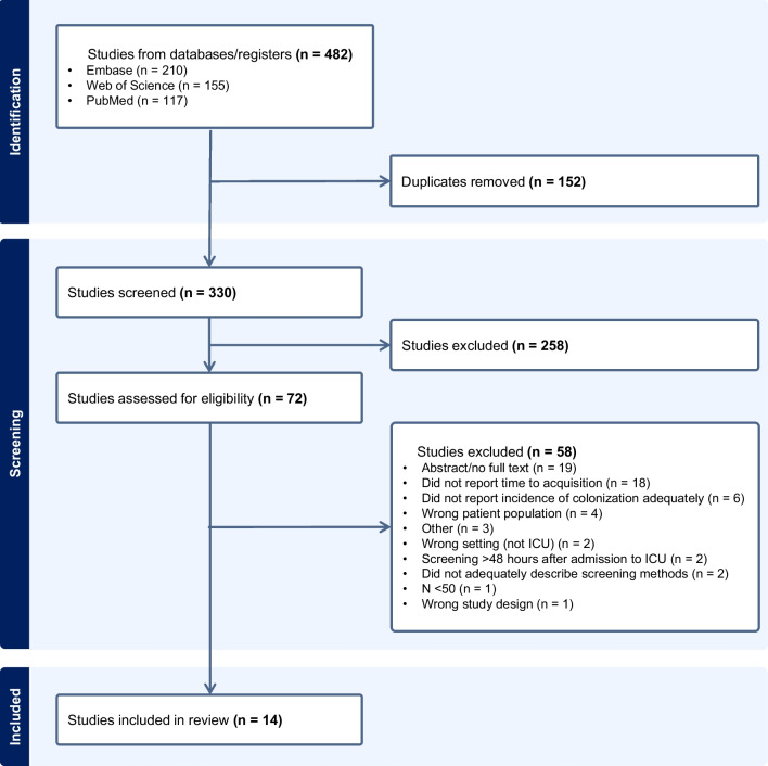
A total of 482 studies were initially identified through the database search. Of these, 152 were removed as duplicates and 258 did not meet criteria based on abstract screening. Full text review was performed for 72 studies, out of which 58 were excluded, most often because they did not report MDRO colonization longitudinally (e.g., reported baseline colonization but did not report a measure of time to acquisition of colonization in the ICU) (Fig. 1).
Fig. 1.
PRISMA flow diagram
Study characteristics
The final meta-analysis comprised 14 studies consisting of a total of 37,305 patients (Table 1). Thirteen studies reported on NP-GN, three reported on Pseudomonas, and one reported on VRE. Most of the studies (11/14) were based in the E.U. or U.S. and most were conducted pre-2013 (10/14). Patient age ranged from 49 to 65.3 with a modest male sex predominance (median 61.5% male). Matrix assisted laser desorption/ionization (MALDI) was the most common method used for organism identification, with or without disk diffusion testing for antimicrobial susceptibility (Supplemental Table S2).
Table 1.
Characteristics of included studies
| Study | Location | Period | Type of ICU | Age (years) | Sex (male, %) |
|---|---|---|---|---|---|
| Ajao [32] | United States | 2001–09 | MICU; SICU | 55.7 ± 15.6 | 55 |
| Alves [33] | France | 2011 | MICU | 64 (51–76) | 56 |
| Boutrot [34] | France | 2015–17 | SICU | 63 (49–72) | 72 |
| Gomez-Zorrilla [35] | Spain | 2012–13 | MICU; SICU | 65.3 ± 13.3† & 62.2 ± 14.3‡ | 64 |
| Grohs [23] | France | 2011 | MICU; SICU | 64.2 ± 18.3 | 59 |
| Jolivet [25] | France | 1997–2015 | MICU; SICU | 59.4 (44.4–72.0) | 64 |
| Marchenay [36] | France | 2011 | MICU; SICU | 65.3 ± 14.4† & 7.4 ± 17.6‡ | 69 |
| Papadimitriou-Olivgeris [37] | Greece | 2010–11 | ICU | 56.4 ± 19.0 | 67 |
| Poignant [38] | France | 2008–10 | MICU; SICU | 63 (49–75) | 63 |
| Qin [39] | China | 2018 | Neuro | 53.7 ± 17.8† & 56.7 ± 16.6‡ | 70 |
| Razazi [40] | France | 2010–11 | MICU | 64 (49–76)† & 64 (50–75)‡ | 60 |
| Sharma [41] | India | 2019–20 | MICU; SICU |
51 (26–62)† & 50 (32–63)‡ |
58 |
| Thiébaut [42] | France | 2005–06 | MICU; SICU | 59 (47–72) | 59 |
| Torres-Gonzalez [43] | Mexico | 2014 | ICU | 49 (32–64) | 44 |
MICU = medical intensive care unit; SICU = surgical intensive care unit; Neuro = Neurological intensive care unit; Age was reported as “Mean ± SD” or “Median (interquartile range)” depending on what was reported in the study. Some studies only reported age based on colonization status: † indicates the subgroup that was colonized with a multidrug resistant organism and ‡ indicates the subgroup and that was not colonized
Gut colonization with MDRO
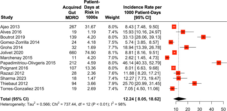
The cumulative prevalence of gut colonization at the time of ICU admission (within 48 h) was 8% (median: 10%, IQR: 7–12%) (Table 2 and Supplemental Figure S1). The cumulative incidence of MDRO gut acquisition during ICU hospitalization (i.e., the proportion of patients who were negative for gut MDRO at admission and subsequently positive) was a mean of 5% (range 1–43%) and a median of 7% (IQR 6–15%). The median time to acquisition of MDRO ranged from 4 to 26 days. The overall incidence rate for acquisition of gut MDRO was 12.2 (95% CI 8.1–18.6) per 1,000 patient-days of ICU stay (Fig. 2). Significant heterogeneity across studies was observed (I2 = 98%, p < 0.01).
Table 2.
Outcomes of interest for included studies
| Study | Organism | N | Admission colonization N (%) |
Acquired colonization N (%) |
Median time to acquisition (days) | ICU length of stay (days) | Acquisition incidence rate per 1000 patient-days (95% CI) | ||
|---|---|---|---|---|---|---|---|---|---|
| Non-colonized | Colonized | All | |||||||
| Ajao [32] | NP-GN | 8437 | 786 (9%) | 267 (3%) | 8 (4–15) | 4 (2–8) | 11 (5–24) | – | 8.4 (7.4–9.5) |
| Alves [33] | NP-GN | 309 | 25 (8%) | 19 (7%) | 7 (4–15) | 4 (3–6) | 12 (8–23) | – | 15.9 (10.2–25.0) |
| Boutrot [34] | NP-GN | 352 | 33 (9%) | 87 (27%) | 14 (6–20) | 13 (8–24) | 25 (16–46) | – | 20.6 (16.7–25.4) |
| PA | 7 (2%) | 52 (15%) | 13 (7–21) | 11.6 (8.8–15.2) | |||||
| Gomez-Zorrilla [35] | PA | 414 | 23 (6%) | 24 (6%) | 9 (7.5–12) | 10.8 ± 9.2 | 15.6 ± 10.5 | – | 5.7 (3.9–8.6) |
| Grohs [23] | NP-GN | 269 | 61 (23%) | 32 (15%) | 5.5 (4–8.3) | – | – | 8.6* | 18.9 (13.4–26.8) |
| Jolivet [25] | NP-GN | 23,423 | 1667 (7%) | 660 (3%) | 8 (5–13) | – | – | 3.3 (1.2–8.8) | 8.8 (8.2–9.5) |
| Marchenay [36] | NP-GN | 347 | 11 (3%)† | 6 (2%) | 15.3 ± 10.7 | 12.4 ± 12.5 | – | – | 1.4 (0.6–3.2) |
| PA | 5 (1%) | 1.2 (0.5–2.9) | |||||||
| Papadimitriou-Olivgeris [37] | NP-GN | 481 | 59 (12%) | 181 (43%) | 9.3 ± 5.5 | – | 12.7 ± 13.8 | 16.4 ± 20.3 | 32.1 (27.8–37.2) |
| VRE | 63 (13%) | 31 (7%) | 16.1 ± 8.9 | 16.6 ± 21.4 | 4.6 (3.2–6.4) | ||||
| Poignant [38] | NP-GN | 1,209 | 24 (2%) | 107 (9%) | 14 (7–21) | 11 (7–17) | 18 (10–30) | – | 8.0 (6.6–9.7) |
| Qin [39] | NP-GN | 243 | 37 (15%)‡ | 39 (19%)‡ | 7 (7–7) | – | – | – | – |
| Razazi [40] | NP-GN | 531 | 82 (15%) | 28 (6%) | 9 (8–20) | 5 (3–10) | 9 (4–19) | – | 11.9 (8.2–17.2) |
| Sharma [41] | NP-GN | 192 | 19 (10%) | 18 (10%) | 4 (4–5.5) | 9 (6–16) | 12 (8–22) | – | 12.3 (7.7–19.5) |
| Thiébaut [42] | NP-GN | 768 | 74 (10%) | 94 (14%) | 7 (3–11) | – | – | 5 (3–11) | 25.7 (21.0–31.5) |
| Torres-Gonzalez [43] | NP-GN | 330 | 36 (11%) | 19 (6%) | 26 (12–46) | 8 (3–16) | 15 (8–28) | – | 7.1 (4.5–11.1) |
NP-GN = non-Pseudomonas gram-negative organism. PA = Pseudomonas aeruginosa. VRE = vancomycin-resistant Enterococcus. ICU length of stay was reported as “Mean ± SD” or “Median (interquartile range)” depending on what was reported in the study and separated by colonization status depending on what was reported. A “–” indicates the value was not reported and/or incalculable. † indicates that the number of cases on admission was not separated by organism. ‡ indicates that the number of cases was not separated by rectal vs nasopharyngeal swab. *indicates a mean with no standard deviation reported. Admission colonization is a prevalence out of the total population while acquired colonization is an incidence out of the population at risk
Fig. 2.
Forest plot of incidence rate of gut acquisition of MDRO per 1000 patient-days
Stratified Analyses
To assess which factors may drive heterogeneity, the studies were stratified based on type of organism (NP-GN vs. Pseudomonas), approach to screening (sampling less vs. more than weekly), study location (Europe vs. elsewhere), data collection period (pre- vs. post-2013), and organism identification (MALDI vs. non-MALDI) (Table 3 & Supplemental Figure S2–S7). An additional analysis was performed excluding the largest study, Jolivet et al., which contained 63% of the total meta-analysis population. Studies which screened for MDRO more than weekly had higher incidence rates for gut acquisition of MDRO compared to those that screened less than weekly (16.8 (95% CI: 12.6–22.6) vs. 10.2 (95% CI: 5.3–19.4) per 1000 patient-days respectively). The incidence rate for acquiring MDRO was also substantially higher for NP-GN than PA (11.8 (95% CI: 7.6–18.2) vs. 4.6 (95% CI: 1.3–16.6) per 1000 patient-days respectively).
Table 3.
Pooled outcomes, stratified by organism and other study factors
| Studies (N) | Population (N) | Admission colonization (N, %) |
Acquired colonization (N, %) |
Incidence rate per 1000 patient-days (95% CI)* |
|
|---|---|---|---|---|---|
| Overall outcomes | 14 | 37,305 | 3,007 (8%) | 1669 (5%) | 12.2 (8.1–18.6) |
| Excluding Jolivet et al | 13 | 13,882 | 1,340 (10%) | 1009 (8%) | 12.6 (8.0–19.8) |
| Organism | |||||
| NP-GN | 13 | 36,891 | 2,914 (8%) | 1557 (5%) | 11.8 (7.6–18.2) |
| PA | 3 | 1113 | 41 (4%) | 81 (8%) | 4.6 (1.3–16.6) |
| VRE | 1 | 481 | 63 (13%) | 31 (7%) | 4.6 (3.2–6.4) |
| Screening approach | |||||
| Weekly | 9 | 35,236 | 2,746 (8%) | 1478 (5%) | 10.2 (5.3–19.4) |
| More than weekly | 5 | 2069 | 261 (13%) | 191 (11%) | 16.8 (12.6–22.6) |
| Continent | |||||
| Europe | 10 | 28,103 | 2,129 (8%) | 1326 (5%) | 13.4 (7.9–22.7) |
| Outside Europe | 4 | 9202 | 878 (10%) | 343 (4%) | 8.8 (6.8–11.4) |
| Data collection period | |||||
| Majority pre-2013 | 10 | 36,188 | 2,875 (8%) | 1454 (4%) | 11.6 (7.1–19.1) |
| Majority post-2013 | 4 | 1,117 | 132 (12%) | 139 (14%) | 14.5 (5.9–35.5) |
| Organism identification method | |||||
| MALDI | 5 | 24,596 | 1,830 (7%) | 889 (4%) | 17.2 (9.9–29.8) |
| Other | 9 | 12,709 | 1177 (9%) | 780 (7%) | 10.5 (6.1–18.0) |
NP-GN = non-Pseudomonas gram-negative organism. PA = Pseudomonas aeruginosa. VRE = vancomycin-resistant Enterococcus
*Incidence rate was calculated without Qin et al. [39] given that incidence rate could not be calculated for that study, while all other columns do include data from Qin et al. [39] Admission colonization is a prevalence out of the total population while acquired colonization is an incidence out of the population at risk
Studies Reporting multiple specific timepoints
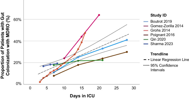
There were six studies which reported the prevalence of gut MDRO colonization at multiple predetermined timepoints (e.g., prevalence of colonization at ICU Day 7, prevalence of colonization at ICU Day 14, etc.) (Fig. 3). Among these six studies, there was evidence of a steadily increasing proportion of patients with MDRO gut colonization through up to 30 days of ICU hospitalization (linear trend with an increase of 1.41% per day, R2 = 0.50, p < 0.01). Only Grohs et al. [23] accounted for the possibility of loss of colonization, while the 5 remaining studies assumed continuous colonization after a positive screening result.
Fig. 3.
Cumulative proportion of multidrug resistant organisms (MDRO) bacteria over time in the ICU, among the six studies which reported the prevalence of gut acquisition of MDRO at multiple timepoints. Each study has been connected with colored lines and a linear regression line with 95% confidence intervals (dotted black lines) has been added which incorporates all of the data
Quality and risk of Bias
Studies were assessed for quality and clarity using the 14-point Quality Assessment Tool of the National Institutes of Health [22]. Overall, quality was rated as good to fair for the majority of studies (Supplemental Figures S8 & S9). Specific areas with poor quality across studies included lack of adequate sample size justification and lack of blinding of outcome, with colonization results generally accessible to the treating clinical team. Only Grohs et al. [23] obtained additional rectal swabs to confirm gut MDRO colonization among patients that tested positive for colonization. Last, while the Egger’s test showed insignificant results for the funnel plot asymmetry (p = 0.805), the funnel plot of the standard error against the log incidence rate suggested possible publication bias against small studies which reported high incidence rates (Supplemental Figure S10).
Discussion
This study reported on the incidence rate of MDRO gut colonization among patients in the ICU who were not colonized with an MDRO at admission. The prevalence of gut colonization at ICU admission and/or discharge has been studied in the past, but exactly when colonization occurs during ICU hospitalization has received less attention. Unlike previous meta-analyses, we were able to report on gut acquisition of MDRO in multiple ways: as a proportion of patients newly colonized during ICU hospitalization, as an incidence rate in patient-days, and in terms of median time to acquisition. Overall, we found 12.2 patients per 1000 patient-days acquired gut colonization with MDRO during ICU hospitalization and the risk of becoming colonized increased linearly with increased time in the ICU. Across studies, the median time to acquisition of colonization after ICU admission ranged from 4 to 26 days. Interestingly, there was less flattening of the curve for acquisition than one might expect, with a quasi-linear relationship between days spent in the ICU and prevalence of colonization. The risk of MDRO gut colonization varied by pathogen, with substantially higher rates observed among non-Pseudomonas Gram negatives compared to Pseudomonas spp. Pathogen colonization rates were also higher when screening was done at least weekly compared to less frequently, and when MALDI was used for pathogen identification compared to alternatives methods. In addition to the observed risk of MDRO gut colonization over the course of their ICU stay, 8% of patients were already gut colonized with MDRO at the time of ICU admission. The findings from this meta-analysis have important implications for the design of future clinical trials that evaluate measures directed at reducing gut colonization and subsequent infections by MDROs. Combined, the results suggest that interventions aiming to prevent or ameliorate MDRO gut colonization may be best deployed early during ICU admission and may need to be continued throughout ICU hospitalization. Our findings also highlight the heterogeneity of prior studies and the need for further research in this area.
Our findings align with prior meta-analyses [9, 12, 24]. Detsis et al. [9] found that the pooled incidence of ICU-acquired extended-spectrum beta-lactamase (ESBL)-producing Enterobacteriaceae colonization was 7%. Another meta-analysis by D’Agata et al. [8] found that the incidence of ICU-acquired colonization ranged from 4 to 29%. This wide range across studies—which was seen in our meta-analysis as well—may be due to baseline differences in populations or to approaches to measuring colonization. Another meta-analysis, focused on VRE, found that approximately 9% of patients were colonized on ICU admission to the ICU and another 9% acquired colonization during ICU hospitalization, similar to our results [10]. Our study found that the prevalence of colonization on ICU admission was higher than colonization acquired in the ICU for NP-GN but not for Pseudomonas. Similarly, Arzilli et al. [12] found that the prevalence of MDR Gram-negative gut colonization on hospital admission was 14% and that 9% acquired colonization, with variability based on organism. In our study and in other studies, the prevalence of MDRO gut colonization is high at ICU admission, with another substantial fraction of patients acquiring MDRO while hospitalized.
In sub-group analyses, we found that the incidence rate of gut colonization was higher among studies conducted within the past ten years, compared to those conducted from ten to 20 years ago. One of our largest included studies, Jolivet et al., reported on 18 years of data and found an increasing prevalence in ESBL-producing organisms from 1997 to 2015 [25]. Arzilli et al. observed that gut colonization prevalence was lowest before 2010, highest during the years 2010–2014, and decreased after 2014 [12]. Detsis et al. also found evidence of increasing MDRO gut colonization over time [9]. This data confirms the prioritization of WHO and other monitoring organizations. Our study also found that more frequent screening was associated with an increased detection of new colonization. Culture-based screening may have high false negative rates [26], and it is possible that studies under-represented the true rates of gut acquisition of MDRO. False positives are possible as well, and only one study in this meta-analysis used a tandem swabbing approach to confirm colonization.
The timing of gut colonization during ICU stay has not been studied as extensively as the prevalence, so it is less obvious how our results fit with previous studies. Detsis et al. found a pooled mean time from ICU admission to colonization of 11 days among three studies [9]; D’Agata et al. similarly found the median time to acquisition of a gut MDRO in the ICU ranged from 6 to 11.5 days [8]. These findings are similar to our result of a median time to acquisition ranging from 4 to 26 days. Future trials in this area may opt for study designs which deliver their interventions early during the ICU stay, which would fit into a broader ICU paradigm of early intervention (e.g. early antibiotic administration for sepsis) [27, 28].
This study has several limitations which should be considered when interpreting the findings. There was significant heterogeneity between included studies which raises questions about the validity of pooling results. We have produced pooled results because we believe that these results will help to better focus future studies and will highlight gaps in knowledge. These pooled results must be interpreted cautiously and they highlight the need for further standardization of reporting in future research. We were also unable to report on patient factors which may influence acquisition of gut MDRO such as prior or cumulative antibiotic exposure, immunodeficiency, or local prevalence of MDRO [9, 29, 30]. Additional research is needed on the timing of colonization acquisition based on these important risk factors in order to develop more targeted interventions. We did not separately analyze results based on perianal vs rectal swabs. While there is evidence to suggest results of these tests are similar [31], it is possible that perianal swabs include skin flora that rectal swabs do not which could affect the results of this study. Because the included studies reported a variety of measures (incidence, prevalence, mean, median, etc.), several assumptions had to be made during data extraction and synthesis in order to pool data together. We believe that these assumptions are reasonable, and that they do not detract from the validity of results. Most of the studies were from Europe or the U.S., and generalizability to other regions is unclear. Finally, assessment of colonization may be affected by a high false negative rate [26] and intermittent colonization as not all patients who are colonized will consistently remain colonized throughout their ICU stay [23].
Conclusion
Acquisition of gut MDRO was common in the ICU and there is a positive linear association between the proportion of patients with gut MDRO colonization and time spent in ICU through 30 days of ICU hospitalization. These data may guide future interventions seeking to prevent or ameliorate colonization. Specifically, such interventions will likely need to intervene early during the ICU stay and be maintained continuously to be effective.
Availability of data and raw materials
All data generated or analyzed during this study are included in this published article [and its supplementary information files].
Supplementary Information
Acknowledgements
Not applicable.
Author contributions
Each included author contributed substantially to this manuscript. Madison Heath and Dr. Daniel Freedberg developed with study design, performed the literature search and screening of included studies, data collection, verification, and interpretation, and writing the original manuscript draft. Weijia Fan and Dr. Cheng-Shiun Leu performed the data analysis, data interpretation, creation of figures, and revising the manuscript draft. Dr. Angela Gomez-Simmonds and Dr. Thomas Lodise assissted with study design, data interpretation, and revising the manuscript draft. All authors contributed equally to critically reviewing and editing the manuscript. All authors approved of the final draft of the manuscript to be published and agree to be accountable for all aspects of the work.
Funding
There was no source of funding for this study.
Declarations
Ethics approval and consent to participate
Not applicable.
Consent for publication
Not applicable.
Competing interests
The authors declare that they have no competing interests.
Footnotes
Publisher's Note
Springer Nature remains neutral with regard to jurisdictional claims in published maps and institutional affiliations.
References
- 1.Russell DL, Flood A, Zaroda TE, Acosta C, Riley MMS, Busuttil RW, et al. Outcomes of colonization with MRSA and VRE Among liver transplant candidates and recipients. Am J Transpl. 2008;8(8):1737–1743. doi: 10.1111/j.1600-6143.2008.02304.x. [DOI] [PubMed] [Google Scholar]
- 2.Freedberg DE, Zhou MJ, Cohen ME, Annavajhala MK, Khan S, Moscoso DI, et al. Pathogen colonization of the gastrointestinal microbiome at intensive care unit admission and risk for subsequent death or infection. Intensive Care Med. 2018;44(8):1203–1211. doi: 10.1007/s00134-018-5268-8. [DOI] [PMC free article] [PubMed] [Google Scholar]
- 3.Willems RPJ, van Dijk K, Vehreschild MJGT, Biehl LM, Ket JCF, Remmelzwaal S, et al. Incidence of infection with multidrug-resistant Gram-negative bacteria and vancomycin-resistant enterococci in carriers: a systematic review and meta-regression analysis. Lancet Infect Dis. 2023;23(6):719–731. doi: 10.1016/S1473-3099(22)00811-8. [DOI] [PubMed] [Google Scholar]
- 4.Campos-Madueno EI, Moradi M, Eddoubaji Y, Shahi F, Moradi S, Bernasconi OJ, et al. Intestinal colonization with multidrug-resistant Enterobacterales: screening, epidemiology, clinical impact, and strategies to decolonize carriers. Eur J Clin Microbiol Infect Dis. 2023;42(3):229–254. doi: 10.1007/s10096-023-04548-2. [DOI] [PMC free article] [PubMed] [Google Scholar]
- 5.Prado V, Hernández-Tejero M, Mücke MM, Marco F, Gu W, Amoros A, et al. Rectal colonization by resistant bacteria increases the risk of infection by the colonizing strain in critically ill patients with cirrhosis Graphical abstract Rectal colonization by multidrug-resistant organism Predominant colonizing strain. J Hepatol. 2022;76:1079–1089. doi: 10.1016/j.jhep.2021.12.042. [DOI] [PubMed] [Google Scholar]
- 6.Prematunge C, MacDougall C, Johnstone J, Adomako K, Lam F, Robertson J, et al. VRE and VSE Bacteremia Outcomes in the Era of Effective VRE Therapy: A Systematic Review and Meta-analysis. Infect Control Hosp Epidemiol. 2016;37(1):26–35. doi: 10.1017/ice.2015.228. [DOI] [PMC free article] [PubMed] [Google Scholar]
- 7.Alevizakos M, Gaitanidis A, Nasioudis D, Tori K, Flokas ME, Mylonakis E. colonization with vancomycin-resistant enterococci and risk for bloodstream infection among patients with malignancy: a systematic review and meta-analysis. Open Forum Infect Dis. 2017;4(1):ofw246. [DOI] [PMC free article] [PubMed]
- 8.D'Agata EMC, Geffert SF, McTavish R, Wilson F, Cameron C. Acquisition of antimicrobial-resistant bacteria in the absence of antimicrobial exposure: a systematic review and meta-analysis. Infect Control Hosp Epidemiol. 2019;40(10):1128–1134. doi: 10.1017/ice.2019.208. [DOI] [PubMed] [Google Scholar]
- 9.Detsis M, Karanika S, Mylonakis E. ICU acquisition rate, risk factors, and clinical significance of digestive tract colonization with extended-spectrum beta-lactamase–producing enterobacteriaceae. Crit Care Med. 2017;45(4):705–714. doi: 10.1097/CCM.0000000000002253. [DOI] [PubMed] [Google Scholar]
- 10.Ziakas PD, Thapa R, Rice LB, Mylonakis E. Trends and significance of VRE colonization in the ICU: a meta-analysis of published studies. PLOS ONE. 2013;8(9):e75658-e. [DOI] [PMC free article] [PubMed]
- 11.Tesfa T, Mitiku H, Edae M, Assefa N. Prevalence and incidence of carbapenem-resistant K. pneumoniae colonization: systematic review and meta-analysis. Systc Rev. 2022;11(1). [DOI] [PMC free article] [PubMed]
- 12.Arzilli G, Scardina G, Casigliani V, Petri D, Porretta A, Moi M, et al. Screening for antimicrobial-resistant Gram-negative bacteria in hospitalised patients, and risk of progression from colonisation to infection: systematic review. J Infect. 2022;84(2):119–130. doi: 10.1016/j.jinf.2021.11.007. [DOI] [PubMed] [Google Scholar]
- 13.Ferrer R, Soriano A, Cantón R, Del Pozo JL, García-Vidal C, Garnacho-Montero J, et al. A systematic literature review and expert consensus on risk factors associated to infection progression in adult patients with respiratory tract or rectal colonisation by carbapenem-resistant Gram-negative bacteria. Rev Esp Quimioter. 2022;35(5):455–467. doi: 10.37201/req/062.2022. [DOI] [PMC free article] [PubMed] [Google Scholar]
- 14.Choy A, Freedberg DE. Impact of microbiome-based interventions on gastrointestinal pathogen colonization in the intensive care unit. Ther Adv Gastroenterol. 2020;13:175628482093944. doi: 10.1177/1756284820939447. [DOI] [PMC free article] [PubMed] [Google Scholar]
- 15.Saha S, Tariq R, Tosh PK, Pardi DS, Khanna S. Faecal microbiota transplantation for eradicating carriage of multidrug-resistant organisms: a systematic review. Clin Microbiol Infect. 2019;25(8):958–963. doi: 10.1016/j.cmi.2019.04.006. [DOI] [PubMed] [Google Scholar]
- 16.Straub TJ, Lombardo M-J, Bryant JA, Diao L, Lodise TP, Freedberg DE, et al. Impact of a purified microbiome therapeutic on abundance of antimicrobial resistance genes in patients with recurrent clostridioides difficile infection. Clin Infect Dis. 2024;78(4):833–841. doi: 10.1093/cid/ciad636. [DOI] [PMC free article] [PubMed] [Google Scholar]
- 17.Munn Z, Moola S, Lisy K, Riitano D, Tufanaru C. Methodological guidance for systematic reviews of observational epidemiological studies reporting prevalence and cumulative incidence data. Int J Evid Based Healthc. 2015;13(3):147–153. doi: 10.1097/XEB.0000000000000054. [DOI] [PubMed] [Google Scholar]
- 18.Tacconelli E, Carrara E, Savoldi A, Harbarth S, Mendelson M, Monnet DL, et al. Discovery, research, and development of new antibiotics: the WHO priority list of antibiotic-resistant bacteria and tuberculosis. Lancet Infect Dis. 2018;18(3):318–327. doi: 10.1016/S1473-3099(17)30753-3. [DOI] [PubMed] [Google Scholar]
- 19.Page MJ, McKenzie JE, Bossuyt PM, Boutron I, Hoffmann TC, Mulrow CD, The PRISMA, et al. statement: an updated guideline for reporting systematic reviews. BMJ. 2020;2021:n71. doi: 10.1136/bmj.n71. [DOI] [PMC free article] [PubMed] [Google Scholar]
- 20.Covidence systematic review software: Veritas Health Innovation; 2023 [updated March 8, 2023]. Available from: www.covidence.org.
- 21.Egger M, Smith GD, Schneider M, Minder C. Bias in meta-analysis detected by a simple, graphical test. BMJ. 1997;315(7109):629–634. doi: 10.1136/bmj.315.7109.629. [DOI] [PMC free article] [PubMed] [Google Scholar]
- 22.National Heart, Lung, and Blood Institute. Quality assessment tool for observational cohort and cross-sectional studies. 2021. Available from: https://www.nhlbi.nih.gov/health-topics/study-quality-assessment-tools.
- 23.Grohs P, Podglajen I, Guerot E, Bellenfant F, Caumont-Prim A, Kac G, et al. Assessment of five screening strategies for optimal detection of carriers of third-generation cephalosporin-resistant Enterobacteriaceae in intensive care units using daily sampling. Clin Microbiol Infect. 2014;20(11):O879–O886. doi: 10.1111/1469-0691.12663. [DOI] [PubMed] [Google Scholar]
- 24.Karanika S, Karantanos T, Arvanitis M, Grigoras C, Mylonakis E. Fecal colonization with extended-spectrum beta-lactamase-producing enterobacteriaceae and risk factors among healthy individuals: a systematic review and metaanalysis. Clin Infect Dis. 2016;63(3):310–318. doi: 10.1093/cid/ciw283. [DOI] [PubMed] [Google Scholar]
- 25.Jolivet S, Lolom I, Bailly S, Bouadma L, Lortat-Jacob B, Montravers P, et al. Impact of colonization pressure on acquisition of extended-spectrum β-lactamase-producing Enterobacterales and meticillin-resistant Staphylococcus aureus in two intensive care units: a 19-year retrospective surveillance. J Hosp Infect. 2020;105(1):10–16. doi: 10.1016/j.jhin.2020.02.012. [DOI] [PubMed] [Google Scholar]
- 26.Lagier JC, Armougom F, Million M, Hugon P, Pagnier I, Robert C, et al. Microbial culturomics: paradigm shift in the human gut microbiome study. Clin Microbiol Infect. 2012;18(12):1185–1193. doi: 10.1111/1469-0691.12023. [DOI] [PubMed] [Google Scholar]
- 27.Kollef MH, Shorr AF, Bassetti M, Timsit J-F, Micek ST, Michelson AP, et al. Timing of antibiotic therapy in the ICU. Crit Care. 2021;25(1). [DOI] [PMC free article] [PubMed]
- 28.Kumar A, Roberts D, Wood KE, Light B, Parrillo JE, Sharma S, et al. Duration of hypotension before initiation of effective antimicrobial therapy is the critical determinant of survival in human septic shock. Crit Care Med. 2006;34(6):1589–1596. doi: 10.1097/01.CCM.0000217961.75225.E9. [DOI] [PubMed] [Google Scholar]
- 29.Massart N, Camus C, Benezit F, Moriconi M, Fillatre P, Le Tulzo Y. Incidence and risk factors for acquired colonization and infection due to extended-spectrum beta-lactamase-producing Gram-negative bacilli: a retrospective analysis in three ICUs with low multidrug resistance rate. Eur J Clin Microbiol Infect Dis. 2020;39(5):889–895. doi: 10.1007/s10096-019-03800-y. [DOI] [PMC free article] [PubMed] [Google Scholar]
- 30.Teerawattanapong N, Kengkla K, Dilokthornsakul P, Saokaew S, Apisarnthanarak A, Chaiyakunapruk N. Prevention and control of multidrug-resistant gram-negative bacteria in adult intensive care units: a systematic review and network meta-analysis. Clin Infect Dis. 2017;64:S51–S60. doi: 10.1093/cid/cix112. [DOI] [PubMed] [Google Scholar]
- 31.Radhakrishnan ST, Gallagher KI, Mullish BH, Serrano-Contreras JI, Alexander JL, Miguens Blanco J, et al. Rectal swabs as a viable alternative to faecal sampling for the analysis of gut microbiota functionality and composition. Sci Rep. 2023;13(1). [DOI] [PMC free article] [PubMed]
- 32.Ajao AO, Johnson JK, Harris AD, Zhan M, McGregor JC, Thom KA, et al. Risk of acquiring extended-spectrum β-lactamase-producing Klebsiella species and Escherichia coli from prior room occupants in the intensive care unit. Infect Control Hosp Epidemiol. 2013;34(5):453–458. doi: 10.1086/670216. [DOI] [PMC free article] [PubMed] [Google Scholar]
- 33.Alves M, Lemire A, Decré D, Margetis D, Bigé N, Pichereau C, et al. Extended-spectrum beta-lactamase–producing enterobacteriaceae in the intensive care unit: acquisition does not mean cross-transmission. BMC Infect Dis. 2016;16:147. doi: 10.1186/s12879-016-1489-z. [DOI] [PMC free article] [PubMed] [Google Scholar]
- 34.Boutrot M, Azougagh K, Guinard J, Boulain T. Antibiotics with activity against intestinal anaerobes and the hazard of acquired colonization with ceftriaxone-resistant Gram-negative pathogens in ICU patients: a propensity score-based analysis. J Antimicrob Chemother. 2019;74(10):3095–3103. doi: 10.1093/jac/dkz279. [DOI] [PubMed] [Google Scholar]
- 35.Gómez-Zorrilla S, Camoez M, Tubau F, Periche E, Cañizares R, Dominguez MA, et al. Antibiotic pressure is a major risk factor for rectal colonization by multidrug-resistant pseudomonas aeruginosa in critically ill patients. Antimicrob Agents Chemother. 2014;58(10):5863–5870. doi: 10.1128/AAC.03419-14. [DOI] [PMC free article] [PubMed] [Google Scholar]
- 36.Marchenay P, Blasco G, Navellou JC, Leroy J, Cholley P, Talon D, et al. Acquisition of carbapenem-resistant Gram-negative bacilli in intensive care unit: predictors and molecular epidemiology. Med Mal Infect. 2015;45(1–2):34–40. doi: 10.1016/j.medmal.2014.12.003. [DOI] [PubMed] [Google Scholar]
- 37.Papadimitriou-Olivgeris M, Spiliopoulou I, Christofidou M, Logothetis D, Manolopoulou P, Dodou V, et al. Co-colonization by multidrug-resistant bacteria in two Greek intensive care units. Eur J Clin Microbiol Infect Dis. 2015;34(10):1947–1955. doi: 10.1007/s10096-015-2436-4. [DOI] [PubMed] [Google Scholar]
- 38.Poignant S, Guinard J, Guigon A, Bret L, Poisson DM, Boulain T, et al. Risk factors and outcomes for intestinal carriage of AmpC-hyperproducing Enterobacteriaceae in intensive care unit patients. Antimicrob Agents Chemother. 2016;60(3):1883–1887. doi: 10.1128/AAC.02101-15. [DOI] [PMC free article] [PubMed] [Google Scholar]
- 39.Qin XH, Wu S, Hao M, Zhu J, Ding BX, Yang Y, et al. The Colonization of carbapenem-resistant klebsiella pneumoniae: epidemiology, resistance mechanisms, and risk factors in patients admitted to intensive care units in China. J Infect Dis. 2020;221:S206–S214. doi: 10.1093/infdis/jiz622. [DOI] [PubMed] [Google Scholar]
- 40.Razazi K, Derde LPG, Verachten M, Legrand P, Lesprit P, Brun-Buisson C. Clinical impact and risk factors for colonization with extended-spectrum β-lactamase-producing bacteria in the intensive care unit. Intensive Care Med. 2012;38(11):1769–1778. doi: 10.1007/s00134-012-2675-0. [DOI] [PubMed] [Google Scholar]
- 41.Sharma K, Tak V, Nag VL, Bhatia PK, Kothari N. An observational study on carbapenem-resistant Enterobacterales (CRE) colonisation and subsequent risk of infection in an adult intensive care unit (ICU) at a tertiary care hospital in India. Infect Prev Pract. 2023;5(4):100312. doi: 10.1016/j.infpip.2023.100312. [DOI] [PMC free article] [PubMed] [Google Scholar]
- 42.Thiébaut AC, Arlet G, Andremont A, Papy E, Sollet JP, Bernède-Bauduin C, et al. Variability of intestinal colonization with third-generation cephalosporin-resistant Enterobacteriaceae and antibiotic use in intensive care units. J Antimicrob Chemother. 2012;67(6):1525–1536. doi: 10.1093/jac/dks072. [DOI] [PubMed] [Google Scholar]
- 43.Torres-Gonzalez P, Cervera-Hernandez ME, Niembro-Ortega MD, Leal-Vega F, Cruz-Hervert LP, García-García L, et al. Factors associated to prevalence and incidence of carbapenem-resistant enterobacteriaceae fecal carriage: a cohort study in a mexican tertiary care hospital. PLoS ONE. 2015;10(10):e0139883. doi: 10.1371/journal.pone.0139883. [DOI] [PMC free article] [PubMed] [Google Scholar]
Associated Data
This section collects any data citations, data availability statements, or supplementary materials included in this article.


