Abstract
Background/Aim
Upper gastrointestinal obstruction is an extremely rare complication of primary ovarian cancer. We present a case of primary advanced ovarian cancer with gastroduodenal obstruction successfully managed with neoadjuvant chemotherapy (NAC) and conservative treatment.
Case Report
A 60-year-old woman was referred to our hospital for advanced ovarian cancer with upper gastrointestinal obstruction. Computed tomography and endoscopy revealed severe duodenal obstruction caused by dissemination. NAC was initiated with conservative management using a nasogastric tube and total parenteral nutrition (TPN). She was able to eat and TPN was stopped after three months. Complete resection was achieved with interval debulking surgery (IDS) not involving pancreatoduodenectomy, which would have been necessary for primary debulking surgery. There were no serious postoperative complications.
Conclusion
NAC with conservative management can improve upper gastrointestinal obstruction in patients with primary advanced ovarian cancer. Furthermore, IDS is expected to allow complete resection, avoiding highly invasive surgeries.
Keywords: Upper gastrointestinal tract, ovarian cancer, neoadjuvant chemotherapy, cytoreductive surgery, pancreaticoduodenectomy
Ovarian cancer often progresses via peritoneal dissemination. Complete resection of intra-abdominal lesions is important for improving the prognosis of patients with advanced ovarian cancer. However, invasive surgical procedures, such as bowel resection, diaphragmatic resection, splenectomy, and peritonectomy, are required to achieve cytoreduction without residual disease. Cytoreduction for advanced ovarian cancer is highly invasive, poses a risk of serious complications, and can be life-threatening.
Gastrointestinal obstruction caused by an intra-abdominal malignancy is described as malignant bowel obstruction (MBO), which is associated with 25-40% of advanced ovarian cancer cases (1-3). The main sites of MBO are the small intestine and colon. Patients with MBO report abdominal fullness, severe nausea, vomiting, and pain; therefore, it is important to control these symptoms. Surgical intervention is suggested for MBO if the patient has a good general condition, with one or two obstruction sites, and when symptomatic relief is expected to improve the prognosis (4). The surgical intervention type (i.e., bypass, diverting ileostomy, or colostomy) depends on the obstruction and lesion location.
Upper gastrointestinal obstruction associated with primary ovarian cancer is extremely rare, and its management has not been established. There are no reported cases of primary ovarian cancer with upper gastrointestinal obstruction treated with neoadjuvant chemotherapy (NAC) and interval debulking surgery (IDS). Herein, we present a case of primary advanced ovarian cancer with gastroduodenal obstruction for which NAC and conservative management include a nasogastric (NG) tube and total parenteral nutrition (TPN), resulting in the release of the obstruction and resumption of oral intake. Complete resection was achieved with IDS to avoid pancreaticoduo-denectomy, which is a highly invasive procedure.
Case Report
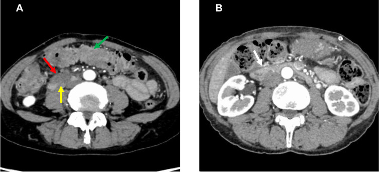
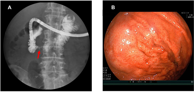
A 60-year-old woman was referred to our gynecology department with suspected advanced ovarian cancer and intraperitoneal dissemination. Her medical and family history were unremarkable. She was admitted to our hospital with vomiting and poor oral intake. Computed tomography (CT) revealed intraperitoneal dissemination causing stenosis in the third portion of the duodenum and adhesions of the omental cake extending to the transverse colon and greater curvature of the stomach (Figure 1A). Enlarged para-aortic lymph nodes were also observed. Diffusion-weighted magnetic resonance imaging of the pelvis revealed a right adnexal mass with a high signal intensity. Upper gastrointestinal endoscopy revealed severe stenosis of the pinhole near the inferior duodenal angulus (Figure 2A). As endoscopy did not reveal neoplastic lesions of the gastrointestinal mucosa, obstruction caused by external compression was considered rather than a primary gastrointestinal tumor (Figure 2B). A central venous port and NG tube were placed for decompression, and TPN was initiated. High-grade serous ovarian carcinoma was pathologically diagnosed based on the results of a percutaneous needle biopsy of the omental cake.
Figure 1. Contrast-enhanced computed tomography (CT) image before treatment revealing dissemination (red arrow) causing obstruction at the third portion of the duodenum (yellow arrow) and the omental cake (green arrow) with adhesions extending to the transverse colon and greater curvature of the stomach (A). CT image after neoadjuvant chemotherapy revealing reduction of the omental cake and improvement of the duodenal obstruction (white arrow) (B).
Figure 2. Upper gastrointestinal endoscopy image revealing severe stenosis (red arrow) near the inferior duodenal angulus of the third portion of the duodenum (A). Endoscopy image demonstrating stenosis of the duodenum caused by external compression but no neoplastic lesions in the gastrointestinal mucosa (B).
Pancreatoduodenectomy as the primary debulking surgery for complete resection was considered too invasive. Other treatments, such as gastrojejunal bypass or duodenal stent placement, have been suggested for upper gastrointestinal obstruction. However, the bypass procedure was difficult to perform because the omental cake involved the gastric wall. Furthermore, the duodenal stent was difficult to place because of severe obstruction. Her symptoms and physical condition were well-controlled with the NG tube and TPN, and the intraperitoneal metastasis of high-grade serous ovarian carcinoma was expected to resolve with chemotherapy. Therefore, we performed exploratory laparotomy and NAC after obtaining informed consent from the patient.
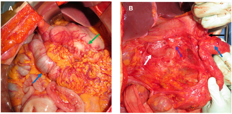
During the exploratory laparotomy, we observed the 15-cm-diameter omental cake invading the transverse colon and greater curvature of the stomach. Furthermore, the tumor adhered strongly to the duodenum on the dorsal side, forming a mass (Figure 3A). The right adnexa had swelled to a diameter of 1.5 cm. We performed right salpingo-oophorectomy, and histopathological examination revealed primary high-grade serous ovarian carcinoma.
Figure 3. Invasion of the greater curvature of the stomach and transverse colon (blue arrow) by the omental cake (green arrow) during exploratory laparotomy. The tumor was strongly adhered to the duodenum on the dorsal side (A). The shrunken omental nodule (2.5 cm; black arrow) was partially adhered to the greater curvature of the stomach and transverse colon (blue arrow) at the time of interval debulking surgery. Both the duodenum (white arrow) and pancreatic head (purple arrow) were exfoliated from the tumor (B).
Chemotherapy comprising paclitaxel and carboplatin was initiated on postoperative day (POD) 7. On POD 8, she was discharged from the hospital with an NG tube and TPN management. The omental cake decreased gradually. On POD 49, after two cycles chemotherapy, the NG tube was removed and she was able to consume water. Subsequently, she was able to consume solid foods. On POD 84, TPN was terminated after three cycles of chemotherapy. CT showed improvement of the duodenal obstruction (Figure 1B).
Thereafter, IDS involving Wertheim hysterectomy, left salpingo-oophorectomy, low anterior resection, right diaphragmatic resection, omentectomy, and retroperitoneal lymphadenectomy was performed. The operative time and blood loss were 475 min and 880 g (without blood transfusion), respectively. During laparotomy, the omental tumor was observed as a 2.5-cm nodule that did not invade the duodenum or pancreatic head. Therefore, the lesion that caused duodenal obstruction was removed along with the omentum by adhesiotomy of the duodenum and pancreatic head (Figure 3B). As the omental nodule adhered strongly to the greater curvature of the stomach and transverse colon, we resected the nodule and serosa of the greater curvature and transverse colon together (Figure 3B). Consequently, surgery without pancreatoduodenectomy was performed. On POD 14, the patient developed chylous ascites. Bowel rest and octreotide administration resulted in rapid improvement of the ascites. The patient did not experience any other complications and was discharged on POD 30. Adjuvant chemotherapy was initiated on POD 37. Thirty months after the initial treatment, there was no evidence of recurrence.
Discussion
To the best of our knowledge, this is the first case of advanced ovarian cancer with upper gastrointestinal obstruction preceded by NAC. Upper gastrointestinal obstruction rarely develops with primary ovarian cancer and intra-abdominal lesions. NAC and conservative management involving an NG tube and TPN can improve severe upper gastrointestinal obstruction caused by advanced ovarian cancer. IDS may result in complete cytoreduction, thus avoiding highly invasive surgery, such as pancreaticoduodenectomy.
During the initial treatment for primary ovarian cancer, severe obstruction of the upper gastrointestinal tract can be successfully managed with NAC and conservative treatment. The most common causes of upper gastrointestinal obstruction are gastric and pancreatic carcinomas, with 15-20% of such cases reportedly experiencing obstruction (5,6). Surgical bypass has been the traditional treatment for upper gastrointestinal obstruction. Recently, endoscopic stent placement has become an established treatment strategy for upper gastrointestinal obstruction symptoms (7). Endoscopic stent placement has a technical failure rate of 3% and clinical success rate of 87% (8). However, severe obstruction sometimes results in difficult stent placement (7). Spencer et al. (9) reported that 11 of 438 patients (2.5%) with ovarian cancer had complicated upper gastrointestinal obstruction; however, all were recurrent cases. Stent placement is effective for treating upper gastrointestinal obstruction in patients with recurrent ovarian cancer (10). However, few cases of primary advanced ovarian cancer complicated by upper gastrointestinal obstruction at the time of the initial treatment have been reported. Chemotherapy resolved the obstruction in our patient within 3 months without complications. Early chemotherapy and conservative treatment may be appropriate options for severe upper gastrointestinal obstruction, especially during the initial treatment of primary ovarian cancer, expected to respond well to chemotherapy.
For advanced ovarian cancer that invades the duodenum, IDS can allow complete resection, thereby avoiding highly invasive procedures such as pancreaticoduodenectomy. Debulking surgery for advanced ovarian cancer requires careful attention to prevent postoperative complications. When the optimal surgical treatment is difficult, IDS can be performed, resulting in no difference in prognosis compared with that associated with primary debulking surgery (PDS) and a lower risk of complications (11-16). Beissel et al. (17) reported a case in which pancreaticoduodenectomy, performed as PDS for a patient with ovarian cancer and upper gastrointestinal obstruction, resulted in a reduction of obstruction symptoms and optimal cytoreduction; however, pancreaticojejunostomy anastomotic leakage developed. After the leakage improved, the patient was readmitted for the treatment of a pancreatico-cutaneous fistula. During fistula treatment, a pelvic abscess was diagnosed and managed with drainage and antibiotics. Approximately two months after PDS, it was possible to administer additional chemotherapy. Cytoreductive surgery, including pancreaticoduodenectomy, could be highly invasive. Our patient had grade 2 chylous ascites as a postoperative complication of IDS and received adjuvant chemotherapy 37 days after surgery. IDS for advanced ovarian cancer can reduce surgical interventions. NAC followed by IDS may be safer and easier for patients with primary ovarian cancer and upper gastrointestinal obstruction.
Conclusion
Severe upper gastrointestinal tract obstruction in patients with primary advanced ovarian cancer may be successfully managed with NAC and conservative treatment. PDS with pancreaticoduodenectomy is associated with a high risk of perioperative complications; however, IDS for primary ovarian cancer is less invasive. As primary ovarian cancer is expected to respond to chemotherapy, NAC with conservative management followed by IDS should be recognized as a viable option for upper gastrointestinal obstruction to avoid pancreaticoduodenectomy.
Conflicts of Interest
The Authors have no conflicts of interest relevant to this article.
Authors’ Contributions
Ayumu Matsuoka: Conceptualization, Methodology, Writing – Original draft preparation. Tomoe Yazaki: Writing – Original draft preparation, Data curation, Visualization. Kaori Koga, Shinichi Tate, Kyoko Nishikimi, Rie Okuya, and Satoyo Otsuka: Supervision. Hirokazu Usui: Writing – Reviewing and Editing.
Funding
None.
References
- 1.Tunca JC, Buchler DA, Mack EA, Ruzicka FF, Crowley JJ, Carr WF. The management of ovarian-cancer-caused bowel obstruction. Gynecol Oncol. 1981;12(2):186–192. doi: 10.1016/0090-8258(81)90148-7. [DOI] [PubMed] [Google Scholar]
- 2.Krebs HB, Goplerud DR. Surgical management of bowel obstruction in advanced ovarian carcinoma. Obstet Gynecol. 1983;61:327–330. [PubMed] [Google Scholar]
- 3.Redman CW, Shafi MI, Ambrose S, Lawton FG, Blackledge GR, Luesley DM, Fielding JW, Chan KK. Survival following intestinal obstruction in ovarian cancer. Eur J Surg Oncol. 1988;14:383–386. [PubMed] [Google Scholar]
- 4.Hope JM, Pothuri B. The role of palliative surgery in gynecologic cancer cases. Oncologist. 2013;18(1):73–79. doi: 10.1634/theoncologist.2012-0328. [DOI] [PMC free article] [PubMed] [Google Scholar]
- 5.Kaw M, Singh S, Gagneja H, Azad P. Role of self-expandable metal stents in the palliation of malignant duodenal obstruction. Surg Endosc. 2003;17(4):646–650. doi: 10.1007/s00464-002-8527-1. [DOI] [PubMed] [Google Scholar]
- 6.Lindsay JO, Andreyev HJN, Vlavianos P, Westaby D. Self-expanding metal stents for the palliation of malignant gastroduodenal obstruction in patients unsuitable for surgical bypass. Aliment Pharmacol Ther. 2004;19(8):901–905. doi: 10.1111/j.1365-2036.2004.01896.x. [DOI] [PubMed] [Google Scholar]
- 7.Kim M, Rai M, Teshima C. Interventional endoscopy for palliation of luminal gastrointestinal obstructions in management of cancer: practical guide for oncologists. J Clin Med. 2022;11(6):1712. doi: 10.3390/jcm11061712. [DOI] [PMC free article] [PubMed] [Google Scholar]
- 8.Dormann AJ, Meisner S, Verin N, Wenk Lang A. Self-expanding metal stents for gastroduodenal malignancies: systematic review of their clinical effectiveness. Endoscopy. 2004;36(6):543–550. doi: 10.1055/s-2004-814434. [DOI] [PubMed] [Google Scholar]
- 9.Spencer JA, Crosse BA, Mannion RA, Sen KK, Perren TJ, Chapman AH. Gastroduodenal obstruction from ovarian cancer: imaging features and clinical outcome. Clin Radiol. 2000;55(4):264–272. doi: 10.1053/crad.1999.0335. [DOI] [PubMed] [Google Scholar]
- 10.Rao A, Land R, Carter J. Management of upper gastrointestinal obstruction in advanced ovarian cancer with intraluminal stents. Gynecol Oncol. 2004;95:739–741. doi: 10.1016/j.ygyno.2004.08.029. [DOI] [PubMed] [Google Scholar]
- 11.Vergote I, Tropé CG, Amant F, Kristensen GB, Ehlen T, Johnson N, Verheijen RH, van der Burg ME, Lacave AJ, Panici PB, Kenter GG, Casado A, Mendiola C, Coens C, Verleye L, Stuart GC, Pecorelli S, Reed NS. Neoadjuvant chemotherapy or primary surgery in stage IIIC or IV ovarian cancer. N Engl J Med. 2010;363(10):943–953. doi: 10.1056/NEJMoa0908806. [DOI] [PubMed] [Google Scholar]
- 12.Kehoe S, Hook J, Nankivell M, Jayson GC, Kitchener H, Lopes T, Luesley D, Perren T, Bannoo S, Mascarenhas M, Dobbs S, Essapen S, Twigg J, Herod J, McCluggage G, Parmar M, Swart AM. Primary chemotherapy versus primary surgery for newly diagnosed advanced ovarian cancer (CHORUS): an open-label, randomised, controlled, non-inferiority trial. Lancet. 2015;386(9990):249–257. doi: 10.1016/S0140-6736(14)62223-6. [DOI] [PubMed] [Google Scholar]
- 13.Onda T, Satoh T, Ogawa G, Saito T, Kasamatsu T, Nakanishi T, Mizutani T, Takehara K, Okamoto A, Ushijima K, Kobayashi H, Kawana K, Yokota H, Takano M, Kanao H, Watanabe Y, Yamamoto K, Yaegashi N, Kamura T, Yoshikawa H. Comparison of survival between primary debulking surgery and neoadjuvant chemotherapy for stage III/IV ovarian, tubaland peritoneal cancers in phase III randomised trial. Eur J Cancer. 2020;130:114–125. doi: 10.1016/j.ejca.2020.02.020. [DOI] [PubMed] [Google Scholar]
- 14.Fagotti A, Ferrandina MG, Vizzielli G, Pasciuto T, Fanfani F, Gallotta V, Margariti PA, Chiantera V, Costantini B, Gueli Alletti S, Cosentino F, Scambia G. Randomized trial of primary debulking surgery versus neoadjuvant chemotherapy for advanced epithelial ovarian cancer (SCORPION- NCT01461850) Int J Gynecol Cancer. 2020;30(11):1657–1664. doi: 10.1136/ijgc-2020-001640. [DOI] [PubMed] [Google Scholar]
- 15.Tate S, Nishikimi K, Matsuoka A, Otsuka S, Shozu M. Highly aggressive surgery benefits in patients with advanced ovarian cancer. Anticancer Res. 2022;42:3707–3716. doi: 10.21873/anticanres.15860. [DOI] [PubMed] [Google Scholar]
- 16.Mitsopoulos V, Innamaa A, Lippiatt J, Plevris N, Biliatis I. Radical surgical procedures in advanced ovarian cancer and differences between primary and interval debulking surgery. Anticancer Res. 2020;40(10):5869–5875. doi: 10.21873/anticanres.14606. [DOI] [PubMed] [Google Scholar]
- 17.Beissel JM, Kendrick ML, Podratz KC, Bakkum-Gamez JN. Pancreaticoduodenectomy in optimal primary cytoreduction of epithelial ovarian cancer: A case report and review of the literature. Gynecol Oncol Rep. 2014;10:25–27. doi: 10.1016/j.gore.2014.09.001. [DOI] [PMC free article] [PubMed] [Google Scholar]


