Abstract
Reverse genetics technology so far established for measles virus (MeV) is based on the Edmonston strain, which was isolated several decades ago, has been passaged in nonlymphoid cell lines, and is no longer pathogenic in monkey models. On the other hand, MeVs isolated and passaged in the Epstein-Barr virus-transformed marmoset B-lymphoblastoid cell line B95a would retain their original pathogenicity (F. Kobune et al., J. Virol. 64:700–705, 1990). Here we have developed MeV reverse genetics systems based on the highly pathogenic IC-B strain isolated in B95a cells. Infectious viruses were successfully recovered from the cloned cDNA of IC-B strain by two different approaches. One was simple cotransfection of B95a cells, with three plasmids each encoding the nucleocapsid (N), phospho (P), or large (L) protein, respectively, and their expression was driven by the bacteriophage T7 RNA polymerase supplied by coinfecting recombinant vaccinia virus vTF7-3. The second approach was transfection with the L-encoding plasmid of a helper cell line constitutively expressing the MeV N and P proteins and the T7 polymerase (F. Radecke et al., EMBO J. 14:5773–5784, 1995) on which B95a cells were overlaid. Virus clones recovered by both methods possessed RNA genomes identical to that of the parental IC-B strain and were indistinguishable from the IC-B strain with respect to growth phenotypes in vitro and the clinical course and histopathology of experimentally infected cynomolgus monkeys. Thus, the systems developed here could be useful for studying viral gene functions in the context of the natural course of MeV pathogenesis.
Measles virus (MeV) belongs to the genus Morbillivirus in the family Paramyxoviridae. The Paramyxoviridae, consisting of enveloped viruses with a linear, nonsegmented negative-strand RNA of approximately 15.5 kb, belongs to the order Mononegavirales with three other families, Rhabdoviridae, Bornaviridae, and Filoviridae. Reverse genetics technology first established for the segmented negative-strand RNA genome of influenza virus, a member of Orthomyxoviridae, has been applied to the nonsegmented viral RNA genomes of the Mononegavirales (21, 22, 24, 26a). The initial success in recovering an infectious virus from the full-length cDNA came in 1994 with rabies virus, a rhabdovirus (33), and many paramyxoviruses representing all four genera have been so far recovered from the cloned cDNAs (2–5, 10–13, 18, 26, 28, 40). Their genomes can now be manipulated at will by site-directed mutagenesis, greatly facilitating detailed studies of viral regulatory elements, protein functions, etc. Thus, paramyxovirus reverse genetics is improving our understanding of viral replication and pathogenesis (20).
The advantage of reverse genetics is to enable us to resolve important questions regarding pathogenicity not only in tissue culture but also in vivo. It is therefore of importance to consider the lineage of parental virus strains that we rely on in our reverse genetics approaches and the availability of appropriate animal models by which the outcomes of mutagenesis are studied (20). As for MeV, reverse genetics technology is currently available only for the Edmonston strain (28), an overattenuated laboratory strain which was isolated several decades ago (6) and extensively adapted to nonlymphoid cell lines no longer producing disease in monkey models (1, 7, 39, 42). Therefore, it is questionable how much of the original nature of MeV this strain retains. Edmonston strain-based technology would certainly help to expand knowledge about MeV (8, 25, 28, 29, 32, 37, 38), but there would likely be a serious gap between experimental findings and observations in human infections.
Wild-type MeVs circulating in nature do not grow efficiently in Vero (African green monkey kidney) cells, most frequently used for isolating MeV from patients, whereas the Edmonston strain has been adapted to grow well in Vero cells (42). Therefore, Vero cell-adapted viruses should have been selected from minor populations of quasispecies in patients, which are not fully pathogenic for monkeys and perhaps for humans (15). Thus, a marmoset B-cell line, B95a, has recently replaced Vero cells, since isolates in this cell line retain full pathogenicity in monkeys, producing clinical signs and histopathology similar to those seen in humans (15). The Ichinose-B (IC-B) strain (referred to as the 84-01 strain in a previous paper [15]), a representative pathogenic MeV, was isolated from a patient with acute measles in 1984, using B95a cells (15, 16). It is pathogenic for cynomolgus monkeys, as described previously in detail (16); Takeuchi et al. recently reported the entire genome structure of this virus, noting only two nucleotide differences in the P and M genes compared with the nonpathogenic counterpart isolated from the same patient in Vero cells (36). Here, we report two systems for virus recovery from the cloned cDNA of the IC-B strain. Reverse genetics technology based on the genetic lineage of this pathogenic isolate would bridge the gap between Edmonston-based MeV analyses and infection induced by wild-type MeV in humans.
Recovery of infectious MeV from the full-length cDNA of the IC-B strain by two different approaches.
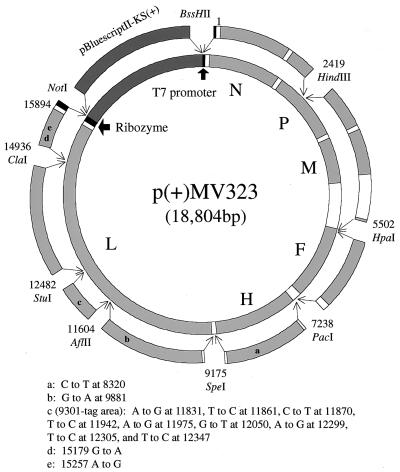
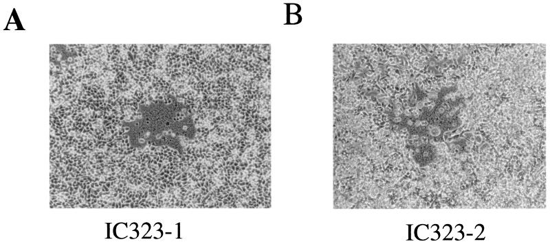
p(+)MV323, carrying the full-length cDNA of the IC-B strain, was constructed (Fig. 1). To distinguish the virus to be recovered from the cDNA from the parental IC-B strain and other field isolates, the genome segment (AflII11604 to StuI12482) in the plasmid was replaced by that of the 9301B strain, another pathogenic MeV isolate in B95a cells (34, 35), as a tag (Fig. 1). Pathogenic MeV isolated in B95a cells will grow only in lymphoid cells such as human peripheral blood mononuclear cells (PBMCs) and a monkey B95a cell line, whereas MeV strains passaged in nonlymphoid cells, such as Vero cells, are remarkably attenuated in pathogenicity for monkeys (15, 34). Thus, B95a cells are desirable for the generation of pathogenic MeV from the cDNA. B95a cells were transfected by using the lipofection reagent DOSPER (Roche Diagnostics GmbH, Mannheim, Germany) and then infected with the T7 RNA polymerase-expressing vaccinia virus vTF7-3 (a kind gift from B. Moss) (9) to express the nucleocapsid (N), phospho (P), and large (L) proteins and the antigenome of MeV from the cotransfected pEMC-Na, pEMC-Pa, pEMC-La (kind gifts from M. A. Billeter), and p(+)MV323, respectively. The cytopathic effect on B95a cells induced by vTF7-3 was negligible. Several syncytia did develop when the extract, which was prepared by three cycles of freezing and thawing of the vTF7-3-infected and plasmid-transfected cells 2 days after incubation, was inoculated into a fresh culture of B95a cells (Fig. 2A). One of the syncytia was picked up and transferred to another culture of B95a cells. After 3 days, the culture supernatant was assayed for 50% tissue culture infectious dose (TCID50) of MeV and PFU of vTF7-3 in B95a cells and Vero cells, respectively. The MeV titer was 1.3 × 10⁵ TCID50/ml, whereas vTF7-3 titer was below 50 PFU/ml. The supernatant, after diluted to 10−5-fold to eliminate vTF7-3 completely, was inoculated into B95a cells and passaged four times in the same manner to obtain a sufficient virus stock. The final stock of MeV was named IC323-1.
FIG. 1.
Using a primer containing a BssHII site and T7 promoter sequence (black area) just upstream of the MeV leader sequence and a genome-specific antisense primer, the fragment covering from the MeV leader sequence to the HindIII site at nucleotide position 2419 [frg(BssHII-T7-HindIII2419)] was generated by NAT with RT. The cDNA fragments, frg(HindIII2419-HpaI5502), frg(HpaI5502-PacI7238), frg(PacI7238-SpeI9175), frg(SpeI9175-StuI12482), and frg(StuI12482-ClaI14936), were generated by NAT. To generate a fragment from ClaI14936 to the end of the MeV genome with the genomic hepatitis delta virus ribozyme sequence (Rz) (27) (black area) and NotI [frg(ClaI14936-Rz-NotI)], NAT was carried out using an MeV genome-specific sense primer and primers containing Rz sequence and a NotI site. All fragments were assembled stepwise and cloned into the pBluescriptII KS(+) (Stratagene, La Jolla, Calif.) (dark gray area), whose multiple cloning site was replaced with BssHII, BamHI, PacI, HindIII, and NotI sites. In addition, the AflII11604 -to-StuI12482 segment of the full-genome cDNA of the IC-B strain was replaced with the corresponding segment from another B95a-isolated pathogenic 9301B strain [9301-tag area (c)]. Compared to the IC-B genome, nine synonymous mutations were accumulated in 9301-tag area (c) and another four synonymous mutations were present at nucleotide positions 8320 (a), 9881 (b), 15179 (d), and 15257 (e). Light gray areas show open reading frames of MeV N, P, M, F, H, and L proteins.
FIG. 2.
Subconfluent B95a cells in dishes 3.5 cm in diameter were infected with vTF7-3 at MOI of 1.0. Then, using the lipofection reagent DOSPER, vTF7-3-infected B95a cells were transfected with 1 μg of p(+)MV323 together with 1, 0.5, and 0.5 μg of N, P, and L protein-expressing plasmids pEMC-Na, pEMC-Pa, and pEMC-La, respectively (kind gifts from M. A. Billeter). Two days after transfection, the transfected B95a cells were scraped into the culture medium. After three cycles of freezing and thawing, the cells and medium were inoculated into fresh cultures of B95a cells. One or two days later, syncytia in B95a cells were observed by microscopy (A). Subconfluent 293-3-46 cells (a kind gift from M. A. Billeter) in dishes 3.5 cm in diameter were transfected with 5 μg of p(+)MV323 and 10 ng of pEMC-La. Two days later, B95a cells were overlaid on the transfected 293-3-46 cells. Two or three days later, syncytia were observed by microscopy in the overlaid B95a cells (B).
We also tried to recover infectious virus from p(+)MV323 by using the human embryonic kidney cell-derived 293-3-46 cells stably expressing MeV N and P proteins and T7 RNA polymerase (a kind gift from M. A. Billeter), because this system was reported to be highly efficient for recovery of the Edmonston strain and advantageous in requiring no helper virus (28). However, preliminary experiments by immunoflourescence staining of infected cells showed that the IC-B strain replicated very poorly in 293-3-46 cells, suggesting that the virus, even recovered from p(+)MV323, would not grow in these cells. As expected, by use of 293-3-46 cells alone, neither syncytium formation indicating replication of MeV (28) nor infectious virus was recovered from p(+)MV323 (data not shown). Thus, a modification that B95a cells were overlaid onto the 293-3-46 cell monolayers was attempted 2 days after transfection. This modification enabled us to recover infectious viruses efficiently from p(+)MV323. Two or 3 days later, several syncytia developed in the overlaid B95a cells (Fig. 2B). One of the syncytia was picked up and passaged three times in B95a cells to obtain a sufficient stock of the recovered MeV. The virus stock was named IC323-2.
Characterization of the recovered viruses.
Viral RNAs were purified from the culture supernatant of B95a cells infected with IC323-1 and IC323-2. To exclude possible contamination with the full-genome cDNA plasmid p(+)MV323, several regions of the MeV genome were amplified by the nucleic acid amplify technique (NAT) without reverse transcription (RT). No DNA fragment was generated by NAT, strongly suggesting that cDNA contamination did not occur. The purified RNAs of each virus were then transcribed into cDNA by reverse transcriptase, and the generated cDNAs were amplified by NAT. The cDNAs of protein-coding regions of N (C-terminal half), hemagglutinin (H), matrix (M), and P genes, as well as the 9301-tag area (nucleotide positions 11604 to 12482) (Fig. 1), were amplified and sequenced. All sequence data so far determined for the two viruses were identical to the p(+)MV323 sequence, supporting the authenticity of our plasmid-based MeV recovery system.
Growth kinetics of IC323-1 and IC323-2 analyzed in B95a and Vero cells showed that the both recovered viruses grew efficiently in B95a cells comparably to the original IC-B strain. The molecular size of each viral polypeptide and its amount synthesized in B95a cells were comparable among IC323-1, IC323-2, and the parental IC-B strains (data not shown). On the other hand, the recovered viruses replicated poorly without synthesis of any viral proteins in Vero cells, as did the original IC-B strain (data not shown). It was therefore demonstrated that both of the recovered viruses retained the in vitro phenotype characteristic of the parental IC-B strain.
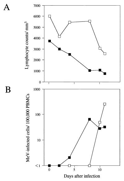
Two 10-year-old cynomolgus monkeys were infected intranasally with 105 TCID50 of IC323-2. MeV-infected PBMC counts increased up to days 8 to 11 after inoculation (Fig. 3B). Lymphopenia appeared in the monkeys as severely as that reported for infection with the original IC-B virus (16) (Fig. 3A) and also in humans (23). Typical manifestations of measles, such as coughing, Koplik spots, and maculopapular rashes (14, 17), developed in one of the monkeys as reported for IC-B-infected monkeys (16). Histopathological examinations on the monkeys autopsied on day 11 demonstrated extensive giant cell formation of lymphoid cells called Warthin-Finkeldey cells (19) as well as a number of MeV-infected mononuclear cells, which were stained intensively by the immunoperoxidase method using anti-MeV N monoclonal antibody, in lymphoid organs (data not shown). Giant cells were also found in the bronchiolar cells, epidermal cells of oral mucosa with mild acanthosis and intraepidermal neutrophil infiltration, and epidermis and hair follicles around skin rashes. These histological findings were also comparable to those with the parental IC-B strain (16) and resemble to those observed in humans (30). Finally, we determined the partial nucleotide sequences including the 9301-tag segment of the viruses isolated from the experimented monkeys and confirmed that these pathogenic viruses were derived from p(+)MV323. These results taken together demonstrated that the MeV recovered from p(+)MV323 reproduced clinical manifestations and histopathology in monkeys similar to findings for human cases (23, 30) and essentially identical to those induced by the original IC-B strain and in contrast to nonpathogenic Vero cell-grown counterparts (16), indicating the neutral effect of the synonymous mutations introduced in the p(+)MV323 sequence as the tag (Fig. 1) on both in vitro and in vivo phenotypes.
FIG. 3.
Two cynomolgus monkeys (monkeys 1 [□] and 2 (■) were inoculated intranasally with 105 TCID50 of the infectious MeV clone IC323-2. Before and at 2, 4, 8, 10, and 11 days after infection, lymphocyte count was assayed (A). PBMCs were isolated on Percoll gradients (Amersham Pharmacia biotech, Uppsala, Sweden), the starting density of which was adjusted to 1.070 g/ml. Aliquots of PBMCs (105/ml) were prepared and divided into twofold serial dilutions. Then, a 1-ml aliquot of each diluted PBMC sample was inoculated into subconfluent B95a cells in 24-well cluster plate, and cells were observed under an inverted microscope. On the assumption that one MeV-infected PBMC was contained in the maximum diluted PBMC sample that induced syncytium, the number of MeV-infected PBMC per 105 PBMCs (MeV-infected/105 PBMCs) was calculated (B).
Although paramyxovirus reverse genetics is improving our understanding of viral replication and pathogenesis, the MeV technology developed so far is based on the Edmonston strain (28), which is no longer pathogenic in the monkey models, possibly due to numerous rounds of passages in nonlymphoid cell lines (7, 42). Thus, it is not clear how useful the Edmonston-based technology will be for answering questions relevant to MeV pathogenesis. We have now succeeded in recovering infectious MeV isolates from cDNA derived from a pathogenic strain isolated in B95a cells (16). The feasibility of this reverse genetics system was demonstrated for MeV pathogenicity studies by the facts that the recovered viruses retained several in vitro and in vivo markers of MeV pathogenicity, including lymphoid cell tropism, and clinical manifestations and histopathology in monkeys. The technology could therefore facilitate comparative studies between genetic structures and pathogenicity. These studies will fill the gap between the classical virology based on the nonpathogenic Edmonston strain and actual pathogenic MeVs.
The T7 polymerase expression system from vTF7-3, which has been most frequently used in virus rescue systems, is often detrimental due to the strong cytopathogenicity of this helper virus. In addition, the helper virus generally replicated efficiently in the rescue process, and elimination of vTF7-3 from the recovered virus stock is laborious. Thus, a more attenuated vaccinia virus strain, such as MVA, has been used to express T7 polymerase (2, 4, 5, 11, 31, 41), or the enzyme is constitutively expressed in the transformed cells (28). However, the high potency of vTF7-3 to express T7 polymerase is still advantageous. Here, we showed that vTF7-3 was less cytopathic to B95a cells, replicated poorly, but still provided the sufficient polymerase activity for MeV rescue. On the other hand, B95a cells are susceptible not only to morbilliviruses but also to respiroviruses and rubulaviruses (data not shown). Therefore, the present system using B95a cells and vTF7-3 will also be applicable for the plasmid-based reverse genetics of other paramyxoviruses, and perhaps of some other nonsegmented negative-strand virus families.
The human embryonic kidney-derived 293-3-46 cells were not susceptible to B95a cell-isolated MeVs including the IC-B strain. Nevertheless, the successful rescue of IC-B virus from the overlaid B95a cells indicated that 293-3-46 cells could be permissive for replication of the transfected cDNA to yield at least one infectious unit or generate the functional RNP template to be transferred to a neighboring B95a cell. Thus, when the reconstitution of progeny virus is not fully supported by any helper cells, overlay of susceptible or natural host cells will be worthy of trial.
Acknowledgments
We thank M. A. Billeter for providing plasmids and H. Yoshikura, T. Kurata, T. Sata, A. Kato, K. Tanabayashi, M. Hishiyama, M. Kohase, S. Saito, W. Sugiura, Z. Matsuda, Y. Yanagi, and R. Cattaneo for helpful discussions.
This work was supported in part by the Ministry of Education, Science, Sports and Cultures, Japan, the Ministry of Health and Welfare, Japan, and the Organization for Pharmaceutical Safety and Research (OPSR), Tokyo.
REFERENCES
- 1.Auwaerter P G, Rota P A, Elkins W R, Adams R J, DeLozier T, Shi Y, Bellini W J, Murphy B R, Griffin D E. Measles virus infection in rhesus macaques: altered immune responses and comparison of the virulence of six different virus strains. J Infect Dis. 1999;180:950–958. doi: 10.1086/314993. [DOI] [PubMed] [Google Scholar]
- 2.Baron M D, Barrett T. Rescue of rinderpest virus from cloned cDNA. J Virol. 1997;71:1265–1271. doi: 10.1128/jvi.71.2.1265-1271.1997. [DOI] [PMC free article] [PubMed] [Google Scholar]
- 3.Buchholz U J, Finke S, Conzelmann K-K. Generation of bovine respiratory syncytial virus (BRSV) from cDNA: BRSV NS2 is not essential for virus replication in acts as a functional BRSV genome promoter. J Virol. 1999;73:251–259. doi: 10.1128/jvi.73.1.251-259.1999. [DOI] [PMC free article] [PubMed] [Google Scholar]
- 4.Collins P L, Hill M G, Camargo E, Grosfeld H, Chanock R M, Murphy B R. Production of infectious human respiratory syncytial virus from cloned cDNA confirms an essential role for the transcription elongation factor from the 5′ proximal open reading frame of the M2 mRNA in gene expression and provides a capability for vaccine development. Proc Natl Acad Sci USA. 1995;92:11563–11567. doi: 10.1073/pnas.92.25.11563. [DOI] [PMC free article] [PubMed] [Google Scholar]
- 5.Durbin A P, Hall S L, Siew J W, Whitehead S S, Collins P L, Murphy B R. Recovery of infectious human parainfluenza virus type 3 from cDNA. Virology. 1997;235:323–332. doi: 10.1006/viro.1997.8697. [DOI] [PubMed] [Google Scholar]
- 6.Enders J F, Peebles T C. Propagation in tissue culture of cytopathic agents from patients with measles. Proc Soc Exp Biol Med. 1954;86:277–286. doi: 10.3181/00379727-86-21073. [DOI] [PubMed] [Google Scholar]
- 7.Enders J F, Katz S L, Milovanovic M V. Studies of an attenuated measles virus vaccine. I. Development and preparation of the vaccine: technics for assay of effects of vaccination. N Engl J Med. 1960;263:153–159. doi: 10.1056/NEJM196007282630401. [DOI] [PubMed] [Google Scholar]
- 8.Escoffier C, Manié S, Vincent S, Muller C P, Billeter M, Gerlier D. Nonstructural C protein is required for efficient measles virus replication in human peripheral blood cells. J Virol. 1999;73:1695–1698. doi: 10.1128/jvi.73.2.1695-1698.1999. [DOI] [PMC free article] [PubMed] [Google Scholar]
- 9.Fuerst T R, Niles E G, Studier F W, Moss B. Eukaryotic transient-expression system based on recombinant vaccinia virus that synthesizes bacteriophage T7 RNA polymerase. Proc Natl Acad Sci USA. 1986;83:8122–8126. doi: 10.1073/pnas.83.21.8122. [DOI] [PMC free article] [PubMed] [Google Scholar]
- 10.Garcin D, Pelet T, Calain P, Roux L, Curran J, Kolakofsky D. A highly recombinogenic system for the recovery of infectious Sendai paramyxovirus from cDNA: generation of a novel copy-back nondefective interfering virus. EMBO J. 1995;14:6087–6094. doi: 10.1002/j.1460-2075.1995.tb00299.x. [DOI] [PMC free article] [PubMed] [Google Scholar]
- 11.He B, Paterson R G, Ward C D, Lamb R A. Recovery of infectious SV5 from cloned DNA and expression of a foreign gene. Virology. 1997;237:249–260. doi: 10.1006/viro.1997.8801. [DOI] [PubMed] [Google Scholar]
- 12.Hoffman M A, Banerjee A K. An infectious clone of human parainfluenza virus type 3. J Virol. 1997;71:4272–4274. doi: 10.1128/jvi.71.6.4272-4277.1997. [DOI] [PMC free article] [PubMed] [Google Scholar]
- 13.Kato A, Sakai Y, Shioda T, Kondo T, Nakanishi M, Nagai Y. Initiation of Sendai virus multiplication from transfected cDNA or RNA with negative or positive sense. Genes Cells. 1996;1:569–579. doi: 10.1046/j.1365-2443.1996.d01-261.x. [DOI] [PubMed] [Google Scholar]
- 14.Katz M. Clinical spectrum of measles. Curr Top Microbiol Immunol. 1995;191:1–12. doi: 10.1007/978-3-642-78621-1_1. [DOI] [PubMed] [Google Scholar]
- 15.Kobune F, Sakata H, Sugiura A. Marmoset lymphoblastoid cells as a sensitive host for isolation of measles virus. J Virol. 1990;64:700–705. doi: 10.1128/jvi.64.2.700-705.1990. [DOI] [PMC free article] [PubMed] [Google Scholar]
- 16.Kobune F, Takahashi H, Terao K, Ohkawa T, Ami Y, Suzaki Y, Nagata N, Sakata H, Yamanouchi K, Kai C. Nonhuman primate models of measles. Lab Anim Sci. 1996;46:315–320. [PubMed] [Google Scholar]
- 17.Koplik H. The diagnosis of the invasion of measles from a study of exanthema as it appears on the buccal mucous membrane. Arch Pediatr. 1896;12:918–920. [PubMed] [Google Scholar]
- 18.Lawson N D, Stillman E A, Whitt M A, Rose J K. Recombinant vesicular stomatitis viruses from DNA. Proc Natl Acad Sci USA. 1995;92:4477–4481. doi: 10.1073/pnas.92.10.4477. [DOI] [PMC free article] [PubMed] [Google Scholar]
- 19.Lightwood R, Nolan R. Epithelial giant cells in measles as an acid in diagnosis. J Pediatr. 1970;77:59–64. doi: 10.1016/s0022-3476(70)80045-2. [DOI] [PubMed] [Google Scholar]
- 20.Nagai Y. Paramyxovirus replication and pathogenesis. Reverse genetics transforms understanding. Rev Med Virol. 1999;9:83–99. doi: 10.1002/(sici)1099-1654(199904/06)9:2<83::aid-rmv244>3.0.co;2-5. [DOI] [PubMed] [Google Scholar]
- 21.Nagai Y, Kato A. Paramyxovirus reverse genetics is coming of age. Microbiol Immunol. 1999;43:613–624. doi: 10.1111/j.1348-0421.1999.tb02448.x. [DOI] [PubMed] [Google Scholar]
- 22.Neumann G, Kawaoka Y. Genetic engineering of influenza and other negative-strand RNA viruses containing segmented genomes. Adv Virus Res. 1999;53:265–300. doi: 10.1016/s0065-3527(08)60352-8. [DOI] [PubMed] [Google Scholar]
- 23.Okada H, Kobune F, Sato T A, Kohama T, Takeuchi Y, Abe T, Takayama N, Tsuchiya T, Tashiro M. Extensive lymphopenia due to apoptosis of uninfected lymphocytes in acute measles patients. Arch Virol. 2000;145:905–920. doi: 10.1007/s007050050683. [DOI] [PubMed] [Google Scholar]
- 24.Palese P, Zheng H, Engelhardt O G, Pleschka S, Garcia-Sastre A. Negative-strand RNA viruses: genetic engineering and applications. Proc Natl Acad Sci USA. 1996;93:11354–11358. doi: 10.1073/pnas.93.21.11354. [DOI] [PMC free article] [PubMed] [Google Scholar]
- 25.Patterson J B, Thomas D, Lewicki H, Billeter M A, Oldstone M B. V and C proteins of measles virus function as virulence factors in vivo. Virology. 2000;267:80–89. doi: 10.1006/viro.1999.0118. [DOI] [PubMed] [Google Scholar]
- 26.Peeters B P, de Leeuw O S, Koch G, Gielkens A L. Rescue of Newcastle disease virus from cloned cDNA: evidence that cleavability of the fusion protein is a major determinant for virulence. J Virol. 1999;73:5001–5009. doi: 10.1128/jvi.73.6.5001-5009.1999. [DOI] [PMC free article] [PubMed] [Google Scholar]
- 26a.Pekosz A, He B, Lamb R A. Reverse genetics of negative-strand RNA viruses: closing the circle. Proc Natl Acad Sci USA. 1999;96:8804–8806. doi: 10.1073/pnas.96.16.8804. [DOI] [PMC free article] [PubMed] [Google Scholar]
- 27.Perrotta A T, Been M D. A pseudoknot-like structure required for efficient self-cleavage of hepatitis delta virus RNA. Nature. 1991;350:434–436. doi: 10.1038/350434a0. [DOI] [PubMed] [Google Scholar]
- 28.Radecke F, Spielhofer P, Schneider H, Kaelin K, Huber M, Dötsch C, Christiansen G, Billeter M A. Rescue of measles viruses from cloned DNA. EMBO J. 1995;14:5773–5784. doi: 10.1002/j.1460-2075.1995.tb00266.x. [DOI] [PMC free article] [PubMed] [Google Scholar]
- 29.Radecke F, Billeter M A. The nonstructural C protein is not essential for multiplication of Edmonston B strain measles virus in cultured cells. Virology. 1996;217:418–421. doi: 10.1006/viro.1996.0134. [DOI] [PubMed] [Google Scholar]
- 30.Sata T, Kurata T, Aoyama Y, Sakaguchi M, Yamanouchi K, Takeda K. Analysis of viral antigens in giant cells of measles pneumonia by immunoperoxidase method. Virchows Arch A. 1986;410:133–138. doi: 10.1007/BF00713517. [DOI] [PubMed] [Google Scholar]
- 31.Schneider H, Spielhofer P, Kaelin K, Dötsch C, Radecke F, Sutter G, Billeter M A. Rescue of measles virus using a replication-deficient vaccinia-T7 vector. J Virol Methods. 1997;64:57–64. doi: 10.1016/s0166-0934(96)02137-4. [DOI] [PubMed] [Google Scholar]
- 32.Schneider H, Kaelin K, Billeter M A. Recombinant measles viruses defective for RNA editing and V protein synthesis are viable in cultured cells. Virology. 1997;227:314–322. doi: 10.1006/viro.1996.8339. [DOI] [PubMed] [Google Scholar]
- 33.Schnell M J, Mebatsion T, Conzelmann K-K. Infectious rabies virus from cloned cDNA. EMBO J. 1994;13:4195–4203. doi: 10.1002/j.1460-2075.1994.tb06739.x. [DOI] [PMC free article] [PubMed] [Google Scholar]
- 34.Takeda M, Kato A, Kobune F, Sakata H, Li Y, Shioda T, Sakai Y, Asakawa M, Nagai Y. Measles virus attenuation associated with transcriptional impediment and a few amino acid changes in the polymerase and accessory proteins. J Virol. 1998;72:8690–8696. doi: 10.1128/jvi.72.11.8690-8696.1998. [DOI] [PMC free article] [PubMed] [Google Scholar]
- 35.Takeda M, Sakaguchi T, Li Y, Kobune F, Kato A, Nagai Y. The genome nucleotide sequence of a contemporary wild strain of measles virus and its comparison with the classical Edmonston strain genome. Virology. 1999;256:340–350. doi: 10.1006/viro.1999.9643. [DOI] [PubMed] [Google Scholar]
- 36.Takeuchi K, Miyajima N, Kobune F, Tashiro M. Comparative nucleotide sequence analyses of the entire genomes of B95a cell-isolated and Vero cell-isolated measles viruses from the same patient. Virus Genes. 2000;20:253–257. doi: 10.1023/a:1008196729676. [DOI] [PubMed] [Google Scholar]
- 37.Tober C, Seufert M, Schneider H, Billeter M A, Johnston I C D, Niewiesk S, ter Meulen V, Schneider-Schaulies S. Expression of measles virus V protein is associated with pathogenicity and control of viral RNA synthesis. J Virol. 1998;72:8124–8132. doi: 10.1128/jvi.72.10.8124-8132.1998. [DOI] [PMC free article] [PubMed] [Google Scholar]
- 38.Valsamakis A, Schneider H, Auwaerter P G, Kaneshima H, Billeter M A, Griffin D E. Recombinant measles virus with mutations in the C, V, or F gene have altered growth phenotypes in vivo. J Virol. 1998;72:7754–7761. doi: 10.1128/jvi.72.10.7754-7761.1998. [DOI] [PMC free article] [PubMed] [Google Scholar]
- 39.von Binnendijk R S, van der Heijden R W, van Amerongen G, UytdeHaag F G, Osterhaus A D. Viral replication and development of specific immunity in macaques after infection with different measles virus strains. J Infect Dis. 1994;170:443–448. doi: 10.1093/infdis/170.2.443. [DOI] [PubMed] [Google Scholar]
- 40.Whelan S P J, Ball L A, Barr J N, Wertz G T W. Efficient recovery of infectious vesicular stomatitis virus entirely from cDNA clones. Proc Natl Acad Sci USA. 1995;92:8388–8392. doi: 10.1073/pnas.92.18.8388. [DOI] [PMC free article] [PubMed] [Google Scholar]
- 41.Wyatt L S, Moss B, Rozenblatt S. Replication-deficient vaccinia virus encoding bacteriophage T7 RNA polymerase for transient gene expression in mammalian cells. Virology. 1995;210:202–205. doi: 10.1006/viro.1995.1332. [DOI] [PubMed] [Google Scholar]
- 42.Yamanouchi K, Egashira Y, Uchida N, Kodama H, Kobune F, Hayami M, Fukuda A, Shishido A. Giant cell formation in lymphoid tissues of monkeys inoculated with various strains of measles virus. Jpn J Med Sci Biol. 1970;23:131–145. doi: 10.7883/yoken1952.23.131. [DOI] [PubMed] [Google Scholar]


