Abstract
Many recent outbreaks of infectious bursal disease in commercial chicken flocks worldwide are due to the spread of very virulent strains of infectious bursal disease virus (vvIBDV). The molecular determinants for the enhanced virulence of vvIBDV compared to classical IBDV are unknown. The lack of a reverse genetics system to rescue vvIBDV from its cloned cDNA hampers the identification and study of these determinants. In this report we describe, for the first time, the rescue of vvIBDV from its cloned cDNA. Two plasmids containing a T7 promoter and either the full-length A- or B-segment cDNA of vvIBDV (D6948) were cotransfected into QM5 cells expressing T7 polymerase. The presence of vvIBDV could be detected after passage of the transfection supernatant in either primary bursa cells (in vitro) or embryonated eggs (in vivo), but not QM5 cells. Rescued vvIBDV (rD6948) appeared to have the same virulence as the parental isolate, D6948. Segment-reassorted IBDV, in which one of the two genomic segments originated from cDNA of classical attenuated IBDV CEF94 and the other from D6948, could also be rescued by using this system. Segment-reassorted virus containing the A segment of the classical attenuated isolate (CEF94) and the B segment of the very virulent isolate (D6948) is not released until 15 h after an in vitro infection. This indicates a slightly retarded replication, as the first release of CEF94 is already found at 10 h after infection. Next to segment reassortants, we generated and analyzed mosaic IBDVs (mIBDVs). In these mIBDVs we replaced the region of CEF94 encoding one of the viral proteins (pVP2, VP3, or VP4) by the corresponding region of D6948. Analysis of these mIBDV isolates showed that tropism for non-B-lymphoid cells was exclusively determined by the viral capsid protein VP2. However, the very virulent phenotype was not solely determined by this protein, since mosaic virus containing VP2 of vvIBDV induced neither morbidity nor mortality in young chickens.
Infectious bursal disease virus (IBDV) is the causative agent of a highly contagious disease among chickens known as Gumboro disease (11). IBDV is a member of the family of Birnaviridae, having a double-stranded RNA (dsRNA) genome divided over two segments (14). The dsRNA genome is covered by a capsid of two viral proteins, which results in a single-shelled naked virus particle (60 nm) with icosahedral (T=13) symmetry (5). The largest dsRNA segment (A segment, about 3,260 bp) contains two partly overlapping open reading frames (ORFs). The first, smaller ORF encodes nonstructural viral protein 5 (VP5; ∼145 amino acids, 17 kDa; see Fig. 1A). The second ORF encodes a polyprotein (1,012 amino acids, 110 kDa; see Fig. 1B) which is autocatalytically cleaved to yield the viral proteins pVP2 (also known as VPX; 48 kDa), VP4 (29 kDa), and VP3 (33 kDa). During in vivo virus maturation pVP2 is processed into VP2 (41 to 38 kDa), probably resulting from site-specific cleavage of the pVP2 by a host cell-encoded protease (19). VP2 and VP3 are the two proteins that constitute the shell of the virion. Neutralizing antibodies are only known for VP2, and these antibodies are conformation dependent. The B segment (about 2,827 bp) contains one large ORF encoding the 91-kDa VP1 protein (see Fig. 1C). This protein contains a consensus RNA-dependent RNA polymerase motif (8). Furthermore, this protein has been reported to be linked to the 5′ ends of the genomic RNA segments (viral protein genome linked; VPg) (12, 29).
FIG. 1.
Amino acid comparison between the products of the different ORFs of the cDNAs of wild-type vvIBDV isolate D6948 and the cell culture-adapted classical isolate CEF94. The complete sequence of the D6948 proteins is given, while only those amino acids which differ from the D6948 sequence are given for CEF94. The nucleotide sequences of the A and B segments which were used to deduce the amino acid sequences can be found in the GenBank database under accession numbers AF240686 and AF194428 (A segments of D6948 and CEF94, respectively) and AF240687 and AF194429 (B segments of D6948 and CEF94, respectively). (A) Amino acid sequence encoded by the first ORF (VP5) of the A segments. (B) Amino acid sequence encoded by the second ORF (polyprotein) of the A segment. The VP4 of the polyprotein (underlined) is preceded by pVP2, while VP3 is located at the C terminus (Fig. 2). The putative cleavage sites between pVP2 and VP4 and between VP4 and VP3 (as suggested by Hudson et al. [18]) have been used. Only recently it was shown that the actual cleavage sites are most likely located between amino acids 512 and 513 (pVP2-VP4) and 755 and 756 (VP4-VP3) (27). (C) Amino acid sequence encoded by the single ORF of the B-segment (VP1/VPg). Dashes show where corresponding amino acids are missing. Amino acid changes which are found in all vvIBDV sequences are in boldface. Amino acid residues that are reported to be involved in adaptation to non-B-lymphoid cells are in italics.
The pathogenic serotype I IBDV isolates are subdivided into classical, antigenic-variant, and very virulent isolates. Antigenic-variant IBDVs have only been reported in the United States (since 1985) and were found to have single amino acid changes in a specific region of the VP2 protein (the hypervariable region), leading to a different pathologic phenotype (28). Later on in Europe (since 1988 [9]), there were reports describing IBDV isolates that had an enhanced virulence (very virulent IBDV [vvIBDV]) while having the same antigenic structure as classical isolates. Amino acid differences between viral proteins of vvIBDV and classical IBDV isolates were found scattered throughout all viral proteins, although most of them were found in the hypervariable region of VP2 (7, 24). It is currently unknown whether all or only a few of these amino acid mutations contribute to the enhanced virulence of the vvIBDV isolates.
Wild-type IBDV replicates specifically in developing B-lymphoid cells in the bursa of Fabricius. During this replication, viral proteins induce apoptosis resulting in a rapid depletion of B cells (31). A common way of producing IBDV vaccines is the adaptation of wild-type virus by propagation in chicken embryos or in cell culture using primary chicken embryo cells, primary chicken embryo fibroblast cells (CEF), or cell lines such as quail-derived cells (QT35, QM5, or QM7) and mammalian cells (Vero cells). Adaptation of wild-type IBDV is always reported to correlate with attenuation (32, 34). Adapted IBDV is able to infect non-B-lymphoid chicken cells, resulting most likely in a reduced viral load in the B-lymphoid cells in the bursas of infected chickens. Several reports which described amino acid mutations resulting from adaptation of wild-type IBDV during propagation on non-B-lymphoid cells have appeared (20, 33, 34). Furthermore, two published studies described amino acid mutations that have been introduced, using a reverse genetics system, in the VP2 region of IBDV (20, 22). These analyses show that important amino acids for propagation on non-B-lymphoid cells are found within the hypervariable region of VP2 (i.e., amino acids at position 253, 279, and 284). The influence of mutations found in regions encoded by other parts of the genome (e.g., in VP1 [7, 34]) is unclear at the moment. Studies focused on determining the influence of site-directed single or multiple amino acid mutations are hampered by the lack of a reverse-genetics system which can generate vvIBDV. In this report we describe such a system. Using the full-length cDNA of a wild-type vvIBDV isolate (D6948) we have successfully rescued recombinant D6948 (rD6948). Furthermore we rescued mosaic IBDV (mIBDV) after transfection of plasmids which contained largely cDNA originating from an attenuated classical isolate (CEF94) and partly cDNA originating from wild-type vvIBDV (D6948).
MATERIALS AND METHODS
Viruses, cells, and antibodies.
The classical IBDV isolate CEF94 is a derivative of PV1 which has been adapted for growth on cell cultures (3, 23). The wild-type vvIBDV isolate D6948 was originally isolated by the Poultry Health Service of The Netherlands (Doorn, The Netherlands; 1989) and was purified by repeated limited-dilution passages in embryonated eggs (five times). It was subsequently passaged twice in specific-pathogen-free (SPF) chickens in our laboratory. Recombinant fowlpox virus containing the T7 polymerase gene (FPV-T7) (6) was received from the laboratory of M. Skinner (Compton Laboratory, Berks, United Kingdom). QM5 cells (1) were maintained by using QT35 medium (Gibco-BRL) supplemented with 5% fetal calf serum (FCS) and 2% antibiotic solution ABII (1,000 U of penicillin [Yamanouchi], 1 mg of streptomycin [Radiumfarma], 20 μg of amphotericin B [Fungizone], 500 μg of polymixin B, and 10 mg of kanamycin/ml) in a CO2 (5%) incubator at 37°C. Primary bursa cells were isolated from 14-day-old SPF embryos and were maintained in Eagle's modified minimal essential medium (EMEM) supplemented with 15% FCS, 0.125% lactoalbumin hydrolysate (Oxoid), 1,000 units of penicillin/ml (Yamanouchi), and 1 mg of streptomycin (Radiumfarma)/ml. Polyclonal rabbit antiserum against VP1 was produced by injecting rabbits with purified recombinant VP1 (30). A polyclonal rabbit serum against VP3 and VP4 of IBDV was produced as follows. A PCR fragment containing nucleotide (nt) 2297 to nt 3192 of the A-segment cDNA (amino acids 722 to 1018 of the polyprotein) of CEF94 was fused to a six-His tag, using Escherichia coli expression plasmid pQE-30 (Qiagen). The expressed fusion protein was purified using Ni-nitrilotriacetic acid resin (Qiagen) according to the supplier's instruction. The purified proteins were separated in a sodium dodecyl sulfate-polyacrylamide gel electrophoresis (SDS-PAGE) gel and the full-length fusion product (∼35 kDa) was isolated from the gel as described by Hardy et al. (16). The recovered protein was subsequently used to immunize rabbits. The specificity (both VP4 and VP3 are recognized) and reactivity of the rabbit serum (PαVP3/4) were confirmed by enzyme-linked immunosorbent assay, radioimmunoprecipitation assay, and immunoperoxidase monolayer assay (IPMA) analysis (data not shown).
Generation of full-length A- and B-segment clones.
To produce full-length single-stranded cDNA of both the A and B segments of the two IBDV isolates (CEF94 and D6948), we used primers specific for the 3′ end of the coding strand for reverse transcription (4). Two primers specific for the 3′ ends of both the coding and noncoding strands were subsequently used to amplify the full-length A- and B-segment cDNAs in a PCR amplification using a mixture of Taq and Pwo enzymes (Expand; Boehringer Mannheim) (4). The two primers, which hybridize with the 3′ terminus of the noncoding strand, contained 5′ extensions carrying the T7 promoter (4). Three independent reverse transcriptase PCRs (RT-PCRs) were performed for each segment, and the resulting PCR fragments were cloned into the pGEM-T vector (Promega) (A segment) or in a pUC19 derivative which contained the antigenomic cis-acting hepatitis delta virus (HDV) ribozyme (10) and a T7 polymerase terminator (see Fig. 2) (3) (B segment). Sequence analysis was performed on both strands using sequence-specific primers in a cycle sequencing reaction (BigDye terminator kit; PE Applied Biosystems) and an ABI310 apparatus (PE Applied Biosystems). An unintended mutation in the A segment of one of the D6948 clones (pHB-22) was restored by exchanging a restriction enzyme fragment from an independent clone (data not shown), yielding pHB-22R. pHB-22R contains the consensus cDNA of the A segment of the D6948 vvIBDV isolate. We subsequently transferred this full-length A-segment sequence into a pUC18-based vector, which contained the HDV ribozyme and a T7 polymerase terminator (see above) by PCR amplification using the same primers as those used in the RT-PCR protocol (4). During this transfer an unintended mutation was introduced in the coding sequence for the VP4 part of the polyprotein (A1817G). This mutation was subsequently used as a genetic tag for virus rescued from this D6948 A-segment plasmid. The A-segment cDNA clone of CEF94 (pHB-36W) contains a 2-nt genetic tag (3172C→T and 3173T→A), thereby introducing a unique KpnI restriction site GGTAAC in the 3′-untranslated region of the A segment (3).
FIG. 2.
Schematic representation of the plasmids containing the full-length cDNA sequences of the A segment (pHB-60) and B segment (pHB-55) of the wild-type vvIBDV isolate D6948. The cDNA sequence is preceded by a T7 promoter sequence and is followed by the HDV ribozyme (HDR) and a T7 terminator. The different ORFs are represented by open boxes. UTR, untranslated region.
Protein sequence comparisons.
Amino acid sequences used for sequence alignments were retrieved from the GenBank database (accession numbers are in parentheses). For the alignment of VP5 and polyprotein (A segment) of the vvIBDV we used the predicted amino acid sequences of isolates D6948 (AF240686), HK46 (AF092944), UK661 (X92760), and OKYM (D49707). For the alignment of the vvIBDV VP1 sequences (B segment) we used the predicted amino acid sequences of D6948 (AF240687), HK46 (AF092944), UK661 (X92761), OKYM (D49707), IL3 (AF083093), and IL4 (AF083092).
In vitro transcription and translation.
Circular plasmids (0.4 μg) containing full-length A or B segments preceded by a T7 promoter were used as templates in a 2.5-μl in vitro transcription/translation reaction mixture (TnT-T7Quick; Promega) in the presence of [35S]methionine. The resulting viral proteins were separated in an SDS-PAGE gel (12%) and visualized by autoradiography.
Infection and transfection of QM5 cells.
QM5 cells were grown to 80% confluency in a 35-mm-diameter culture dish (M-6; 9.6 cm2/well) and infected with FPV-T7 (multiplicity of infection [MOI] = 3). After 1 h, the cells were washed twice with 3 ml of QT-35 medium and covered with 3 ml of Optimem 1 (Gibco-BRL). In the meantime, DNA (1.0 μg) was mixed with 12.5 μl of Lipofectamine (Gibco-BRL) in 250 μl of Optimem 1 and kept at room temperature for at least 30 min. The QM5 cells were subsequently covered with 2 ml of fresh Optimem 1, and the DNA-Lipofectamine mixture was added. The transfection was performed overnight (18 h) in a 37°C incubator (5.0% CO2). The transfected monolayer was rinsed once with phosphate-buffered saline (PBS), fresh QM5 medium (supplemented with 5% FCS and 2% ABII) was added, and the plates were further incubated in the 37°C (5.0% CO2) incubator. After 24 h of incubation the plates were freeze-thawed once and the supernatant was filtered through a 200-nm-pore-size filter (Acrodisc; Gelman Sciences) to remove the FPV-T7 and cellular debris. The cleared supernatant was either stored at −70°C or used directly for further analysis.
Detection of infectious rIBDV.
After transfection, infectious rescued IBDV (rIBDV) was detected by inoculating a nearly confluent monolayer of QM5 cells with part of the cleared transfection supernatant. IBDV-specific proteins were visualized after 24 h in an IPMA (3). rIBDV (e.g., rD6948) which is unable to replicate on QM5 cells was also used to inoculate a monolayer of primary bursa cells which had been grown in vitro for 24 h in a 35-mm-diameter tissue culture dish in a CO2 incubator (5%) at 39°C. IBDV-specific proteins in infected B-lymphoid cells were detected in an IPMA after 48 h.
Single-step IBDV growth curves.
To assess the replication ability of the rCEF94 and segment-reassorted IBDV (srIBDV)-CADB isolates (see Table 1), we determined single-step growth curves. QM5 cells (2 × 10⁶) were grown overnight (16 h) in a 60-mm-diameter cell culture dish. The medium was subsequently removed, and 1 ml of medium containing IBDV (50% tissue culture infective dose [TCID50] = 107.0; MOI = 5) was used to cover the cells. After 1 h the medium was removed, the cells were rinsed three times with PBS, and 5 ml of fresh medium was added. At different time points (5, 10, 15, and 25 h postinfection [p.i.]) samples were taken from the medium and stored at −20°C. The amount of IBDV (TCID50) in each sample was determined by infecting 96-well tissue culture plates containing nearly confluent QM5 cells with 10-fold dilutions of the IBDV samples. The 96-well plates were incubated for 48 h, and infective centers of IBDV (IBDV antigen-positive areas) were detected by an IPMA (see “Detection of infectious rIBDV”).
TABLE 1.
Description of plasmids and viruses
| Plasmid or virus | Based on plasmid | Main feature |
|---|---|---|
| Plasmids | ||
| pUC18-Ribo | pUC18 | Contains the HDV ribozyme |
| pHB-22R | pGEM-T | A-segment cDNA of D6948 (consensus sequence) |
| pHB-60 | pUC18-Ribo | A-segment cDNA of D6948, including a genetic tag |
| pHB-36W | pUC18-Ribo | A-segment cDNA of CEF94, including a genetic tag |
| pHB-34Z | pUC18-Ribo | B-segment cDNA of CEF94 (consensus sequence) |
| pHB-55 | pUC18-Ribo | B-segment cDNA of D6948 (consensus sequence) |
| pHB36-vvVP2 | pHB-36W | pVP2 of CEF94 cDNA is replaced by pVP2 of D6948 cDNA (453 amino acids) |
| pHB36-vvVP3 | pHB-36W | VP3 of CEF94 cDNA is replaced by VP3 of D6948 cDNA (289 amino acids) |
| pHB36-vvVP4 | pHB-36W | VP4 of CEF94 cDNA is replaced by VP4 of D6948 cDNA (270 amino acids) |
| Viruses | ||
| CEF94 | n.r.a | Classical, cell culture-adapted IBDV isolate (derivative of PV1) |
| D6948 | n.r. | vvIBDV field isolate (The Netherlands, 1989) |
| rCEF94 | pHB-36W + pHB-34Z | rIBDV; classical, cell culture-adapted IBDV |
| rD6948 | pHB-60 + pHB-55 | rIBDV; vvIBDV |
| srIBDV-CADB | pHB-36W + pHB-55 | Rescued srIBDV (A segment, CEF94; B segment, D6948) |
| srIBDV-DACB | pHB-60 + pHB-34Z | Rescued srIBDV (A segment, D6948; B segment, CEF94) |
| mCEF94-vvVP2 | pHB36-vvVP2 + pHB-34Z | Rescued mIBDV; pVP2 originates from D6948, the remaining parts from CEF94 |
| mCEF94-vvVP3 | pHB36-vvVP3 + pHB-34Z | Rescued mIBDV, VP3 originates from D6948, the remaining parts from CEF94 |
| mCEF94-vvVP4 | pHB36-vvVP4 + pHB-34Z | Rescued mIBDV, VP4 originates from D6948, the remaining parts from CEF94 |
n.r., not relevant.
Construction of mosaic A-segment plasmid pHB36-vvVP2.
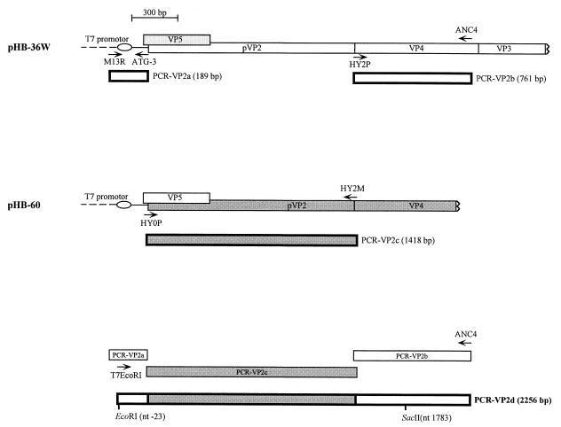
To replace the coding sequence for the pVP2 part of the CEF94 polyprotein with that for the corresponding part of the D6948 polyprotein, we generated three PCR fragments (i.e., VP2a, VP2b, and VP2c; see Fig. 6). PCR fragment VP2a (189 bp) was generated by using primers M13R (TCACACAGGAAACAGCTATGAC) and ATG3 (CATCGCTGCGATCGTTTGTCTGATCTCTAC), with pHB-36W as the template. PCR fragment VP2b (761 bp) was generated by using primers (ATCCGGGCCCTAAGGAGG) and ANC4 (GCCAAGTCGGTGTGCAG), with pHB-36W as the template. PCR fragment VP2c (1,418 bp) was generated by using primers HY0P (TATCATTGATGGTCAGTAGAG) and HY2M (CACCGGCACAGCTATCC), with pHB-22R as the template. These three PCR fragments were agarose gel purified using a Qiaex gel extraction kit (Qiagen) and used as templates (50 ng of each fragment) in a fusion PCR using primers T7EcoRI (GGAATTCTAATACGACTCACTATAGG) and ANC4. This PCR fragment (2,256 bp) was subsequently digested with EcoRI and SacII (resulting in a 1,806-bp fragment), agarose gel purified (Qiaex gel extraction kit), ligated into pHB-36W (which had been digested with the same restriction enzymes), and then used to transform E. coli DH5α cells. Several plasmids were analyzed, and one plasmid having the intended sequence was selected (pHB36-vvVP2).
FIG. 6.
Schematic representation of the construction of a mosaic PCR fragment having a middle part originating from D6948 cDNA (shaded boxes) and flanking parts originating from CEF94 cDNA (open boxes). For the construction of pHB-36-vvVP2 we first generated PCR fragments VP2a, VP2b, and VP2c. These PCR fragments were purified and subsequently used as templates in a fusion PCR, yielding PCR fragment VP2d (17). This PCR fragment was subsequently purified and used to replace the corresponding part of the CEF94 A-segment cDNA, by using the indicated EcoRI and SacII restriction sites. pHB36-vvVP3 and -vvVP4 were generated by the same approach, using different primers to generate the PCR fragments and different restriction sites to introduce the PCR fragments into the full-length A-segment clone (see Materials and Methods and Table 1).
Construction of mosaic A-segment plasmid pHB36-vvVP3.
To replace the coding sequence for the VP3 part of the CEF94 polyprotein with the corresponding part of the D6948 polyprotein, we generated two PCR fragments. PCR fragment VP3a (1,622 bp) was generated by using primers AC3 (GGTAGCCACATGTGACAG) and HY3M (CCAGTCcCGcGGATTGTGAGG), with pHB-36W as the template. PCR fragment VP3b (1,252 bp) was generated by using primers HY3P (AACGTTTTCCTCACAATCCgCGgGACTGGG) and M13F (GTAAAACGACGGCCAGT), with pHB-22R as the template. These two PCR fragments were agarose gel purified and used as templates (50 ng of each fragment) in a fusion PCR using primers AC4 (ACCCAGCCAATCACATCC) and AGTM (GAGACTCCCAGGtaCCTCACT). This PCR fragment (2,154 bp) was subsequently digested with EagI and KpnI (resulting in a 1,857-bp fragment) and used to exchange the corresponding part of pHB-36W in the same way as described for pHB36-vvVP2, yielding pHB36-vvVP3.
Construction of mosaic A-segment plasmid pHB36-vvVP4.
To replace the coding sequence of the VP4 part of the CEF94 polyprotein with the corresponding part of the D6948 polyprotein, we generated three PCR fragments. PCR fragment VP4a (796 bp) was generated by using primers AC3 and HY4M (CCGGCACAGCTATCCT), with pHB-36W as the template. PCR fragment VP4b (644 bp) was generated by using primers HY3P and ANC2 (CTGCCTGTCCTGGAGCC), with pHB-36W as the template. PCR fragment VP4c (864 bp) was generated by using primers HY4P (ACATAATCCGGGCCATAAGG) and HY3M, with pHB-22R as the template. These three PCR fragments were agarose gel purified and used as templates (50 ng of each fragment) in a fusion PCR using primers AC4 and ANC3 (CGATGGGCGTTCGGGTC). This PCR fragment (2,154 bp) was subsequently digested with EagI and DraIII (resulting in a 1,189-bp fragment) and used to exchange the corresponding part of pHB-36W in the same way as described for pHB36-vvVP2, yielding pHB36-vvVP4.
Virulence of rIBDV in young SPF chickens.
The virulence of the rIBDV, srIBDV, and mIBDV (see Table 1) isolates was evaluated in SPF layer type chickens. The IBDV isolates were first propagated on embryonated eggs by inoculating the QM5 cell supernatant from a transfection experiment into 11-day-old embryonoted eggs via the chorioallantoic membrane route. After 5 days of incubation the embryos (dead or alive) were recovered, homogenized in a Sorval Omni-mixer (three times for 10 s each at maximum speed), subsequently clarified by centrifugation (6,000 × g, 10 min), and stored in aliquots at −70°C. The virus titers (50% embryo lethal dose [ELD50]) in these samples were determined using 11-day-old embryonated eggs. In the virulence test, nine groups of chickens (21 days old) were inoculated intranasally and by eye drop with 1,000 ELD50s of IBDV in PBS. In addition one group of 10 chickens received PBS only (negative-control group). The groups of chickens were housed separately in isolators. The animals were monitored for clinical signs daily, and dead chicks were removed and necropsied. At 9 days p.i., all chicks from the negative-control groups and all remaining chicks from groups in which mortality had occurred were bled (5 ml) and euthanized for necropsy. From the other groups, six chicks were bled (5 ml) and taken for necropsy at day 9 p.i., whereas the remaining four were bled (5 ml) and taken for necropsy at day 15 p.i. Bursa and body weights of all chicks that had been euthanized at day 9 p.i. were determined. The bursa weight/body weight ratios were analyzed by a one-way analysis of variance with a factor for groups. Ratios were log transformed prior to analysis. Pairwise comparisons between groups were based on Fisher's least significant difference method (pairwise t tests with a pooled-variance estimator). At necropsy at days 9 and 15 p.i. samples from the bursa of Fabricius were fixed in 10% neutrally buffered formalin for histopathology. Bursa samples were snap-frozen in liquid nitrogen and preserved at −70°C for immunohistochemical examination. IBDV antibody titers in serum, taken just before euthanasia, were determined using the infectious bursal disease antibody detection kit (Idexx, Westbrook, Maine).
Histopathology and immunohistochemistry.
Formalin-fixed bursa samples were dehydrated, embedded in paraffin wax, sectioned, and stained with hematoxylin-eosin. The histopathologic bursal lesion score (HBLS) was determined by microscopic analysis of the bursa. The HBLS was determined on a scale of 1 to 5 as described in reference 2: 1, normal bursa; 2, scattered or partial follicle damage; 3, 50% or less follicle damage; 4, 50 to 75% follicle damage; 5, 75 to 100% follicle damage. Frozen bursa samples were sectioned for immunohistology on a cryostat at 8-μm thickness and taken up on glass slides (Superfrost). The sections were fixed with acetone for 10 min, air dried, and stored at −20°C until used. An immunoperoxidase staining was performed as described previously (25). Briefly, endogenous peroxidase in sections was eliminated by 0.01% H2O2. Nonspecific reactions were blocked by 0.2% bovine serum albumin. A 1:100 dilution of PαVP3/4 was used as the primary antibody, whereas a 1:100 dilution of goat anti-rabbit immunoglobulin G antibody conjugated to peroxidase (Dako) was used as the secondary antibody, and diaminobenzidine was used as the substrate.
Detection of genetic tag.
Bursas of chickens originating from the groups in which mortality had occurred during the animal experiment (see above) were homogenized, and viral dsRNA was extracted using a QiaAmp tissue kit (Qiagen, Hilden, Germany). The dsRNA was concentrated by standard ethanol precipitation and subsequently used as the template in a reverse transcription reaction using primer ANC1 (GGGGACCCGCGAACG). PCR (Taq polymerase) was performed on this template using primers AC3 and ANC5 (CCCATCTGGAGCATATCC) by cycling 35 times between 94 (15 s), 55 (15 s), and 72°C (90 s). The obtained PCR products (1,266 bp) were gel purified (Qiaex gel extraction kit; Qiagen) and used as template for sequence analysis, by using primer AC6 (TTCACCTGGGGTACTCCG).
RESULTS
Cloning of full-length cDNA of vvIBDV.
To study molecular determinants that are responsible for the very virulent phenotype of vvIBDV isolates, we cloned and sequenced the full-length A and B segments of the vvIBDV isolate D6948 three times independently. The cDNA sequence of the D6948 A segment (3,260 bp) differs at 122 positions from the sequence of the classical, attenuated CEF94 IBDV isolate (data not shown). These nucleotide differences result in an N-terminal extension of four amino acids (MLSL) for the predicted VP5 of D6948 and five additional amino acid differences between the two VP5 proteins (Fig. 1A). Furthermore, there were 18 amino acid differences between the two polyproteins, of which 11, 5, and 2 are located in the pVP2, VP4, and VP3 parts, respectively, of the polyprotein (Fig. 1B). Between the two B segments (2,827 bp) we found 288 nt differences (data not shown), resulting in 17 amino acid differences between the two VP1s (Fig. 1C), the only protein encoded by the B segment.
By combining sequences of different full-length cDNA clones we constructed plasmids which contained the consensus cDNA sequences of the D6948 A and B segments. The A- and B-segment cDNAs, including an artificially introduced T7 promoter sequence, were subsequently transferred to a pUC19-based vector which contained a cis-acting HDV ribozyme (10), yielding pHB-60 (A segment) and pHB-55 (B segment) (Table 1 and Fig. 2). These pUC-19-based transcription plasmids are the basis for rescuing rIBDV from cloned cDNA, using a recombinant helper virus (fowlpox) that expresses T7 polymerase (6). We have used this in vivo T7 expression system previously to rescue infectious IBDV from cloned cDNA of the cell culture-adapted, classical IBDV isolate CEF94 (3).
In vitro transcription/translation.
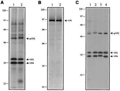
The A- and B-segment cDNA clones of vvIBDV isolate D6948 were used as templates in an in vitro transcription/translation reaction (TnT-T7Quick; Promega). The protein products of these cDNA clones appeared to be identical to those of the classical attenuated CEF94 cDNA plasmids (i.e., pVP2, VP3, and VP4 for the A segment and VP1 for the B segment) in an SDS-PAGE analysis (Fig. 3A and B). The pVP2 from D6948 is found at a slightly higher position than the pVP2 protein from CEF94. Plasmid pHB-60 (D6948 A segment) contains a single nucleotide substitution at position 1817 (A1817G), resulting from the PCR-based cloning strategy. This mutation leads to a conserved amino acid substitution in VP4 (I563V). As this mutation did not affect the processing of the polyprotein (Fig. 3A) (unpublished results), we have subsequently used this mutation as a tag for virus rescued from this D6948 A-segment plasmid.
FIG. 3.
Autoradiogram of an SDS-PAGE analysis of a coupled in vitro transcription/translation reaction. (A) Full-length A-segment plasmids of attenuated classical IBDV isolate CEF94 (pHB-36W, lane 1) and wild-type vvIBDV isolate D6948 (pHB-60, lane 2). (B) Full-length B-segment plasmids of CEF94 (pHB-34Z, lane 1) and D6948 (pHB-55, lane 2). (C) Full-length A-segment plasmid of CEF94 (pHB-36W, lane 1) and full-length A-segment plasmid of the classical attenuated IBDV isolate in which either pVP2 (pHB36-vvVP2; lane 2), VP3 (pHB36-vvVP3; lane 3), or VP4 (pHB36-vvVP4; lane 4) has been exchanged. The positions of the viral proteins are on the right. The sizes of the marker proteins (Rainbow marker; Amersham) are on the left.
Transfection of QM5 cells.
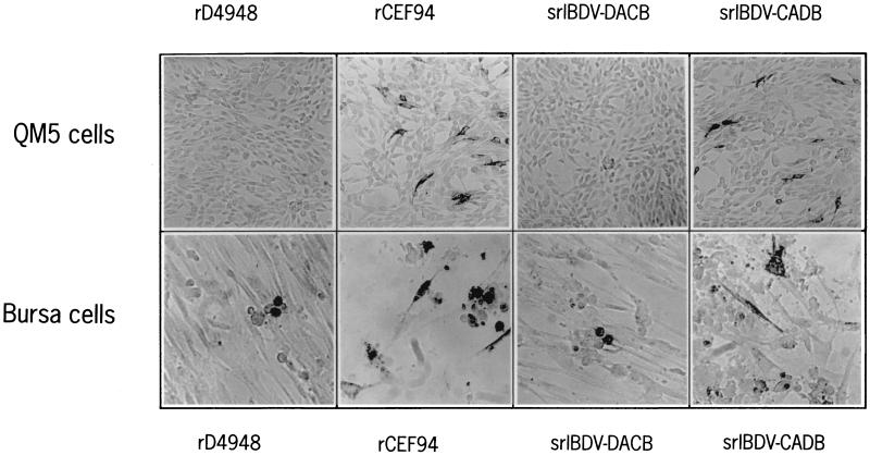
Plasmids containing the A- and B-segment cDNA of either CEF94 or D6948 were used to cotransfect QM5 cells. Transient expression of the viral proteins originating from both the A and B segments was observed in all examined cases in an IPMA by using antibodies specific for either VP3 and VP4 (A segment) or VP1 (B segment) (data not shown). To assess the production of infectious IBDV originating from cloned cDNA (rIBDV), we transferred part of the supernatant onto a fresh monolayer of QM5 cells (first passage) and analyzed the expression of viral proteins in these cells after 24 h in an IPMA. This analysis showed that no infectious rIBDV could be detected in QM5 cells treated with the supernatant from a cotransfection of the A- and B-segment plasmids of vvIBDV isolate D6948 (Fig. 4). In contrast, infectious rIBDV was present in QM5 cells treated with supernatant from the cotransfection with plasmids containing the A and B segments of the attenuated CEF94 isolate (Fig. 4). The cotransfection of the A-segment plasmid of D6948 with the B-segment plasmid of CEF94 yielded no infectious srIBDV-DACB (Table 1). However, the reciprocal combination srIBDV-CADB (Table 1) did yield infectious srIBDV (Fig. 4). Wild-type IBDV isolates such as D6948 are unable to grow on fibroblast cells such as QM5 cells (see the introduction). Therefore we assessed the presence of infectious rD6948 originating from cloned cDNA by transferring supernatants of transfected QM5 cells onto primary bursa cells which had been grown in vitro for 24 h. Similar to what was found for wild-type D6948, which can infect only the lymphoid cells present in this monolayer (data not shown), we were able to detect rD6948 in several lymphoid cells after 2 days of incubation with the transfection supernatant (Fig. 4, lower panels). Incubation of primary bursa cells with the transfection supernatant of A- and B-segment cDNAs from CEF94 showed that not only lymphoid cells but also fibroblast cells were infected, leading to a destruction of the monolayer within 48 h of incubation. Virus present in the supernatant from a transfection with the A-segment cDNA of D6948 and B-segment cDNA of CEF94 was only able to infect lymphoid cells, and no destruction of the monolayer was observed after 48 h. Similar to rD6948, srIBDV-DACB is apparently unable to infect fibroblast cells. Again, infection of fibroblast cells was found with virus originating from transfection of the A-segment cDNA of CEF94 and B-segment cDNA of D6948, although the monolayer showed less destruction after 48 h than was produced by rCEF94 (Fig. 4).
FIG. 4.
Detection of IBDV using the VP3 and VP4 polyclonal antiserum. Samples of supernatant from different plasmid transfections were used to infect QM5 or primary bursa cells. After infection the QM5 cells were incubated for 24 h, while the primary bursa cells were incubated for 48 h. IBDV proteins were visualized by performing an IPMA.
Single-step growth curves.
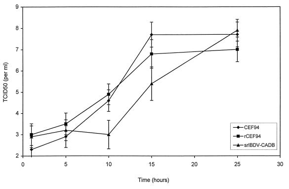
To determine whether the rescued rCEF94 and srIBDV-CADB had the same replication properties on QM5 cells as wild-type CEF94, we performed single-step growth curves (in triplicate) for each of these viruses. QM5 cells were infected during 1 h with IBDV (MOI = 5), after which time the cells were rinsed three times and incubated in complete medium. Part of the supernatant of the infected QM5 cultures was removed at different time points. The amount of IBDV (TCID50 per milliliter) was determined in each sample (Fig. 5). Release of infectious IBDV was found at 10 h p.i. for both wild-type CEF94 and rCEF94, while the first release of srIBDV-CADB was only found at 15 h p.i. Final titers (24 h p.i.) were about the same for wild-type CEF94, rCEF94, and the rescued srIBDV containing the A segment of CEF94.
FIG. 5.
Single-step growth curves of CEF94, rCEF94, and srIBDV-CADB. QM5 cells were infected with IBDV (MOI = 5) for 1 h beginning at time zero, washed three times, and covered with fresh medium. At different time points samples were taken from the supernatant and the amount of IBDV (TCID50 per milliliter) was determined. The TCID50 at each time point is the mean of three independent experiments; error bars represent standard deviations.
Construction of mosaic A-segment plasmids.
To analyze the viral determinants responsible for the very virulent phenotype of isolate D6948, we constructed three different plasmids containing full-length mosaic IBDV A-segment cDNA. Using a PCR-based method (Fig. 6) we constructed DNA fragments of which the middle part consisted of cDNA originating from D6948 (i.e., the pVP2-, VP3-, or VP4-encoding part), while the flanking cDNA originated from CEF94. The transition positions between the different cDNAs were at the putative cleavage sites between the pVP2-VP4 and VP4-VP3 parts of the polyprotein (i.e., behind amino acids 453 and 723, respectively [Fig. 1B]) (18). The mosaic PCR fragments were subsequently used to replace the corresponding part of CEF94 A-segment cDNA in plasmid pHB-36W, using specific restriction endonucleases. The exchanged parts of the generated plasmids, named pHB36-vvVP2, -vvVP3, and -vvVP4, were sequenced, and plasmids containing the intended mosaic cDNA sequence were subsequently used for in vitro transcription/translation and analyzed for the production of viral proteins by means of SDS-PAGE (Fig. 3C). No apparent difference in the autocatalytic cleavage was observed in the polyproteins encoded by these mosaic cDNA A segments.
Rescue of IBDV from mosaic A-segment cDNA.
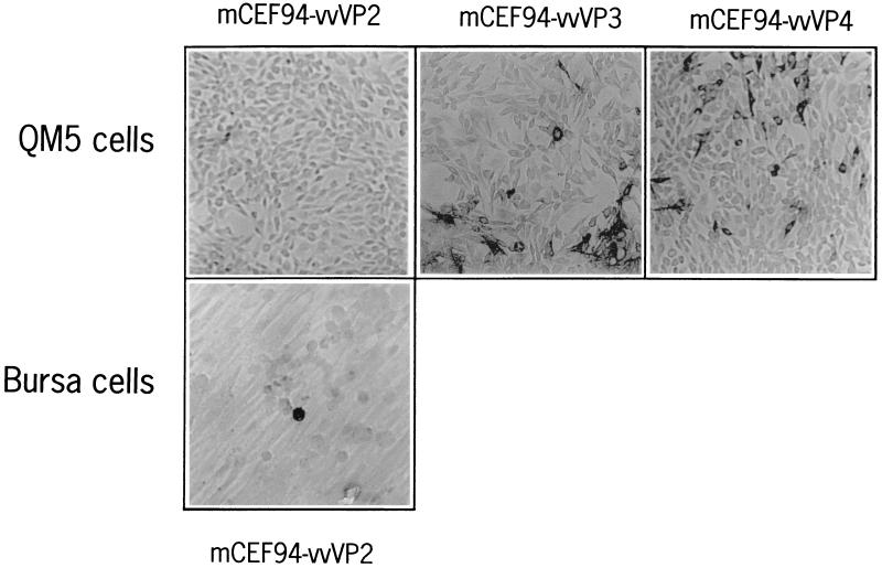
Plasmids pHB36-vvVP2, -vvVP3, and -vvVP4 were used to transfect QM5 cells together with the B-segment cDNA of CEF94 (pHB-34Z). The presence of mIBDV (Table 1) was analyzed by transferring aliquots of the transfection supernatant to a fresh monolayer of QM5 cells. After 1 day of incubation we fixed the cells and used an IBDV-specific antibody assay (IPMA) to test for the presence of mIBDV. No mIBDV could be detected in the QM5 monolayer when supernatant of the pHB36-vvVP2 plasmid was used, while for pHB36-vvVP3 and pHB36-vvVP4 mIBDV was clearly present (Fig. 7). The fact that we did not detect mCEF94-vvVP2 in the QM5 monolayer could result from the difference in cell tropism between the rCEF94 and mCEF94-vvVP2. To check for this possibility, we transferred part of the supernatant from the transfection with pHB36-vvVP2 onto a monolayer of primary bursa cells. After 2 days we were able to show the presence of mCEF94-vvVP2 virus in some of the B-lymphoid cells (Fig. 7, lower panel). Apparently, mCEF94-vvVP2 had the same cell tropism as D6948, rD6948, and srIBDV-DACB, since it was only able to grow on B-lymphoid cells and not on fibroblast cells.
FIG. 7.
Detection of mosaic IBDV using VP3 and VP4 polyclonal antiserum. Samples of supernatant of different plasmid transfections were used to infect QM5 and primary bursa cells. After infection, the QM5 cells were incubated for 24 h, while the primary bursa cells were incubated for 48 h (Fig. 4). In contrast to what was found for the negative control (mock infected), in which no B-lymphoid cells stained positive (data not shown), we found several stained B-lymphoid cells scattered throughout the culture tissue dish in the IPMA shown in the lower panel.
Animal experiment.
To compare the virulence of rIBDV, srIBDV, and mIBDV isolates with that of the parent isolates, we inoculated 21-day-old SPF chickens (10 chickens per virus) intranasally and by eye drop in one eye with 1,000 ELD50s each. Clinical signs occurred only in the groups that had received D6948, rD6948, and srIBDV-DACB (data not shown). In these groups the mortality was 3 out of 10 for D6948, 5 out of 10 for rD6948, and 2 out of 10 for srIBDV-DACB (Table 2). Neither morbidity nor mortality was found in the other groups. At day 9 p.i. all remaining chickens of the groups in which mortality had occurred (D6948 [n = 7], rD6948 [n = 5], and srIBDV-DACB [n = 8]), and all chickens from the control group were bled and necropsied. From the remaining groups six chickens were bled and necropsied, while the remaining four were left until 15 days p.i. to determine the IBDV antibody levels. The bursa weight/body weight ratio in all groups in which mortality occurred was very much reduced (<2.0). Fisher's least significant difference test showed that the bursa weight/body weight ratios for groups receiving D6948, rD6948, srIBDV-DACB, and mCEF94-vvVP2 were indeed significantly lower (P < 0.01) than the values for the control group and for the groups which received the other viruses. That reduction in bursa weight/body weight ratio does not always correlate with the occurrence of mortality is clear from inoculation with mCEF-vvVP2. Although the bursa weight/body weight ratio is severely reduced (<2.0), no morbidity (data not shown) or mortality (Table 2) occurred in this group. The mCEF94-vvVP3 and -vvVP4 isolates induced neither mortality nor morbidity nor a large reduction in the bursa weight/body weight ratio. Only in those groups where the bursa weight/body weight ratio was severely reduced did we find that all chicks possessed antibodies against IBDV 9 days p.i., while several chickens in the groups where no mortality had occurred possessed no IBDV antibodies (Table 2). All birds which were examined at 15 days p.i. possessed IBDV antibodies, except for three chickens (of the four examined) which had received wild-type CEF94 virus (data not shown).
TABLE 2.
Experimental data for 21-day-old chickens challenged with 1,000 ELD50s of wild-type, rIBDV, srIBDV, or mIBDV isolates
| Virusg | Mortalitya | Bursa wt (g)b | Bursa wt/body wt (10−3)c | HBLSd | IBDV antigense | Antibody titersf |
|---|---|---|---|---|---|---|
| None (PBS) | 0/10 | 1.9 (0.4, 10) | 6.1 (1.2) | 1.0 (0.0, 9) | − | 0 (0, 10, 0) |
| CEF94 | 0/10 | 2.0 (0.6, 6) | 6.0 (1.8) | 1.6 (1.1, 10) | + | 207 (507, 6, 1) |
| D6948 | 3/10 | 0.4 (0.1, 7) | 1.7 (0.6) | 5.0 (0.0, 7) | nd | 1,626 (690, 7, 7) |
| rCEF94 | 0/10 | 1.3 (0.5, 6) | 4.2 (1.3) | 1.9 (0.7, 10) | + | 260 (290, 6, 3) |
| rD6948 | 5/10 | 0.4 (0.1, 5) | 1.7 (0.2) | 5.0 (0.0, 4) | + | 1,549 (791, 4, 4) |
| srIBDV-DACB | 2/10 | 0.4 (0.1, 8) | 1.5 (0.3) | 5.0 (0.0, 6) | + | 833 (415, 8, 8) |
| srIBDV-CADBh | 0/10 | 1.5 (0.4, 5) | 4.7 (1.0) | 1.9 (1.3, 10) | + | 400 (619, 6, 2) |
| mCEF94-vvVP2 | 0/10 | 0.6 (0.2, 6) | 1.9 (0.4) | 5.0 (0.0, 10) | + | 953 (186, 6, 6) |
| mCEF94-vvVP3 | 0/10 | 2.0 (0.3, 6) | 6.2 (0.7) | 1.4 (0.5, 10) | nd | 454 (399, 6, 4) |
| mCEF94-vvVP4 | 0/10 | 1.5 (0.5, 6) | 4.4 (1.3) | 1.5 (0.5, 10) | + | 431 (356, 6, 4) |
Accumulated number of mortalities during the first 9 days p.i./total number of challenged birds.
Average bursa weights (9 days p.i.) of chickens (standard deviation, number of animals).
The bursa weight/body weight ratio for each animal was calculated, and the mean value (standard deviation) for each group is given.
Average HBLS of birds who survived the challenge (standard deviation, number of bursas scored).
Presence of VP4 and VP3 antigens in bursa coupes was determined. nd, not done.
IBDV antibody titers (Idexx) of the chickens euthanized at day 9 p.i. (standard deviation, number of chickens used, number of positive chickens).
Viruses in boldface are unable to grow on QM5 cells.
The values of one chick of this group have been left out, as this chick had an unusually large bursa (3.4 g).
Detection of genetic tag.
The bursas of several chickens of the groups in which mortality had occurred (D6948, rD6948, and srIBDV-DACB) were recovered. Viral RNA derived from the homogenized bursas was used as the template in an RT-PCR (see Materials and Methods). The resulting PCR fragments, spanning the A segment from nt 709 to 1975, were used for the nucleotide sequence determination. The PCR fragments derived from the groups inoculated with rD6948 and srIBDV-DACB contained the genetic tag (A1817G), which was present in the A-segment cDNA plasmid (pHB-60) (data not shown). The PCR fragment originating from dsRNA of bursas infected with wild-type D6948 did not contain this genetic tag (data not shown).
DISCUSSION
There are several published studies in which amino acid changes of vvIBDV isolates, resulting from adaptation to non-B-lymphoid cell cultures, have been mapped (20, 22, 33). The reverse genetic IBDV systems described so far (3, 20, 22) are only capable of rescuing IBDV from cloned cDNA originating from adapted (i.e., being able to grow on non-B-lymphoid cells) IBDV isolates. As a result of this limitation no mutagenesis studies can be performed with rescued IBDV possessing the original B-lymphoid cell tropism. To overcome this limitation, we have modified our method of rescuing IBDV from cloned cDNA.
The possibility of using primary bursa cells for rescuing vvIBDV was explored. Therefore we first transfected primary bursa cells with the A- and B-segment cDNA-containing plasmids. We found that transfection of B-lymphoid bursa cells was very inefficient. The cells are very fragile, and both the isolation and maintenance of these B-lymphoid cells are laborious. Furthermore, fibroblast cells, isolated from the bursa along with the B-lymphoid cells, grow rapidly in vitro, in contrast to the lymphoid cells. This difference in growth rate results in a monolayer consisting mostly of non-B-lymphoid cells after 42 h of incubation, the time required for transfection using the FPV-T7 in vivo expression system.
In search for a more efficient system of rescuing vvIBDV, we modified subsequently our method of rescuing cell culture-adapted IBDV (CEF94) (3) from transfected QM5 cells by harvesting supernatant of transfected QM5 cells (nonpermissive for propagation of wild-type IBDV) and transferring this to a monolayer of primary bursa cells (containing B-lymphoid cells which are permissive for propagation of wild-type IBDV). This method allowed us to rescue vvIBDV from its cloned cDNA (rD6948). Rescued D6948 was subsequently used in an animal experiment, along with wild-type D6948, to assess its virulence. Chicks (21 days old; SPF) were infected with different IBDV isolates. A comparison of all analyzed parameters (mortality, bursa weight/body weight ratio, HBLS, immunohistochemistry, antibody titers [Table 2], and morbidity [data not shown]) collected during 9 days p.i. showed identical virulences for wild-type and rescued D6948. This showed that the rD6948 is indeed very virulent. The dsRNA of both D6948 and rD6948 was isolated from the bursas of infected chickens and used in an RT-PCR, followed by direct sequence determination. The genetic tag in the VP4 part of the polyprotein encoded by the D6948 A-segment cDNA plasmid was as expected present in rD6948-derived dsRNA (data not shown). The single mutation in VP4 (I563V), which results from the genetic tag in the A-segment cDNA, does not result in a difference in phenotype (in vitro and in vivo) of rD6948, compared to that of D6948. This is the first report describing the rescue of a wild-type vvIBDV isolate from cloned cDNA.
After having verified that the deduced cDNA sequence of D6948 represents indeed a true vvIBDV sequence, we compared the deduced amino acid sequences of D6948 proteins with those of other vvIBDV isolates. One striking difference between the VP5 sequences of the classical attenuated isolate CEF94 and the vvIBDV isolate D6948 is that the vvIBDV VP5 has an N-terminal extension of four amino acids (Fig. 1A). This extension is also predicted for the two other vvIBDV isolates whose sequences in this region have been published (HK46 [20] and UK661 [7]). All classical isolates lack this N-terminal extension, while the only antigenic variant (GLS) whose cDNA sequence in this region is available has the same extension. Whether the AUG codon at position 85 is indeed used as a start codon for the production of VP5 of the very virulent and the GLS isolates remains to be determined. It would be interesting to see if the N-terminally extended VP5 results in a different phenotype. Of the other amino acid differences in VP5 (Fig. 1A), only R49G and W137R are found for all known vvIBDV VP5 sequences (UK661, OKYM [33], and HK46). Several reports have focused on differences between the pVP2 sequences of IBDV isolates of different origin (e.g., reference 15). The amino acid differences at positions 253, 279, 284, and 330 (Fig. 1B) are most likely the result of adaptation of CEF94 on CEF cells (20, 22, 33). Of the remaining amino acid differences only eight are conserved (Fig. 1B) among all the four vvIBDV isolates (D6948, UK661, OKYM, and HK46) relative to the published sequences of classical and antigenic-variant isolates.
The product of the ORF for VP1/VPg of CEF94 is two amino acids longer than the product of the same ORF of D6948. Heterogeneity in the length of the VP1/VPg ORF has also been found by others (34). All VP1/VPg proteins of vvIBDV isolates appear to have a size of 879 amino acids, while some of the VP1 proteins of classical isolates have a size of only 877 amino acids. The 881 amino acids found for the VP1/VPg of CEF94 make it the largest VP1 whose sequence has been published so far. Furthermore, we identified 17 amino acid differences between the VP1/VPg sequences of CEF94 and D6948 (Fig. 1C). Although it has been found that attenuation of IBDV results in amino acid changes in the VP1 sequence, no direct correlation between the three changes identified by Yehuda et al. (34) and attenuation has been proven. In fact we show that srIBDV-DACB (Table 1), which contains the B segment of a classical, attenuated IBDV isolate, induces the same mortality and histopathologic lesions as wild-type vvIBDV. This indicates that the amino acid changes found in VP1 after adaptation (34) are not related to the attenuation of the virus. Six of the 17 amino acid differences are found in all of the vvIBDV isolates whose cDNA sequences have been published (Fig. 1C).
To assess the influence of the amino acid differences between the viral proteins from different isolates, we constructed plasmids which contained cDNA from both CEF94 and D6948. A small difference in molecular weight between the pVP2 proteins encoded by the cDNA of CEF94 and D6948 was found in an SDS-PAGE analysis (Fig. 3A). Differences in position in the SDS-PAGE gel were also found for the pVP2 proteins encoded by pHB36W and pHB36-vvVP2 (Fig. 3C). A small difference between the position of VP4 encoded by pHB36-vvVP3 and pHB36-vvVP4 and that of VP4 encoded by pHB-36W was present. The first amino acid difference between the two VP3 proteins of CEF94 and D6948 (H751D; Fig. 1B) is found 28 amino acids downstream of the proposed cleavage site (behind amino acid 722 [Fig. 1B] 18). The difference in the molecular weights of VP4 can be explained if the actual cleavage site of VP4-VP3 is not behind the two basic amino acid residues, but rather at least 28 amino acids farther downstream. Alternative cleavage sites between VP4 and VP3 have been proposed to be located behind amino acid 736, after Tyr-Leu (13) or in the region 752 to 756 (A-X-A-A-S [18]). Recently it was shown, using a site-directed mutagenesis approach, that the cleavage site between VP4 and VP3 is most likely located between amino acid residues 755 and 756 (27). We observed that polyclonal antibodies raised against part of the polyprotein starting at position 722 are also able to react strongly with VP4, adding further proof to the assumption that the actual cleavage site is located downstream of amino acid 722 (H. J. Boot, unpublished data).
The viruses derived from the mosaic A-segment plasmids were checked for growth on QM5 cells and primary bursa cells and used to challenge SPF chickens. Segment-reassorted virus containing the A segment of D6948 and the B segment of CEF94 (srIBDV-DACB; Table 1) induced the same signs and lesions as wild-type and recombinant D6948 (Table 2). The differences found within the VP1/VPg proteins of classical (adapted) and very virulent (nonadapted) isolates have apparently no major influence on virulence. The reciprocal combination (srIBDV-DACB; Table 1) induced less CPE in the monolayer of primary bursa cells than rCEF94 (Fig. 4). We also found a delay in the release of new viral particles by srIBDV-CADB in comparison with CEF94 and rCEF94 (Fig. 5). Despite the difference in in vitro virus release between these isolates, we found no difference in the in vivo challenge using SPF chickens. IBDV antibody titers of rCEF94 and srIBDV-CADB are comparable at both 9 (Table 2) and 15 days p.i. (data not shown). It has been suggested that segment reassortment between B segments of serotype I and II isolates occurs in the field (7). Naturally occurring segment reassorting is frequently found for highly segmented dsRNA viruses (reovirus and rotavirus [26]). We did not find any phylogenetic evidence for a natural reassortment of IBDV segments. All VP1 proteins of vvIBDV have distinct amino acid changes compared to those of classical serotype I isolates and serotype II isolates (Fig. 1C) (data not shown). Although no proof for IBDV reassortments in the field exists, Müller has succeeded in producing segment-reassorted virus in the laboratory by cocultivation of strains belonging to the different serotypes (21).
Exchange of either pVP2 (including a large part of VP5), VP3, or VP4 of CEF94 with the corresponding proteins of D6948 yielded in all cases viable virus. Both mCEF94-vvVP3 and mCEF94-vvVP4 are able to replicate on QM5 cells, while mCEF94-vvVP2 is only able to replicate on B-lymphoid cells. No differences in the single-step growth curves of mCEF94-vvVP3, mCEF94-vvVP4, CEF94, and rCEF94 on QM5 cells were found (data not shown). It has been shown previously that the factor responsible for propagation in non-B-lymphoid cells is located on VP2 (20, 22, 32). In this study we show that wild-type vvIBDV is able to replicate in QM5 cells once it is artificially (transfection of cDNA) introduced in these cells. Thus the inability to replicate on non-B-lymphoid cells is apparently associated with the inability to recognize the receptor and/or the inability to enter the cell. Two possible explanations for this change in cell tropism, resulting from mutations in wild-type VP2, exist. (i) These mutations lead to a (conformational) change in VP2, resulting in recognition of a receptor present on a wide range of cells (general IBDV receptor) and, at the same time, loss of recognition of the specific B-lymphoid cell receptor (B-lymphoid IBDV receptor). The domain responsible for interacting with the general IBDV receptor and with the B-lymphoid IBDV receptor is in this case located in the same region of VP2 (i.e., the hypervariable region). (ii) These mutations lead to a (conformational) change in VP2, resulting in recognition of the general IBDV receptor, but leave the recognition of the B-lymphoid IBDV receptor (either by VP2, VP3, or a combination of both) intact. The phenomenon that cell culture-adapted IBDV is still able to enter and replicate in B-lymphoid cells is explained by the assumption that the general IBDV receptor is also present on B-lymphoid cells (entry models 1 and 2). Entry model 2 even predicts replication of cell culture-adapted isolates in B-lymphoid cells when the general IBDV receptor is missing on B-lymphoid cells, by usage of the genuine B-lymphoid IBDV receptor.
Although the introduction of the pVP2 of D6948 into CEF94 (mCEF94-vvVP2) resulted in highly increased damage of the bursa, it did not induce mortality in a challenge experiment using SPF chickens. Thus, VP2 is apparently not the sole determinant for the observed difference in virulence between cell culture-adapted and non-cell-culture adapted IBDV isolates.
ACKNOWLEDGMENTS
We thank Egbert Mundt for hospitality (H.J.B.) and for useful suggestions for obtaining infectious cDNA clones. We thank Ingrid Wiggers for construction of the mosaic A-segment plasmids, Arie Kant for assistance in the animal experiment, and Jan Pol, Gerard Kok, and Ralph Kok for assistance at postmortem, histopathology, and immunohistology. Furthermore, we thank Erwin Claassen for production of polyclonal rabbit antisera against VP1 and VP3 and VP4.
REFERENCES
- 1.Antin P B, Ordahl C P. Isolation and characterization of an avian myogenic cell line. Dev Biol. 1991;143:111–121. doi: 10.1016/0012-1606(91)90058-b. [DOI] [PubMed] [Google Scholar]
- 2.Bayyari G R, Story J D, Beasley J N, Skeeles J K. Pathogenicity studies of an Arkansas variant infectious bursal disease virus. Avian Dis. 1996;40:516–532. [PubMed] [Google Scholar]
- 3.Boot H J, ter Huurne A A, Peeters B P, Gielkens A L. Efficient rescue of infectious bursal disease virus from cloned cDNA: evidence for involvement of the 3′-terminal sequence in genome replication. Virology. 1999;265:330–341. doi: 10.1006/viro.1999.0042. [DOI] [PubMed] [Google Scholar]
- 4.Boot H J, ter Huurne A H, Peeters B P. Generation of full-length cDNA of the two genomic dsRNA segments of infectious bursal disease virus. J Virol Methods. 2000;84:49–58. doi: 10.1016/s0166-0934(99)00132-9. [DOI] [PubMed] [Google Scholar]
- 5.Bottcher B, Kiselev N A, Stel'Mashchuk V Y, Perevozchikova N A, Borisov A V, Crowther R A. Three-dimensional structure of infectious bursal disease virus determined by electron cryomicroscopy. J Virol. 1997;71:325–330. doi: 10.1128/jvi.71.1.325-330.1997. [DOI] [PMC free article] [PubMed] [Google Scholar]
- 6.Britton P, Green P, Kottier S, Mawditt K L, Penzes Z, Cavanagh D, Skinner M A. Expression of bacteriophage T7 RNA polymerase in avian and mammalian cells by a recombinant fowlpox virus. J Gen Virol. 1996;77:963–967. doi: 10.1099/0022-1317-77-5-963. [DOI] [PubMed] [Google Scholar]
- 7.Brown M D, Skinner M A. Coding sequences of both genome segments of a European 'very virulent‘ infectious bursal disease virus. Virus Res. 1996;40:1–15. doi: 10.1016/0168-1702(95)01253-2. [DOI] [PubMed] [Google Scholar]
- 8.Bruenn J A. Relationships among the positive strand and double-strand RNA viruses as viewed through their RNA-dependent RNA polymerases. Nucleic Acids Res. 1991;19:217–226. doi: 10.1093/nar/19.2.217. [DOI] [PMC free article] [PubMed] [Google Scholar]
- 9.Chettle N J, Stuart J C, Wyeth P J. Outbreak of virulent infectious bursal disease in East Anglia. Vet Rec. 1989;125:271–272. doi: 10.1136/vr.125.10.271. [DOI] [PubMed] [Google Scholar]
- 10.Chowrira B M, Pavco P A, McSwiggen J A. In vitro and in vivo comparison of hammerhead, hairpin, and hepatitis delta virus self-processing ribozyme cassettes. J Biol Chem. 1994;269:25856–25864. [PubMed] [Google Scholar]
- 11.Cosgrove A S. An apparently new disease of chickens: avian nephrosis. Avian Dis. 1962;6:385–389. [Google Scholar]
- 12.Dobos P. In vitro guanylylation of infectious pancreatic necrosis virus polypeptide VP1. Virology. 1993;193:403–413. doi: 10.1006/viro.1993.1137. [DOI] [PubMed] [Google Scholar]
- 13.Dobos P. The molecular biology of infectious pancreatic necrosis virus (IPNV) Annu Rev Fish Dis. 1995;5:25–54. [Google Scholar]
- 14.Dobos P, Hill B J, Hallett R, Kells D T C, Becht H, Teninges D. Biophysical and biochemical characterization of five animal viruses with bisegmented double-stranded RNA genomes. J Virol. 1979;32:593–605. doi: 10.1128/jvi.32.2.593-605.1979. [DOI] [PMC free article] [PubMed] [Google Scholar]
- 15.Eterradossi N, Arnauld C, Tekaia F, Toquin D, Le Coq H, Rivallan G, Guitet M, Domenech J, Van den Berg T P, Skinner M A. Antigenic and genetic relationships between European very virulent infectious bursal disease viruses and an early West African isolate. Avian Pathol. 1999;28:36–46. [Google Scholar]
- 16.Hardy E, Santana H, Sosa A, Hernandez L, Fernandez-Patron C, Castellanos-Serra L. Recovery of biologically active proteins detected with imidazole-sodium dodecyl sulfate-zinc (reverse stain) on sodium dodecyl sulfate gels. Anal Biochem. 1996;240:150–152. doi: 10.1006/abio.1996.0343. [DOI] [PubMed] [Google Scholar]
- 17.Higuchi R. Recombinant PCR. In: Innis M A, Gelfand D H, Sninsky J J, White T J, editors. PCR protocols. San Diego, Calif: Academic Press; 1990. pp. 177–183. [Google Scholar]
- 18.Hudson P J, McKern N M, Power B E, Azad A A. Genomic structure of the large RNA segment of infectious bursal disease virus. Nucleic Acids Res. 1986;14:5001–5012. doi: 10.1093/nar/14.12.5001. [DOI] [PMC free article] [PubMed] [Google Scholar]
- 19.Kibenge F S, Qian B, Cleghorn J R, Martin C K. Infectious bursal disease virus polyprotein processing does not involve cellular proteases. Arch Virol. 1997;142:2401–2419. doi: 10.1007/s007050050251. [DOI] [PubMed] [Google Scholar]
- 20.Lim B L, Cao Y, Yu T, Mo C W. Adaptation of very virulent infectious bursal disease virus to chicken embryonic fibroblasts by site-directed mutagenesis of residues 279 and 284 of viral coat protein VP2. J Virol. 1999;73:2854–2862. doi: 10.1128/jvi.73.4.2854-2862.1999. [DOI] [PMC free article] [PubMed] [Google Scholar]
- 21.Müller H. Effect of viral structure and replication characteristics on the pathogenesis of infectious bursal disease. Berl Muench Tieraerztl Wochenschr. 1991;104:113–117. [PubMed] [Google Scholar]
- 22.Mundt E. Tissue culture infectivity of different strains of infectious bursal disease virus is determined by distinct amino acids in VP2. J Gen Virol. 1999;80:2067–2076. doi: 10.1099/0022-1317-80-8-2067. [DOI] [PubMed] [Google Scholar]
- 23.Petek M, D’Aprile P N, Cancellotti F. Biological and physico-chemical properties of the infectious bursal disease virus (IBDV) Avian Pathol. 1973;2:135–152. doi: 10.1080/03079457309353791. [DOI] [PubMed] [Google Scholar]
- 24.Pitcovski J, Goldberg D, Levi B Z, Di-Castro D, Azriel A, Krispel S, Maray T, Shaaltiel Y. Coding region of segment A sequence of a very virulent isolate of IBDV—comparison with isolates from different countries and virulence. Avian Dis. 1998;42:497–506. [PubMed] [Google Scholar]
- 25.Pol J M A, Gielkens A L J, van Oirschot J T. Comparative pathogenesis of three strains of pseudorabies virus in pigs. Microb Pathog. 1989;7:361–371. doi: 10.1016/0882-4010(89)90039-9. [DOI] [PubMed] [Google Scholar]
- 26.Ramig R F. Genetics of the rotaviruses. Annu Rev Microbiol. 1997;51:225–255. doi: 10.1146/annurev.micro.51.1.225. [DOI] [PubMed] [Google Scholar]
- 27.Sanchez A B, Rodriguez J F. Proteolytic processing in infectious bursal disease virus: identification of the polyprotein cleavage sites by site-directed mutagenesis. Virology. 1999;262:190–199. doi: 10.1006/viro.1999.9910. [DOI] [PubMed] [Google Scholar]
- 28.Snyder D B. Changes in the field status of infectious bursal disease virus. Avian Pathol. 1990;19:419–423. doi: 10.1080/03079459008418695. [DOI] [PubMed] [Google Scholar]
- 29.Spies U, Müller H. Demonstration of enzyme activities required for cap structure formation in infectious bursal disease virus, a member of the birnavirus group. J Gen Virol. 1990;71:977–981. doi: 10.1099/0022-1317-71-4-977. [DOI] [PubMed] [Google Scholar]
- 30.Tacken M G, Rottier P J, Gielkens A L, Peeters B P. Interactions in vivo between the proteins of infectious bursal disease virus: capsid protein VP3 interacts with the RNA-dependent RNA polymerase, VP1. J Gen Virol. 2000;81:209–218. doi: 10.1099/0022-1317-81-1-209. [DOI] [PubMed] [Google Scholar]
- 31.Vasconcelos A C, Lam K M. Apoptosis in chicken embryos induced by the infectious bursal disease virus. J Comp Pathol. 1995;112:327–338. doi: 10.1016/s0021-9975(05)80014-3. [DOI] [PubMed] [Google Scholar]
- 32.Yamaguchi T, Kondo T, Inoshima Y, Ogawa M, Miyoshi M, Yanai T, Masegi T, Fukushi H, Hirai K. In vitro attenuation of highly virulent infectious bursal disease virus: some characteristics of attenuated strains. Avian Dis. 1996;40:501–509. [PubMed] [Google Scholar]
- 33.Yamaguchi T, Ogawa M, Inoshima Y, Miyoshi M, Fukushi H, Hirai K. Identification of sequence changes responsible for the attenuation of highly virulent infectious bursal disease virus. Virology. 1996;223:219–223. doi: 10.1006/viro.1996.0470. [DOI] [PubMed] [Google Scholar]
- 34.Yehuda H, Pitcovski J, Michael A, Gutter B, Goldway M. Viral protein 1 sequence analysis of three infectious bursal disease virus strains: a very virulent virus, its attenuated form, and an attenuated vaccine. Avian Dis. 1999;43:55–64. [PubMed] [Google Scholar]


