Abstract
Purpose
To describe the Versatile Teaching Eye (VT Eye), a 3D-printed model eye designed to provide an affordable examination simulator, and to report the results of a pilot program introducing the VT Eye and an ophthalmic training curriculum at a teaching hospital in Ghana.
Methods
TinkerCAD was used to design the VT Eye, which was printed with ABS plastic. The design features an adapter that permits use of a smartphone as a digital fundus. We developed a set of digital flashcards allowing for an interactive review of a range of retinal pathologies. An analog fundus was developed for practicing traditional slit lamp and indirect examinations as well as retinal laser practice. The model was used for a period of 2 weeks by ophthalmic trainees at Komfo Anokye Teaching Hospital, Kumasi, Ghana, to practice indirect ophthalmoscopy, slit lamp biomicroscopy, smartphone funduscopy, and retinal image drawing. Results were assessed at by means of a pre-/post-training survey of 6 residents.
Results
The VT Eye accommodates diverse fundus examination techniques. Its 3D-printed design ensures cost-effective, high-quality replication. When paired with a 20 D practice examination lens, the digital fundus provides a comprehensive, interactive training environment for <$30.00 (USD). This device allows for indirect examination practice without requiring an indirect headset, which may increase the amount of available practice for trainees early in their careers. In the Ghana pilot program, the model’s use in indirect examination training sessions significantly boosted residents’ confidence in various examination techniques. Comparing pre- and post-session ratings, average reported confidence levels rose by 30% for acquiring clear views of the posterior pole, 42% for visualizing the periphery, and 141% for capturing important pathology using personal smartphones combined with a 20 D lens (all P < 0.05).
Conclusions
The VT Eye is readily reproducible and can be easily integrated into ophthalmic training curricula, even in regions with limited resources. It offers an effective and affordable training solution, underscoring its potential for global adoption and the benefits of incorporating innovative technologies in medical education.
Introduction
Proficiency in clinical examination is integral to timely diagnoses and effective learning among ophthalmic trainees. For over a century, ophthalmic examination simulators, such as model eyes, have served as valuable tools in teaching the skills necessary for ophthalmic examination.1,2 Current commercial examination simulators are either affordable but limited in functionality or feature-rich and expensive.3 These barriers may limit usefulness, because trainees can only practice a specific set of examination skills, and programs may purchase only a small number of expensive models for a large training cohort. Moreover, obtaining both commercial and do-it-yourself models can prove particularly difficult in low- and middle-income countries (LMICs), where cost and the availability of specialized components present significant challenges.
The increasing accessibility of 3D printing technology offers a viable solution to these challenges in both high-income countries and LMICs.4 We developed the Versatile Teaching Eye (VT Eye), an affordable, readily 3D-printable model eye that facilitates the practice of all common fundus examination techniques in scalable teaching steps (Video 1).
Video 1.

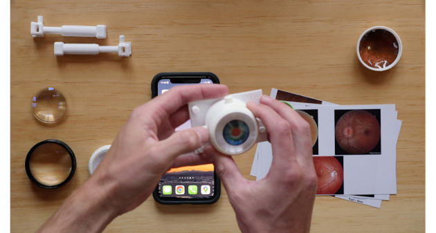
Demonstration of the Versatile Teaching Model Eye (VT Eye) being used with the smartphone-based digital fundus for review of fundus flashcards and the analog back for slit lamp and laser practice. [LINK TO VIDEO]
Methods
Construction
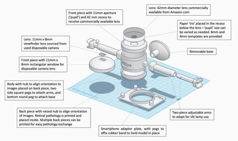
The VT Eye is an enlarged, simplified schematic eye that was designed using TinkerCAD (Autodesk TinkerCAD) (Figure 1). The design was converted to G-code using Cura (Ultimaker Cura, version 4.13; https://ultimaker.com/software/ultimaker-cura/) and 3D-printed with acrylonitrile butadiene styrene (ABS) plastic at the University of Vermont’s academic makerspace at a cost of $16.00 (USD) per model. Additional models were printed for <$3.00 on affordable hobby printers in Kumasi, Ghana, demonstrating the usefulness of 3D printing in the global health setting. Clear plastic lenses used for the refractive component were acquired from Amazon.com for <$1.00 each. A standard viewfinder lens from a spent disposable camera may also be used and obtained free of charge from many camera shops. Anterior and posterior segment pieces are attached with pressure-fitted junctions. They can be easily interchanged during learning sessions. Anterior segment images with varying pupil sizes are combined into image files designed to be printed on 4 × 6 photographic paper at a pharmacy or camera store, standardizing the physical size of the images and ensuring high-quality reproduction.
Figure 1.
Computer-aided design schematic of the VT Eye, showcasing components for smartphone-based digital fundus examinations, traditional indirect ophthalmoscopy, and slit lamp examination practice.
We received permission to use over 40,000 images from the Retina Rocks online image library (www.retinarocks.org) for our models. Each fundus image file has an ophthalmologist-provided diagnosis, and many have accompanying optical coherence tomography (OCT) images. The images were formatted to be printed on 4 × 6 photographic paper and displayed on a smartphone as a “digital fundus.”
Operation
Our 3D-printed smartphone attachment allows trainees to use their personal smartphones to display a wide array of pathology at the back of the model eye. Light from the backlit screen negates the need for an external light source, allowing trainees to practice inverted-image ophthalmoscopy without a slit lamp or indirect headset. Trainees can obtain a monocular view of the digital display through an indirect hand lens, practicing hand positioning and lens movements identical to those used in binocular examinations.5 Developing these skills independent of the need to concomitantly project light into the eye fosters an efficient, stepwise learning process. To further increase accessibility for trainees without their own examination lenses, plastic 20 D practice lenses can be purchased online for as little as $14.00 each, enabling a complete, interactive practice environment for a total cost of approximately $30.00.
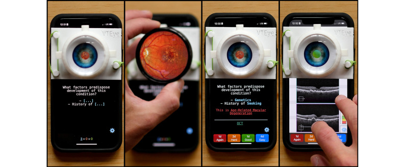
We created a set of flashcards for the digital fundus using the open-source Anki application (Anki, version 2.1). Anki’s interactive response buttons and built-in spaced-repetition algorithm allow for engaging and efficient study sessions (Figure 2). When available, accompanying OCT images are included on the “answer” side of the flashcards, which are also available as JPEG image files organized into shareable albums, facilitating practice examinations and self-quizzing using a smartphone’s native photo-viewing application. The current flashcard deck includes 160 cards grouped into three categories: retina, uveitis, and optic nerve. Flashcards include both pathology identification questions and multistep questions requiring critical thinking based on examination findings. Additionally, trainees and educators can create their own flashcards using the digital template included with the deck.
Figure 2.
Image sequence demonstrating the smartphone-based digital fundus and flashcards created using the open-source Anki application. This sequence demonstrates a trainee conducting an examination to determine the condition depicted, then answer a question about risk factors for this condition. Once the question has been answered, the learner can click the “OCT” button to view the corresponding OCT image.
Once trainees gain proficiency with the digital fundus, they can progress to the analog back, with printed fundus images for practicing with external light sources, such as an indirect headset or slit lamp. The introduction of glare from an external light source makes these examinations more challenging and realistic. The difficulty can be increased further by reducing the pupil size.
When the model is printed using light-colored plastic, an observer can watch the beam of light as it transilluminates the plastic and provide real-time feedback to help the trainee obtain proper focus. The observer can also ask questions specific to the part of the retina being examined.
Additional Uses
The VT Eye is a versatile tool, allowing for the practice of additional techniques, such as direct ophthalmoscopy, retinal image drawing, smartphone funduscopy, and retinal lasering. Direct ophthalmoscopy can be performed using the model in either the digital fundus or analog back configurations. For retinal drawing, the body of the model is easily removable for comparing a trainee’s drawings with the fundus image. In smartphone fundus photography with a 20 D lens, the configuration of the camera and flashlight varies between smartphone models, making it essential that trainees practice using their own smartphones to determine optimal imaging settings.6 The VT eye provides an ideal platform for perfecting smartphone fundus photography settings and technique.
The VT Eye can also be used for indirect and slit lamp laser practice. Standard photographic paper absorbs laser energy at levels similar to those used during retinal laser photocoagulation, providing an affordable practice medium before working with live patients.7
Global Health
Low-cost examination simulators, such as the VT Eye, can promote health equity in ophthalmic training, particularly in regions with limited access to training resources.8 VT Eye models were used in an institutional review board–approved pilot program for residents at Komfo Anokye Teaching Hospital, Kumasi, Ghana, for teaching indirect ophthalmoscopy to trainees, slit lamp biomicroscopy, smartphone funduscopy, and retinal image drawing from May 1, 2023, to May 5, 2023.
Surveys were administered to 6 ophthalmology residents before and 2–3 days after 1-hour examination training sessions to assess self-rated skills using a 5-point scale, where 1 indicated “not confident” and 5 indicated “very confident.” The 2- to 3-day period was intended to allow residents time to acquire facility with the device and practice. Mean and standard deviation of survey responses were calculated. Statistical significance was assessed using a two-sided paired t test. Percent increase was calculated as the increase in confidence rating divided by the initial confidence rating.
Results
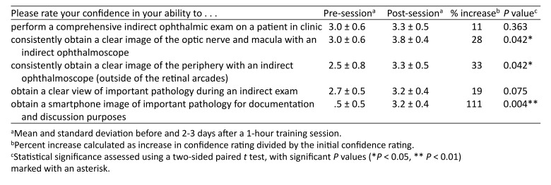
Pre- and post-training survey results revealed significant improvements in self-rated confidence. Average confidence levels rose by 30% for acquiring clear views of the posterior pole, 42% for visualizing the periphery, and 141% for capturing important pathology using personal smartphones combined with a 20 D lens. Compared to pre-test ratings, all of these values showed significance (P < 0.05), based on a two-sided paired t test conducted in Excel 2023 (Microsoft, Redmond, WA). See Table 1.
Table 1.
Self-reported changes in examination skills confidence after using the VT Eye models among residents, who were asked to rate their own confidence level on a scale from 1 (“not confident”) to 5 (“very confident”)
Discussion
The VT Eye represents a cost-effective solution for both education and practice of essential ophthalmic examination techniques. Within the scope of the Ghana pilot program, notable improvements in self-reported confidence levels underscore the utility of these devices in international contexts. Their affordability and open-access design render them especially advantageous for use in resource-limited settings. The production cost per unit of the devices in Ghana is approximately $2.00 USD.
TinkerCAD, the entry-level software used to design the models, is a free online browser-based application. It allows for easy modification of our design based on individual needs, such as adjustments to fit uncommon slit lamp biomicroscope designs. This accessible platform further reduces barriers to local and international collaboration.
To implement the pilot program, a 3D printer was acquired locally and installed at the teaching hospital to manufacture the required number of devices. Going forward, the models will be integrated into the residency curriculum. The greatest barrier to implementation of this novel component of the residency training program is the lack of a standardized method for assessing resident examination competence, which would allow for individual residents to track their progress and provide motivation for improvement. Going forward, there are plans to integrate an assessment-style module into the VT Eye training sessions so that residents can establish a performance baseline, identify weaknesses, and monitor their progress. Objective performance measured by accuracy and time required to complete the module will be compared with self-reported confidence and preceptor-evaluated clinical performance in order to further validate the models and training program.
This study is not without limitations. Participation in the study was voluntary, not mandatory for all residents, suggesting that those who participated were likely more motivated to improve their examination skills, leading to a potential self-selection bias. Additionally, the use of the VT Eye by residents after the training session was not monitored, so we cannot confirm that all survey respondents continued to practice independently. However, it was observed that more than half of the participants engaged in practice on their own following the session. Despite these limitations, the increases in self-reported confidence, as indicated by the survey responses, suggest that the current education program, when paired with unstructured, independent practice time, facilitates valuable improvements in examination skills.
In conclusion, the VT Eye is an economical 3D-printed model eye that provides a practical and adaptable platform for the practice of a variety of fundus examination techniques. The digital fundus facilitates efficient review of an extensive array of pathologies without necessitating an indirect headset or slit lamp biomicroscope, substantially increasing available practice time for trainees early in their careers. Taking advantage of the adaptability of this training platform, we plan to enhance the current training program to include more structured practice and assessment modules. The effectiveness of the VT Eye in enhancing resident education in Ghana provides a strong foundation for its broader application, both locally and globally.
References
- 1.Troncoso MU. New model of schematic eye for skiascopy (retinoscopy) and ophthalmoscopy. Am J Ophthalmol. 1922;5:436–41. [Google Scholar]
- 2.Chou J, Kosowsky T, Payal AR, Gonzalez Gonzalez LA, Daly MK. Construct and face validity of the Eyesi Indirect Ophthalmoscope Simulator. Retina. 2017;37:1967. doi: 10.1097/IAE.0000000000001438. [DOI] [PubMed] [Google Scholar]
- 3.Lee R, Raison N, Lau WY, et al. A systematic review of simulation-based training tools for technical and non-technical skills in ophthalmology. Eye. 2020;34:1737–59. doi: 10.1038/s41433-020-0832-1. [DOI] [PMC free article] [PubMed] [Google Scholar]
- 4.Javaid M, Haleem A, Singh RP, Suman R. 3D printing applications for healthcare research and development. Global Health J. 2022;6:217–26. [Google Scholar]
- 5.Lantz PE, Adams GGW. Postmortem monocular indirect ophthalmoscopy. J Forensic Sci. 2005;50:1450–52. [PubMed] [Google Scholar]
- 6.Iqbal U. Smartphone fundus photography: a narrative review. Int J Retina Vitreous. 2021;7:44. doi: 10.1186/s40942-021-00313-9. [DOI] [PMC free article] [PubMed] [Google Scholar]
- 7.Kylstra JA, Diaz JD. A simple eye model for practicing indirect ophthalmoscopy and retinal laser photocoagulation. Digit J Ophthalmol. 2019;25:1–4. doi: 10.5693/djo.01.2018.11.001. [DOI] [PMC free article] [PubMed] [Google Scholar]
- 8.Lewallen S. A simple model for teaching indirect ophthalmoscopy. Br J Ophthalmol. 2006;90:1328–9. doi: 10.1136/bjo.2006.096784. [DOI] [PMC free article] [PubMed] [Google Scholar]


