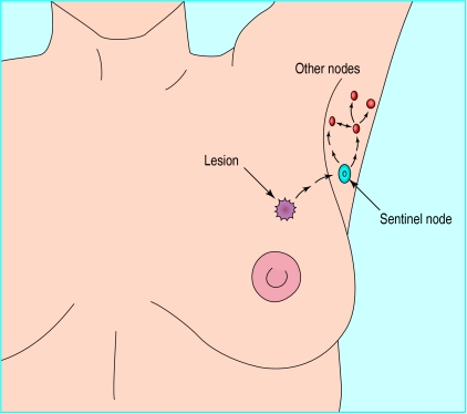
Figure 1.
Sentinel node biopsy. Lymph fluid from the lesion drains to the sentinel node and then on to other nodes in the axillary group. The region of the lesion is injected with a blue dye and radioisotope tracer. Gamma camera images show the location of the sentinel node, and a small incision is made directly over it. The blue dye outlines the lymphatics and the sentinel node

