

Magnetic resonance imaging (MRI) uses the body's natural magnetic properties to produce detailed images from any part of the body. For imaging purposes the hydrogen nucleus (a single proton) is used because of its abundance in water and fat.
The hydrogen proton can be likened to the planet earth, spinning on its axis, with a north-south pole. In this respect it behaves like a small bar magnet. Under normal circumstances, these hydrogen proton “bar magnets” spin in the body with their axes randomly aligned.
When the body is placed in a strong magnetic field, such as an MRI scanner, the protons' axes all line up. This uniform alignment creates a magnetic vector oriented along the axis of the MRI scanner. MRI scanners come in different field strengths, usually between 0.5 and 1.5 tesla.
When additional energy (in the form of a radio wave) is added to the magnetic field, the magnetic vector is deflected. The radio wave frequency (RF) that causes the hydrogen nuclei to resonate is dependent on the element sought (hydrogen in this case) and the strength of the magnetic field.
The strength of the magnetic field can be altered electronically from head to toe using a series of gradient electric coils, and, by altering the local magnetic field by these small increments, different slices of the body will resonate as different frequencies are applied.
When the radiofrequency source is switched off the magnetic vector returns to its resting state, and this causes a signal (also a radio wave) to be emitted. It is this signal which is used to create the MR images. Receiver coils are used around the body part in question to act as aerials to improve the detection of the emitted signal. The intensity of the received signal is then plotted on a grey scale and cross sectional images are built up.
Multiple transmitted radiofrequency pulses can be used in sequence to emphasise particular tissues or abnormalities. A different emphasis occurs because different tissues relax at different rates when the transmitted radiofrequency pulse is switched off. The time taken for the protons to fully relax is measured in two ways. The first is the time taken for the magnetic vector to return to its resting state and the second is the time needed for the axial spin to return to its resting state. The first is called T1 relaxation, the second is called T2 relaxation.
An MR examination is thus made up of a series of pulse sequences. Different tissues (such as fat and water) have different relaxation times and can be identified separately. By using a “fat suppression” pulse sequence, for example, the signal from fat will be removed, leaving only the signal from any abnormalities lying within it.
Most diseases manifest themselves by an increase in water content, so MRI is a sensitive test for the detection of disease. The exact nature of the pathology can be more difficult to ascertain: for example, infection and tumour can in some cases look similar. A careful analysis of the images by a radiologist will often yield the correct answer.
There are no known biological hazards of MRI because, unlike x ray and computed tomography, MRI uses radiation in the radiofrequency range which is found all around us and does not damage tissue as it passes through.
Pacemakers, metal clips, and metal valves can be dangerous in MRI scanners because of potential movement within a magnetic field. Metal joint prostheses are less of a problem, although there may be some distortion of the image close to the metal. MRI departments always check for implanted metal and can advise on their safety. Safety information is also available on the internet on
http://kanal.arad.upmc.edu/MR_Safety/
Figure.

The hydrogen proton can be likened to the planet earth, spinning on its axis, with a north-south pole. In this respect it behaves like a small bar magnet. Under normal circumstances, these hydrogen proton “bar magnets” spin in the body with their axes randomly aligned.
Figure.

When the body is placed in a strong magnetic field, such as an MRI scanner, the protons' axes all line up. This uniform alignment creates a magnetic vector oriented along the axis of the MRI scanner.
Figure.
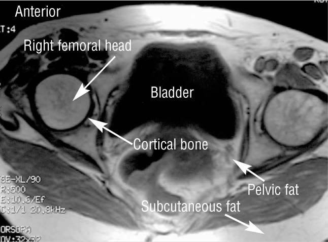
T1 weighted axial pelvis
Figure.
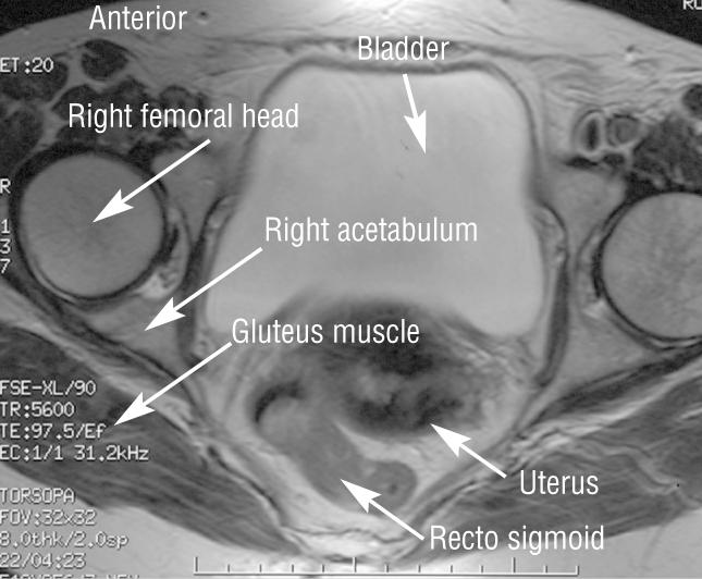
T2 weighted axial pelvis