Abstract
Adult degenerative scoliosis (ADS) is a coronal plane deformity often accompanied by sagittal plane malalignment. Surgical correction may involve the major and/or distally-located fractional curves (FCs). Correction of the FC has been increasingly recognized as key to ameliorating radicular pain localized to the FC levels. The present study aims to summarize the literature on the rationale for FC correction in ADS. Three databases were systematically reviewed to identify all primary studies reporting the rationale for correcting the FC in ADS. Articles were included if they were English full-text studies with primary data from ADS (≥ 18 years old) patients. Seventy-four articles were identified, of which 12 were included after full-text review. Findings suggest FC correction with long-segment fusion terminating at L5 increases the risk of distal junctional degeneration as compared to constructs instrumenting the sacrum. Additionally, circumferential fusion offers greater FC correction, lower reoperation risk, and shorter construct length. Minimally invasive surgery (MIS) techniques may offer effective radiographic correction and improve leg pain associated with foraminal stenosis on the FC concavity, though experiences are limited. Open surgery may be necessary to achieve adequate correction of severe, highly rigid deformities. Current data support major curve correction in ASD where the FC concavity and truncal shift are concordant, suggesting that the FC contributes to the patient’s overall deformity. Circumferential fusion and the use of kickstand rods can improve correction and enhance the stability and durability of long constructs. Last, MIS techniques show promise for milder deformities but require further investigation.
Keywords: Adult spinal deformity, Fractional curve, Radiography, Adult degenerative scoliosis, Spine surgery, Neurosurgery
INTRODUCTION
Adult spinal deformity (ASD) comprises both degenerative spinal deformity (de novo deformity) and residual deformity from adolescent idiopathic scoliosis. One type of de novo ASD – adult degenerative scoliosis (ADS) affects over 30% of elderly Americans [1-3]. Patients with ADS most commonly present with lumbosacral radicular pain caused by stenosis of the foramina on the concavity of the lumbosacral curve (present in up to 97% of patients) [1,2]. For these patients with isolated radicular pain, limited neural element decompression via a laminoforaminotomy may represent a reasonable and effective treatment approach. However, for those patients whose symptoms can be attributed to their global spinal malalignment (e.g., decreased physical activity tolerance and chronic fatigability of the paraspinal and proximal leg musculature), definitive therapy involves surgical correction of the sagittal and coronal alignments via long-segment instrumented fusion [3-7].
When correcting the coronal deformity in these patients, restoration of overall coronal alignment may require treatment of both the major and fractional curves (FCs) [8]. The FC is defined as the compensatory curve located caudal to the major curve, most commonly at the lumbosacral junction (L4–S1) [8]. The L4, L5, and S1 nerve roots exiting on the FC concavity are the most common precipitants of radicular pain in patients with ADS; therefore, many experts recommend correction of any FCs exceeding 15° [9,10]. In fact, prior work suggests that failure to correct a coexisting FC in ADS patients reduces the likelihood that global coronal balance will be successfully restored and maintained in the postoperative period [8,11-13].
Related to the concept of FC correction is whether the fusion construct should extend to the sacrum. For patients with adolescent idiopathic scoliosis (which becomes adult idiopathic scoliosis later in life), termination at L3 or L4 is common as an attempt to preserve motion segments [14]. However, in de novo (degenerative) ADS, the lumbar FC generally contributes to radicular pain generation and may contribute to the overall deformity. In such cases, the construct must include the FC, necessitating instrumentation to the sacrum. However, there remains debate regarding the degree to which the FC drives the overall deformity, and consequently whether it needs to be included in the fusion construct. The present systematic review aims to address these questions, specifically focusing on: (1) characterizing the importance of preoperative FC assessment as it relates to surgical planning, (2) comparing surgical outcomes for ADS patients undergoing deformity correction surgery according to whether or not the FC was corrected, and (3) to discuss future directions relevant to ongoing investigations regarding the importance of FC correction in patients with ADS.
MATERIALS AND METHODS
1. Search Strategy
In accordance with PRISMA (Preferred Reporting Items for Systematic Reviews and Meta-Analyses) guidelines, the PubMed, Ovid and Cochrane databases were queried for studies published between January 2010 and June 2023 focusing on FC management in patients with ADS [15]. All 3 databases were searched using the following keywords: (“spine deformity” OR “complex spinal deformity” OR “scoliosis” OR “degenerative scoliosis”) AND (“fractional curve” OR “fractional lumbosacral curve” OR “lumbosacral hemi curve” OR “compensatory lumbosacral curve” OR “secondary lumbosacral curve” OR “minor lumbosacral curve”) AND (“measurement” OR “analysis” OR “radiography” OR “imaging” OR “assessment”).
2. Study Selection Process
Two investigators (SCR, NJB) independently screened each article according to title and abstract. When points of disagreement regarding study inclusion arose, they were resolved by a third reviewer (MHP). Full-text English translations of each article identified on title and abstract screen were obtained and screened for inclusion in the final analysis. Studies were included if they: (1) presented primary data, (2) described a coherent methodology for assessing the FC or FC and deformity-related symptomatology, (3) included only adults (age≥ 18 years) with degenerative scoliosis, and (4) reported relevant data enabling characterization of the role that FC management plays in corrective surgery for patients with double curves (comprised of the major and FCs) in the setting of ADS.
3. Data Extraction and Analysis
The following information was extracted from included studies (when available): year of publication and surname of first author, study sample size (number of patients), method of FC quantification, surgical details (when surgery was performed), relevant patient inclusion and exclusion criteria implemented by each study, findings relevant to FC correction (and level of statistical significance when indicated), and any conclusions made regarding the importance of correcting the FC in patients with double curves secondary to ADS. Although descriptive statistics were reported, data were too heterogeneous to allow for a quantitative meta-analysis.
4. Quality and Risk of Bias Assessment
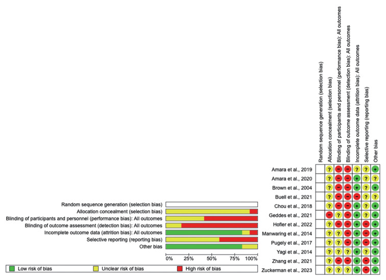
Given the retrospective, nonrandomized nature of the included studies, assessment of potential sources of bias was conducted in order to provide a fair assessment of the strength of the evidence informing the conclusions made in the present study. Accordingly, we assessed the quality and Risk of Bias (ROB) of each study using the Cochrane Risk Of Bias In Non-randomized Studies-of Interventions (ROBINS-I) tool. Overall, studies were mostly in the moderate-to-high ROB category, with 5 studies comprising high ROB and 7 studies qualifying as moderate ROB. Finally, one study was assigned low ROB.
RESULTS
1. Study Selection and Characteristics of Included Studies
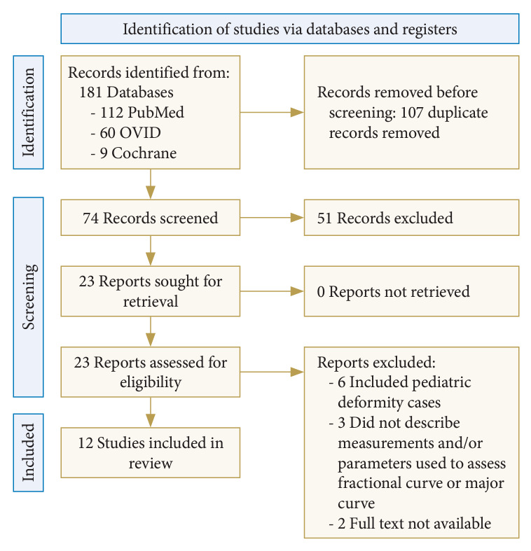
Seventy-four unique studies were identified, of which 23 met criteria for full-text review (Fig. 1). After exclusion criteria were applied, only 12 studies remained: 10 single-institution retrospective cohort studies and 2 multicenter retrospective cohort studies. Details of the included studies are presented in Tables 1 and 2. Furthermore, the results of the ROB assessment, which was conducted using Cochrane ROBINS-I tool, are presented as a traffic light plot in Fig. 2.
Fig. 1.

PRISMA (Preferred Reporting Items for Systematic Reviews and Meta-Analyses) diagram outlining study selection process.
Table 1.
Design and sample description of 12 studies assessing preoperative and/or postoperative fractional curve of adult degenerative scoliosis patients
| Study | Type | Method of FC measurement (Cobb angle) | Surgery type | Inclusion criteria | Exclusion criteria | Study sample size |
|---|---|---|---|---|---|---|
| Zhang et al. [16] 2021 | Single-institution retrospective | Angle between superior endplate of L4 and the line formed by the pedicles of S1 | PSIF of > 5 segments ending at L5–S1 with facetectomy and osteotomy (decompression and TLIF were performed if anterior support was needed or to relieve spinal stenosis) | 1. Primary spinal deformity correction | 1. Fusion levels < 5; 2. history of hip or knee arthroplasty; 3. absolute discrepancy of leg length > 20 mm | N = 101 |
| 2. Instrumented fusion via posterior-only approach | ||||||
| Amara et al. [23] 2020 | Single-institution retrospective | The curve below the major curve of thoracolumbar or lumbar scoliosis. Inclusion criterion: Cobb angle between L3–S1 > 10° | PSIF with 1–3 interbody fusions (ALIF/LLIF/TLIF) at the FC | 1. FC > 10° | NR | N = 78 (1 level = 19; 2 levels = 36; 3 levels = 23) |
| 2. Low back or extremity pain ipsilateral to FC concavity | ||||||
| 3. Treatment of FC with interbody fusion | ||||||
| 4. Preop and postop long-standing radiographs | ||||||
| 5. > 1-year follow-up | ||||||
| Amara et al. [20] 2019 | Single-institution retrospective | The curve below the major curve of a lumbar or thoracolumbar scoliosis measured via Cobb angle; only Cobb angle > 10° considered FC | PSIF of L4–S1 (FC) versus T10-pelvis (LT) versus T2–4 to pelvis (UT) | 1. FC from L4–S1 > 10° | 1. Previous lumbar fusion surgery | N = 99 (FC = 27; LT = 46, UT = 26) |
| 2. Radiculopathy ipsilateral to the concavity of FC | ||||||
| 3. Pre and postop radiography studies | ||||||
| 4. > 1-year follow-up | ||||||
| Chou et al. [22] 2018 | Multicenter retrospective study | Coronal Cobb angle of fractional curve | PSIF vs. cMIS | 1. > 18 years of age | 1. Hybrid open posterior surgery with interbody fusion | N = 118 (open = 79; cMIS = 39) |
| 2. Minimum of 3 levels fused | ||||||
| 3. Minimum 2-year follow-up | ||||||
| 4. FC > 10° | ||||||
| 5. At least one of the following: SVA ≥ 5 cm, PT ≥ 20°, lumbar Cobb angle ≥ 20°, or a PI-LL ≥ 10° | ||||||
| Brown et al. [17] 2004 | Single-institution retrospective | Angle between the line connecting the superior iliac alae and the line formed by the pedicles of l4 | PSIF to L5 | 1. Fusion extending above T12 | 1. Need for decompression at L5–S1 | N = 16 |
| 2. Pre-exisitng L5–S1 deformity (not including isolated degeneration at L5–S1) | ||||||
| Yagi et al. [21] 2014 | Single-institution retrospective | Coronal Cobb method | Combined single-rod anterior fusion and short PSIF to sacrum (hybrid) versus long PSIF with anterior release (control) | 1. Thoracic and thoracolumbar/lumbar curves (> 80°) | 1. Osteoporosis | N = 66 (33 per group) |
| 2. Nonprogressive thoracic deformity (> 30° flexibility) | 2. Revision surgery | |||||
| 3. Fractional curve (with segmental instability, stenosis or facet arthrosis) or degenerative disc disease | ||||||
| Manwaring et al. [24] 2014 | Single-institution retrospective | NR | Staged cMIS with versus without L5–S1 TLIF | 1. Treatment of ADS with at least 2-level MIS LLIF procedure | 1. Hybrid construct involving posterior osteotomies | N = 15 (TLIF = 11; control = NR) |
| 2. Delayed second stage procedure with MIS PLIF | ||||||
| Pugely et al. [18] 2017 | Single-institution retrospective | Coronal Cobb method | No surgery performed | 1. Coronal Cobb angle > 30° | 1. Central stenosis | N = 48 (group B = 14°; group F = 16°; group S = 18°) |
| 2. > 40 years of age | 2. Lateral recess stenosis | |||||
| 3. Standing scoliosis radiographs | 3. Disk herniation | |||||
| 4. Preop CT spine | ||||||
| Buell et al. [25] 2021 | Multicenter retrospective study | Coronal L4–S1 Cobb angle | L4–S1 TLIF vs. ALIF | Index operation that involved TLIF or ALIF at L4–5 and/or L5–S1. Minimum 2-year postoperative follow-up | Any patient with active infection, malignancy, diagnosis of scoliosis other than adult degenerative | N = 106 (TLIF = 47, ALIF = 59) |
| Geddes et al. [26] 2021 | Single-institution retrospective | Coronal Cobb method | ALIF+PSF+S2AI screws versus PSF+S2AI screws for thoracolumbar fusion | 1. Posterior lumbar fusion to the pelvis using S2AI screws | 1. Patients who had posterior 3-column osteotomies | N = 59 (ALIF+PSF = 31, PSF alone = 28) |
| 2. Presence of fractional curve | 2. Those lacking adequate pre- and/or postoperative imaging | |||||
| Hofler et al. [43] 2022 | Single-institution retrospective | Cobb angle method for lumbar fractional curve. The magnitude of the major lumbar coronal curve and fractional lumbar coronal curve caudal to it was measured on preoperative and follow-up anteroposterior imaging | T3-ilum fusion +/- kickstand placement | 1. Deformity correction with fusion from upper thoracic spine to pelvis | NR | N = 15 (kickstand = 7, nonkickstand = 8) |
| 2. Associated coronal deformity | ||||||
| 3. Intraoperative APLCRs performed | ||||||
| Zuckerman et al. [27] 2023 | Single-institution retrospective | Cobb angle between the sacrum and most tilted lower lumbar vertebra (either L3/4/5) | Instrumentation to pelvis/fusion to sacrum and TLIF | 1. ≥ 6-level fusion | NR | N = 243 |
| 2. At least 1 of the following radiographic criteria (Cobb angle > 30˚, SVA > 5 cm, CVA > 3 cm, PT > 25˚, or TK > 60˚) |
PSIF, open posterior spinal instrument fusion; TLIF, transforaminal lumbar interbody fusion; ALIF, anterior lumbar interbody fusion; LLIF, lateral lumbar interbody fusion; FC, fractional curve; LT, lower thoracic; UT, upper thoracic; cMIS, circumferential minimally invasive surgery; SVA, sagittal vertical axis; PT, pelvic tilt; PI-LL, pelvic incidence-lumbar lordosis; NR, not reported; MIS, minimally invasive surgery; CT, computed tomography; PSF, posterior spinal fusion; S2AI, S2 alar iliac screw; APLCR, anteroposterior long cassette radiograph; CVA, coronal vertical axis; TK, thoracic kyphosis.
Table 2.
Results of 12 studies assessing preoperative and/or postoperative fractional curve of adult degenerative scoliosis patients
| Study | Preoperative FC | Postoperative FC | FC correction | FC radiographic predictors | Conclusions |
|---|---|---|---|---|---|
| Zhang et al. [16] 2021 | 13.6° ± 8.2° | 5.9 ± 5.1° | p < 0.001 | Preoperative FC with L4 coronal tilt toward C7 plumbline is associated with postoperative coronal imbalance | Directionality of preoperative FC toward C7 plumbline increasing risk of postoperative coronal imbalance |
| Amara et al. [23] 2020 | 1 Level = 15.3° ± 8.2°, 2 levels = 117.9°, 3 levels = 16.3° | 13.6° ± 8.2° | Group 1 vs. 2 = 0.0062; group 1 vs. 3 = 0.017; group 2 vs. 3 = 0.99 | None | Additional interbody fusion levels at the FC resulted in more fractional curve correction, more major curve correction, increasing lordosis without increasing morbidity |
| Amara et al. [23] 2019 | FC = 15.7°, LT = 16.7°, UT = 16.9° | NR | NR | None | Treatment of only the FC was associated with lower complication rates, shorter hospital LOS and reduced blood loss than fusion to UT or LT levels; FC group had higher rates of re-extension UT or LT levels |
| Chou et al. [23] 2018 | FC > 10° Matched cohort: preop FC–cMIS: 18 and open: 18 | Unmatched cohort: postop FC – cMIS: 17 and open: 19.6 | cMIS = 6.9°; Open = 8.5° | None | cMIS achieved similar reduction in leg pain and correction of fractional curve as traditional open surgery, despite significantly fewer cMIS patients undergoing direct decompression |
| Brown et al. [17] 2004 | 21° | 10.6° | NR | Less postoperative FC decreased risk of L5–S1 degeneration | Patients with good postop FC achieved better outcomes with posterior fusion to L5, avoiding sacral fusion |
| Yagi et al. [21] 2014 | Hybrid = 23° ± 9°, control = 24° ± 10° | Hybrid = 7 ± 4°, control = 15 ± 8° | Percent correction of lumbosacral curve significantly better in hybrid versus control (p < 0.001) | None | Hybrid patients had improved curve correction, fewer levels fused, decreased blood loss and fewer revision procedure when compared to control |
| Manwaring et al. [24] 2014 | TLIF = 9.2°, control = NR | TLIF = 4.1°, control = NR | NA | None | Significant fractional curve correction in staged cMIS is achieved through 2 stage TLIF treatment of L5–S1 |
| Pugely et al. [18] 2017 | Group B = 19.4°; group F = 25.5°; group S = 17.7° | NA | NA | None | Sciatic nerve pain in setting of lumbar structural curves is associated with foraminal stenosis at the concavity of the caudal fractional curve; femoral nerve pain likely caused by stenosis at concavity of main structural curve (L3 or below) |
| Buell et al. [25] 2021 | All = 20.2° ± 7.0°, TLIF = 19.4° ± 7.2°, ALIF = 20.8° ± 6.9° | All = 6.9° ± 5.2°, TLIF = 7.1° ± 5.4°, ALIF = 6.8° ± 5.1° | Multiple regression demonstrated 1-mm increase in L4–5 TLIF cage height led to 2.2° reduction in L4 coronal tilt (p = 0.011), and 1° increase in L5–S1 ALIF cage lordosis led to 0.4° increase in L5–S1 segmental lordosis (p=0.045). Matched analysis demonstrated comparable fractional correction (TLIF = -13.6° ± 6.7° vs. ALIF = -13.6° ± 8.1°, p = 0.982). | None | Results demonstrate comparable fractional curve correction (66.7% for TLIF patients versus 64.8% for ALIF patients), despite the use of significantly larger and more lordotic cages in ALIF |
| Geddes et al. [26] 2021 | PSF = 13.4° ± 7.1°, ALIF+PSF = 18.3 ± 9.3° | PSF = 8.6 ± 4.4°, ALIF+PSF = 6.1° ± 5.3° | PSF = 4.8 ± 4.5° (27% curve correction), ALIF+PSF = 12.1 ± 6.0° (68% correction), p = 0.053 | NR | ALIF+PSF achieves greater correction of the fractional curve than PSF alone. Though not the primary indication of ALIF, this may help facilitate overall deformity correction and pelvic balance |
| Hofler et al. [43] 2022 | Kickstand = 4.3-cm coronal deviation, 43° major lumbar curve, 23° fractional lumbar curve | Kickstand group = 4.3-cm intraoperative coronal deviation, 1.8-cm postoperative coronal deviation | Preoperative lumbar FC was greater in patients requiring a kickstand (23° vs. 35°, p = 0.02) | NR | Intraoperative kickstand rod placement guided by intraoperative APLCR allows for satisfactory reduction in cases where the fractional coronal curve persists without loss of sagittal plane correction |
| Nonkickstand = 2.2-cm coronal deviation, 35° major lumbar curve, 14° fractional lumbar curve | Nonkickstand group = 0.6-cm intraoperative coronal deviation, 2.1 cm postoperative coronal deviation | ||||
| Zuckerman et al. [27] 2023 | Qiu type A=11.1° | Qiu type A=5.3° | Type C patients had the most LSF curve correction (p = 0.023 for change in LSF curve by 9.2°) | NR | Greater correction of LSF curve was seen in Qiu type C patients compared to type A and type B. More TLIFs were associated with greater amount of LSF curve correction. No clear trends seen regarding LSF curve change and postoperative outcomes |
| Qiu type B=12.7° | Qiu type B=7.6° | ||||
| Qiu type C=15.6° | Qiu type C=6.4° |
PSIF, open posterior spinal instrument fusion; TLIF, transforaminal lumbar interbody fusion; ALIF, anterior lumbar interbody fusion; LLIF, lateral lumbar interbody fusion; FC, fractional curve; LOS, length of stay; LT, lower thoracic; UT, upper thoracic; cMIS, circumferential minimally invasive surgery; NR, not reported; PSF, posterior spinal fusion; APLCR, anteroposterior long cassette radiograph.
Fig. 2.
ROB (Results of Risk of Bias) assessment conducted using the ROBINS-I (Risk Of Bias In Non-randomized Studies-of Interventions) tool.
2. Radiographic Quantification of the FC
All but one of the included studies described the methodology for radiographic determination of the FC Cobb angle [16-26]. The one study utilized preoperative radiographic parameters to localize ASD pain to specific sources within structural FC [16]. Examples of methodologies used to determine FC included that of Zhang et al. [16], who defined FC as the angle inscribed by lines through the superior endplate of L4 and pedicles of S1. Similarly, Brown measured FC as the Cobb angle inscribed by the line connecting the superior edge of the iliac alae and a horizontal line through the pedicles of the proximal vertebra of the FC (L4 or L5) [17].
3. FC as a Radiographic Predictor of Outcomes
Seven of the studies presented data correlating FC curve size with the severity of the major curve, extent of operative correction required for a successful outcome, global coronal imbalance, and the severity of lumbosacral radiculopathy. Pugely et al. [18] examined 48 patients evaluated for ADS at a single center. The cohort was divided into those presenting with isolated back pain, those presenting with combined low back and anterior thigh/knee (femoral) pain, and those presenting with combined low back pain and sciatica. Those presenting with sciatica more commonly had symptomatic foraminal stenosis within the concavity of the FC, unless the structural curve lay in the lower lumbar spine (apex below L3), in which case symptoms derived from foraminal stenosis in the structural (main) curve concavity. In both patients with sciatica and femoral pain, foraminal size was significantly lower than in patients with lumbago alone. It thus follows that radicular pain down the back of the leg could suggest foraminal stenosis within the FC concavity as the driver of clinical presentation [18]. The authors concluded that precise localization of the source of leg pain in ADS patients is a critical component of operative planning as it affects decisions related to determining which specific levels should be targeted by operative intervention.
4. FC Analysis and Surgical Planning
Eight articles focused on the impact of including the major/minor curve in devising the construct and the need for pelvic fixation. Amara et al. [20] described their single-institutional experience in which they treated 99 patients with ADS. All included patients demonstrated lumbar radiculopathy ipsilateral to any FCs > 10° at the L4–S1 levels, specifically. Patients were divided into 3 groups—those undergoing correction of the FC only (N = 27), those undergoing T10-pelvis fusion with correction of the both the fractional and major coronal curves (n = 46), and those undergoing fusion to the upper thoracic spine with correction of the major coronal curve only (n = 26). Those undergoing fusion to the upper thoracic spine were noted to have significantly larger major curve angles than patients who were not fused to the upper thoracic levels. Furthermore, the authors found that patients in the group undergoing FC correction alone had significantly lower surgical morbidity, reduced complications, and the lowest likelihood to require inpatient rehabilitation facility placement relative to the other groups. Across all 3 groups, there was no significant difference in coronal balance postoperatively. This suggests that FC correction may be indicated for select patients, namely those presenting with a primary complaint of radiculopathy localizing to the level of the FC. However, these data suggest it is not necessary to incorporate FC correction in the surgical plan for all ASD patients. Although this appeared to be the case, the authors did acknowledge one caveat: patients who underwent FC correction alone exhibited higher rates of reoperation.
The same group subsequently described a subset of 78 patients who underwent surgery for FCs (L3–S1) > 10° involving ≥ 1 interbody fusion within each patient’s FC [23]. All patients included in the study presented with primary complaints of lumbar radiculopathy that could be localized to the level of the FC. With respect to the specifics of FC correction, interbody placement at 2–3 levels (versus 1-level interbody placement) was associated with significantly reduced fractional and major curves without a commensurate increase in complications.
More recently, in an expanded multicenter cohort, Chou et al. [22] examined outcomes in 118 ADS patients who underwent correction of their FCs (79 underwent open correction, while 39 underwent circumferential minimally invasive surgery [cMIS] correction). Despite the fact that direct decompression was performed less frequently in the cMIS group, pain outcomes—including postoperative visual analogue scale (VAS) leg pain scores—were similar between the cMIS and open groups. This suggests foraminal stenosis on the FC concavity drives the radicular leg pain and the indirect decompression afforded by FC correction may be sufficient to offer symptomatic improvement. To clarify, neural element decompression appears to be the key process driving pain relief. While for patients with an exclusively radicular pain picture, direct decompression (such as hemilaminectomy or foraminotomy) may be sufficient. However, for those with concurrent axial pain attributable to the deformity, indirect decompression with interbody placement via either open or cMIS techniques may offer effective symptom relief without the need for concurrent direct decompression. Said strategy helps to maintain fusion surfaces that would be removed with direct decompression, and so theoretically improve the odds of successful radiographic fusion. Finally, one advantage of concurrent FC correction is that the placement of instrumentation helps to maintain the nerve root decompression and so may allow for better long-term symptom relief, though this remains a point of ongoing investigation.
Data from Brown et al. [17] suggested that correction of the FC was associated with more favorable patient-reported outcomes in a series of 16 ADS patients who underwent posterior fusion to L5. Average FC correction was 10.4°and postoperative L5–S1 degeneration correlated significantly with the magnitude of residual FC. For example, patients requiring revision had significantly larger residual FCs (21° vs. 8.7°, p < 0.05) and those with subsequent L5–S1 disc degeneration had significantly larger residual FCs than those without such degeneration (15° vs. 8.7°, p < 0.05). As distal segment degeneration is a driver of surgical revision, these data suggest FC correction may reduce the odds of revision surgery for adjacent segment disease.
Yagi et al. [21] compared the efficacy of a hybrid anterior-posterior surgical approach to conventional anterior column release (ACR) with posterior fusion in 33 patient pairs undergoing corrective surgery for ADS. Each pair was matched by age and coronal curve magnitude for analysis. Patients underwent either “conventional” anterior release followed by long-segment posterior instrumentation, or a “hybrid” multilevel anterior interbody fusion followed by posterior segmental instrumentation. The combined anteroposterior procedure has been previously associated with improved correction of severe deformity curves, lower likelihood for postoperative progression, and reduced rates of pseudarthrosis. In this study, the hybrid group demonstrated significantly lower rates of both postoperative coronal imbalance (38% vs. 56%, p = 0.03), complications (18% vs. 39%, p = 0.01), and rates of surgical revision (12 of 33 patients vs. 6 of 33 patients, p = 0.03), though quality of life (QoL) outcomes were similar. The major complications in the control group, which exhibited the higher overall rate of complications, included proximal junctional kyphosis (PJK) (n = 7) and deep infection. Interestingly, although one of the touted advantages of the anterior-posterior hybrid approach was a decreased risk for pseudoarthrosis, the latter was not demonstrated by Yagi and colleagues. Specifically, only 3 cases of pseudarthrosis were reported, and 2 of them occurred in the hybrid group.
Both Chou et al. [22] and Manwaring et al. [24] compared the ability of cMIS techniques to achieve FC correction relative to open techniques. Manwaring et al. [24] examined the impact of ACR with concomitant extreme lateral interbody fusion (XLIF) on sagittal and coronal balance in 36 patients treated for ADS. Both patients who underwent XLIF without ACR and those who underwent ACR demonstrated improvement in their coronal Cobb angle; there was significant correction in the ACR group (8.2° vs. 4.2°, p < 0.002). As mentioned previously, in their comparison of patients treated via open (n = 79) versus cMIS (n = 39) approaches, Chou et al. [22] observed no differences in postoperative pain, change in coronal Cobb angle, change in pelvic incidence-lumbar lordosis (PI-LL) mismatch, Oswestry Disability Index (ODI) improvement or VAS back pain scores. However, cMIS techniques achieved these results with fewer direct decompression procedures.
Bridwell [10] and Cho et al. [11] have reported that correction of the FC via decompression alone or short-segment fusion both represent reasonable options, even though they may accelerate degeneration of the residual curve. Both TLIF and anterior lumbar interbody fusion (ALIF) approaches can be entertained and allow for curve correction and indirect decompression of the nerve roots ipsilateral to the FC concavity. Ultimately, a less-invasive decompression alone may be preferable in frail patients with predominately radicular symptoms and no evidence of instability on radiographic evaluation.
Buell et al. [25] recently presented the results of a multicenter analysis of 106 patients with ≥ 30° coronal main curves and ≥ 10° lumbosacral FCs who underwent L4–5 TLIF (n = 47) versus L4–5 and/or L5–S1 (lateral) ALIF (n = 59) for FC correction. Matched analysis of 28 pairs of patients showed no significant difference in FC correction between TLIF and ALIF (55.7% vs. 64.8%, respectively). Notably, patient-reported outcomes on the ODI and 36-item Short-Form health survey were similar between groups, as were overall complication rates. It was noted that ALIF offered greater restoration of lordosis at L5–S1, suggesting that ALIF may be preferable when concomitant sagittal plane correction is desired.
Next, in their 59 patient retrospective series, Geddes et al. [26] sought to determine whether ALIF can improve the FC in deformity surgery as compared to posterior surgery alone. The authors found the addition of ALIF led to significantly greater FC correction (12.1° vs. 4.8°, p < 0.01) and smaller postoperative FC (6.1° vs. 8.6°, p = 0.023). Major curve correction (23.5° vs. 14.9°, p = 0.006) was also greater and multivariable analysis showed the addition of ALIF to be independently predictive of FC correction even after accounting for the use of TLIFs in the posterior-only cohort.
Most recently, Zuckerman et al. [27] re-examined the multi-institutional International Spine Study Group (ISSG) dataset previously analyzed by Buell et al. Their analysis focused on the relative importance of correcting the major versus FCs in ADS patients. All 243 patients included in their analysis underwent ≥ 6-level fusion for coronal Cobb angles > 30° and C7 coronal vertical axes > 3 cm. Improvement in patient-reported outcomes assessed with ODI were correlated with both lumbosacral FC and major coronal curve correction; they also correlated preoperative coronal alignment with FC magnitude. They noted that FCs were largest in patients wherein the coronal imbalance lay ipsilateral to the major curve convexity, suggesting the FC was the driver of overall coronal imbalance [27].
DISCUSSION
An ongoing question pertaining to ADS is whether the FC is a driver of coronal malalignment or merely a compensatory curve for the major scoliotic curve. In general, the present literature suggests that the presence of the FC concavity ipsilateral to side of truncal shift is a risk factor for persistent coronal malalignment. It also suggests that FC correction may help to alleviate ASD-associated radiculopathy without requiring direct decompression.
An additional advantage of FC correction is a potential reduction in the odds a patient will require surgical revision, indicating that FC correction in addition to the major curve can optimize surgical outcomes. This finding coincides with the proposal recently advanced by Plais and the International Spine Study Group [28]. Through investigating a multicenter cohort of 404 patients (age> 45 years) with thoracolumbar major coronal curves (> 15°; apex at T11–L3) and FCs > 5°, the authors found that among patients with global coronal malalignment, those with truncal shift ipsilateral to the FC concavity had significantly larger FCs (22.28° vs. 14.84°, p < 0.001) relative to those with truncal shift contralateral to the FC concavity. Additionally, they had greater pelvic obliquity angled towards the side of the truncal shift. This led the authors to suggest that while FCs may be compensatory in cases where truncal shift is contralateral to the side of the FC concavity, where the truncal shift is ipsilateral, the concavity likely plays a role as the primary driver of coronal malalignment. According to this paradigm, restoration of coronal balance depends upon treatment of the FC, as has been reported.
With this paradigm in mind, Obeid et al. [29] released an expert’s consensus, treatment-oriented guideline for correcting coronal imbalance in ASD. Their guideline defines concave and convex coronal malalignment as types 1 and 2, respectively. For convex (type 2) coronal malalignment, Obeid et al. [29] suggests avoiding correction of the main curve as doing so would risk worsening coronal malalignment. Instead, the lumbosacral curve—which may be the FC in type 2A patients—should be corrected. When the main curve is located within the lumbosacral spine and associated with a compensatory lumbar curve (type 2B), 3-column osteotomies at the apex (usually between L4–S1, most commonly at L5) of the lumbosacral curve will generally suffice for correction of the short, main curve. However, most patients will have their major curve in the lumbar or thoracolumbar spine (type 2A) and the FC will lie at the lumbosacral junction. When this is the case, FC correction should be dictated by the degree of flexibility present at the lumbosacral junction. For example, a previously fused interbody space at the lumbosacral junction merits performance of a 3-column osteotomy with L5 pedicle subtraction osteotomy to achieve FC correction. Furthermore, it is important to keep in mind that ending a construct at L4 or L5 in an ASD patient with a preexisting FC predisposes them to subsequent distal segment degeneration. The indications for excluding the sacrum from a construct include: (1) the presence of normal L5–S1 disc and facets; (2) the UIV lies at or below T10; (3) the patient has normal bone mineral density; or (4) the patient is young or relatively less active [30]. Indications for extending the construct to the sacrum are prior surgical decompression at L5–S1, signs and symptoms consistent with L5–S1 radiculopathy, and/or the presence of L5–S1 spondylolisthesis.
When concave coronal malalignment (type 1) is present, on the other hand, correction of the main curve only will result in indirect correction of the FC, which is compensatory, not structural [29]. If the main curve is flexible, a posterior column osteotomy at its apex is likely sufficient for correction, whereas a rigid or fused curve will require a 3-column osteotomy at the apex to achieve correction (type 1 coronal malalignment) [29].
1. Coronal Realignment and Kickstand Rod Technique
For patients in whom the FC appears to contribute significantly to coronal imbalance, there remains debate regarding the magnitude of major curve correction that is required. Prior work by Deviren et al. [31], among others, has suggested that the coronal curve magnitude inversely correlates with curve flexibility. The flexibility of the major curve is best assessed using preoperative lateral bending radiographs. In patients with truncal shift ipsilateral to the FC concavity (Qiu type C curves), major curve correction in the absence of FC correction will exacerbate the truncal shift and result in poorer postoperative coronal alignment. To this end, Bao et al. [32] noted that patients with type C curves are most likely to have coronal malalignment postoperatively. Consequently, for these patients, adequate FC correction is paramount; it may also be beneficial to use less aggressive correction of the major curve, especially for those with flexible major curves preoperatively.
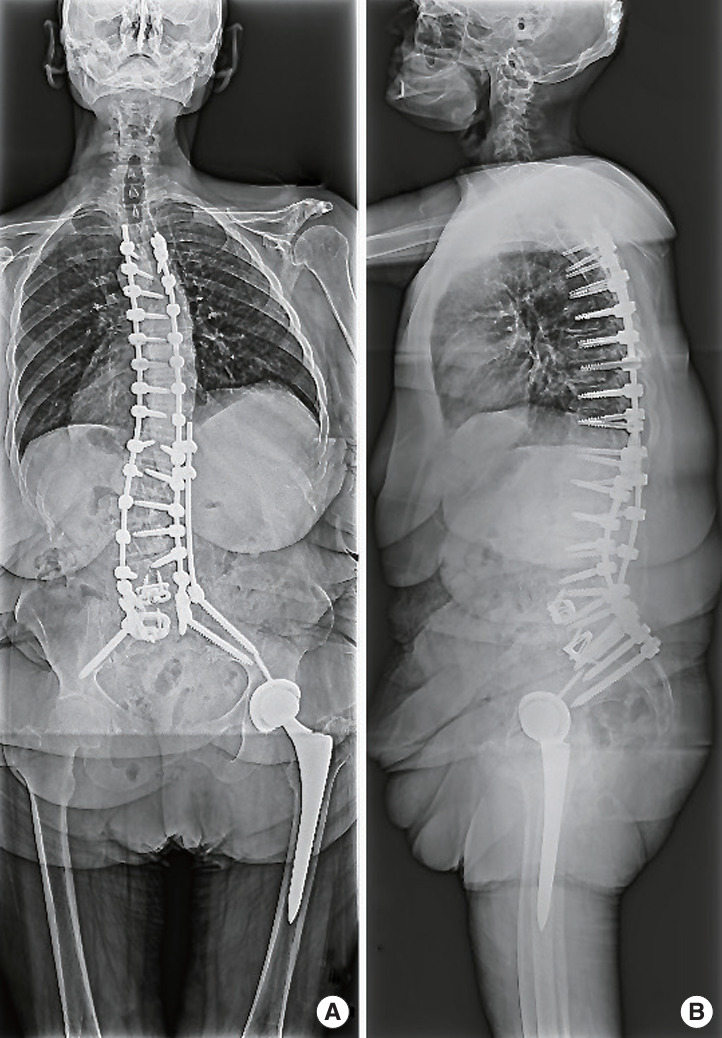
One increasingly popular technique for coronal realignment is the kickstand rod technique, which employs a rod with distal fixation in the ilium and proximal fixation to the thoracolumbar spine [33]. The kickstand rod can provide significant coronal correction due to the distracting force and torque it provides (Fig. 3). These forces are greater than those achieved using rod bending maneuvers (distraction/compression) alone. It is therefore a powerful tool for FC correction. Buell et al. [33] described the successful use of this technique in 17 adult patients with thoracolumbar/lumbar degenerative scoliosis. Highlighting the strength of correction provided by the kickstand rod, they reported coronal overcorrection in one patient, though the authors indicated this is a rare complication when the technique is properly executed. Puvanesarajah et al. [34] and Mundis et al. [35] similarly found the kickstand rod technique to facilitate good coronal correction in their series of 20 and 21 patients treated for adult scoliosis, respectively. Interestingly, Mundis et al. [35] from the ISSG compared coronal correction with the kickstand rod technique to correction achieved when using conventional accessory rods alone. They found that the kickstand technique can produce better overall postoperative coronal balance. However, this did not translate to improved patient-reported outcomes on the Scoliosis Research Society-22 Questionnaire or ODI assessments, suggesting that there may be a point beyond which additional coronal plane correction is no longer meaningful.
Fig. 3.

Anteroposterior (A) and lateral films (B). Case example of patient who underwent T3-pelvis instrumented fusion for likely adult idiopathic scoliosis with a T12-pelvis kickstand rod on her right side (the side of the fractional curve [FC] concavity). The patient was a 63-year-old female with past medical history of depression, prior bariatric surgery, tobacco use (10+ pack years), sensorineural hearing loss (bilateral hearing aid), and obesity (body mass index 36.60 kg/m2) who underwent elective, staged anterior-posterior thoracolumbar instrumented fusion for adult spinal deformity. The patient had presented to clinic with multiple years of chronic back and leg pain. Upright scoliosis films showed a positive sagittal balance (10 cm), degenerative thoracolumbar levoscoliosis of 65° with a second upper thoracic curve (45°) and fractional lumbosacral curve, along with 14° pelvic incidence (PI)-lumbar lordosis mismatch with a PI of 70°. The patient underwent circumferential anterior/posterior surgery for correction of scoliosis, with postoperative radiographs shown here.
2. Radicular Pain and the FC
In approximately 90% of cases, back and/or leg pain is the primary reason for the initial hospital/clinic visit in patients with ADS [36]. The FC appears to play a significant role in this symptomatology for thoracic and thoracolumbar major curves, as it has been demonstrated that radiculopathy at presentation localizes most commonly to the L4, L5, and S1 spinal roots on the side of the FC concavity [23]. Ultimately, many patients will have endured chronic back pain over many years prior to presentation [23]. As long as relative sagittal and coronal balance are maintained; however, the scoliotic deformity may not necessarily be disabling. On the other hand, the radicular pain cause by foraminal stenosis within the FC concavity often leads patients to seek surgical intervention [22]. Furthermore, progression of the scoliotic deformity with multilevel lateral listhesis that can further augment the radicular pain. To this end, the work of Chou et al. [22] found that correction of the FC alone (without an accompanying direct decompression procedure) was sufficient to alleviate lumbar radicular pain. Of note, Chou et al. [22] reported that laminectomy alone is insufficient to address this pain, which results from compression of the dorsal root ganglia within the neural of the FC concavity.
In the case of lumbar structural curves, the relative contribution of the FC to patient symptomatology is less clear, though the distribution of the radicular pain can help to localize the pain to either foraminal stenosis within the structural curve or FC concavity. As suggested by the results from Pugely et al. [18], pain in the sciatic distribution (predominately L5, S1, and S2 dermatomes) is most likely attributable to foraminal stenosis at the concavity of the FC, whereas radicular pain in the femoral distribution (predominately L2–4 dermatomes) is more easily attributed to foraminal stenosis within the structural curve concavity. Accurate assessments of: (1) the concordance of the FC convexity and the directionality of the global coronal malalignment, and (2) the degree to which the patient’s symptomatology is attributable to the FC are key to designing the optimal surgical plan [18]. Finally, these findings mark a paradigm shift from previous beliefs that the FC was always the source of radicular pain in ADS patients [24].
3. Coronal and Sagittal Balance
Achieving coronal and sagittal balance are primary goals of ADS surgery. A recent investigation by Zhang et al. [16] suggests the concordance of FC concavity and global coronal malalignment predicts the odds of residual malalignment following surgical correction. The authors examined the relation of FC orientation and preoperative coronal imbalance to predict restoration of coronal balance in 101 patients (74 instrumented to pelvis) [16]. Those 27 patients who achieved postoperative coronal balance were more likely to have a FC concavity opposite to the preoperative net coronal imbalance (66% vs. 19.4%, p < 0.001). They found on logistic regression that the best predictors of postoperative coronal imbalance were consistency pattern (preoperative coronal imbalance and FC concavity on the same side) and preoperative coronal C7 plumbline > 30 mm towards the convex side of the major curve [16]. This led the authors to argue that greater attention must be paid to the directionality and treatment of the FC.
These results echoed earlier findings by Bao et al. [37], who examined the prevalence and impact of preoperative coronal imbalance on outcomes in 284 patients who underwent surgery for degenerative lumbar scoliosis. They found 34.8% of patients presented with coronal imbalance; those with preoperative coronal imbalance > 30 mm shifted towards the convexity of the major curve were significantly more likely to have persistent coronal imbalance postoperatively. However, preoperative coronal imbalance did not impact patient-reported outcomes such as ODI or VAS for back pain. They, like Zhang et al. [16], consequently argued that asymmetric osteotomies intended to reduce the major curve had also exacerbated truncal shift and coronal instability. Consequently, the authors argued in favor of a TLIF within the FC concavity to restore neutral alignment to the L4 and L5 vertebrae, thereby restoring overall coronal alignment [16]. Similarly, Bao et al. [37] concur as they argued for performing a TLIF within the FC to restore neutral alignment to the L4 and L5 vertebrae and thus the overall coronal alignment.
Additional surgical measures may be warranted in these patients to ensure postoperative sagittal balance, which data suggests to have the greatest influence on long-term patient outcomes [5,38]. However, significant alignment correction often involves the use of long-segment posterior constructs, which are high risk for mechanical failure in elderly osteoporotic/osteopenic patients [36].
4. Surgical Approaches
At present, data does not support a single optimal approach for correction of coronal plane deformities [38]. In general, approaches can be divided into purely posterior approaches versus combined anterior-posterior approaches, and into open versus MIS approaches. None of the included studies directly compared MIS and open approaches for ADS alone. However, a recent article by Chou et al. [39] using the ISSG database reported a propensity-matched cohort study of 154 patients (77 pairs) who underwent cMIS with anterior interbody fusion and posterior percutaneous instrumentation or open posterior fusion for ASD. Patients were matched on multiple metrics, including construct length, age, body mass index, and baseline spinopelvic parameters. Those treated with cMIS surgery had grossly similar QoL outcomes, radiographic outcomes, and surgical revision rates but lower intraoperative blood loss. These same authors also described results within a subset of patients from the ISSG cohort with adult scoliosis, characterized as those > 18 years old with a FC > 10° [22]. As described in the results section, they found cMIS and open posterior fusion achieved similar changes in coronal Cobb angle, PI-LL mismatch, ODI, and VAS back pain. However, this was achieved with fewer decompressive procedures.
Further expounding on the advantages of ACR, Yagi et al. [21] showed in their small experience that multilevel anterior interbody placement afforded superior coronal plane correction in patients with thoracolumbar (61° ± 21° vs. 45° ± 25°, p < 0.01) or lumbosacral curves (67° ± 21° vs. 41° ± 19°, p < 0.001) while reducing intraoperative blood loss (mean reduction 1.5 L, p < 0.001) and the average number of fused segments (6.7 ± 1.2 vs. 14.6 ± 1.3, p < 0.001).
The final extant question concerns the necessity of including the FC within the fusion construct [40,41]. In the adolescent idiopathic scoliosis population, construct termination at L3 or L4 is considered acceptable for most curve types as there appears to be limited potential for distal segment degeneration and it spares additional motion segments [10-12,14,42]. These motion segments may allow for pelvic mobility to compensate for reduced motion within the stiff long-segment construct immediately cephalad to the lumbosacral junction [42]. However, the biomechanics of deformity in the adolescent idiopathic scoliosis population is quite different than the biomechanics of adult deformity. In the ADS population, the lumbar FC is a common source of radicular pain and thus requires treatment at the time of surgery. When it comes to correcting the lumbar FC, multiple studies have shown that ending the construct at S1 as opposed to terminating at L4 or L5 decreases the rate of adjacent segment degeneration [9,10,41]. Based upon the small series of Brown and colleagues [17] distal junctional kyphosis (DJK) may occur in over one-third of patients treated with constructs terminating at L5 [17]. Using the ISSG multicenter database, Yao et al. [42] showed that terminating ASD constructs distally at L4–5 versus the sacrum was associated with poorer sagittal alignment restoration at 6-week follow-up, though pelvic fixation was associated with higher rates of PJK. Coronal balance restoration was similar in both groups both at 6-week and 2-year follow-up. QoL outcomes were also similar between groups, and DJK requiring surgical revision was only noted in one of 28 included patients in the matched groups analysis. However, as illustrated by Brown et al. [17], failure to correct the FC can lead to L5–S1 segment breakdown, ultimately requiring surgical revision. Therefore, optimum FC correction is critical to a good overall outcome irrespective of the distal instrumented segment; patients with inadequate FC correction are at increased risk of adjacent segment disease and coronal decompensation at the L5–S1 level [20,23]. However, treatment of the FC alone appears insufficient to produce an optimal outcome. As shown by Amara et al. [20], who compared outcomes between patients receiving long-construct fusion to the upper or lower thoracic spine to those undergoing treatment of the FC alone, long-segment constructs invite higher risk profiles, but lower the risk of surgical revision.
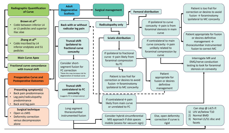
In summation, we used the findings of the present study to synthesize a summary “mindmap” diagram that illustrates an algorithmic approach to surgical management of the FC in patients with ADS (Fig. 4).
Fig. 4.
Mindmap demonstrating algorithmic approach to adult degenerative scoliosis diagnosis, characterization, and correction. cMIS, circumferential minimally invasive surgery; FC, fractional curve; MC, major curve; BMD, bone mineral density; EMG, electromyography.
5. Limitations
There are several limitations to acknowledge regarding the present study. In general, there is currently a paucity of studies comparing cMIS and open approaches for FC management, in part due to the recent advent of cMIS techniques. Consequently, it is unclear whether cMIS techniques offer similar levels of coronal plane realignment, pain relief, or functional improvement relative to traditional open techniques. Additionally, the present review is retrospective by design and utilizes data reported by previously published studies. As we did not directly collect or report the data within each study it is difficult to determine the extent to which particular studies may be biased. The present review is therefore limited by the quality of the included studies and is subject to reporting and sampling biases at baseline. Furthermore, the degree of heterogeneity presents across studies included in this review made quantitative synthesis challenging; nonetheless, we were deliberate and meticulous in our screening and enforced strict application of inclusion and exclusion criteria to ensure that, despite differences in study designs and potential differences in participating patient populations, the focus of each study was the corrective ability and role of the FC in the operative management of ADS.
CONCLUSION
Current literature suggests that the FC—defined as the compensatory lumbosacral distal to the major curve in ADS—may be a key structural contributor to malalignment and symptoms in patients with ADS. When coronal plane malalignment is ipsilateral to the FC concavity, the FC likely contributes to deformity progression and therefore must be corrected in the final construct. By contrast, when truncal shift is contralateral to the FC concavity, the FC is likely compensatory and ending constructs proximal to the sacrum may be reasonable, allowing for preservation of an additional motion segment, albeit at the cost of increased risk of adjacent segment disease. For curves requiring significant coronal plane realignment, kickstand rods appear to be the most effective technique when combined with conventional rod bending maneuvers and osteotomy work. Preliminary data suggests that cMIS techniques targeting ACR for deformity correction may help to preserve motion segments by enabling similar degrees of coronal plane correction as open posterior-only constructs. Nevertheless, the utility of cMIS techniques can be limited, especially for severe, flexible curves. Additional investigation into the strengths and weaknesses of cMIS techniques relative to open approaches is necessary, as is an improved understanding of the extent to which the FC is a precipitant of pathology in ADS as opposed to a compensatory mechanism for the underlying degenerative major curve.
Footnotes
Conflict of Interest
The authors have nothing to disclose.
Funding/Support
This study received no specific grant from any funding agency in the public, commercial, or not-for-profit sectors.
Author Contribution
Conceptualization: SCR, ZP, NJB, ALM, NL, LDDA, JAO, BDE, MHP; Data curation: SCR, ZP, NJB, SS, JR, AS, JCH; Formal analysis: SCR, NJB, SS, JR, JCH; Methodology: SCR, NJB, JCH, ALM, NL, LDDA, JAO, MHP; Project administration: LDDA, JAO, BDE, MHP; Visualization: SCR, ZP, NJB, ALM, NL, LDDA, JAO, BDE, MHP; Writing original draft: SCR, ZP, NJB, SS, JR, AS; Writing – review & editing: SCR, ZP, NJB, JR, JCH, ALM, NL, LDDA, JAO, BDE, MHP.
REFERENCES
- 1.Hong JY, Suh SW, Modi HN, et al. Centroid method: an alternative method of determining coronal curvature in scoliosis. A comparative study versus Cobb method in the degenerative spine. Spine J. 2013;13:421–7. doi: 10.1016/j.spinee.2012.11.051. [DOI] [PubMed] [Google Scholar]
- 2.Hong JY, Suh SW, Modi HN, et al. Reliability analysis for radiographic measures of lumbar lordosis in adult scoliosis: a case-control study comparing 6 methods. Eur Spine J. 2010;19:1551–7. doi: 10.1007/s00586-010-1422-x. [DOI] [PMC free article] [PubMed] [Google Scholar]
- 3.Carter OD, Haynes SG. Prevalence rates for scoliosis in US adults: results from the first National Health and Nutrition Examination Survey. Int J Epidemiol. 1987;16:537–44. doi: 10.1093/ije/16.4.537. [DOI] [PubMed] [Google Scholar]
- 4.Hong JY, Suh SW, Modi HN, et al. The prevalence and radiological findings in 1347 elderly patients with scoliosis. J Bone Joint Surg Br. 2010;92:980–3. doi: 10.1302/0301-620X.92B7.23331. [DOI] [PubMed] [Google Scholar]
- 5.Schwab F, Dubey A, Gamez L, et al. Adult scoliosis: prevalence, SF-36, and nutritional parameters in an elderly volunteer population. Spine (Phila Pa 1976) 2005;30:1082–5. doi: 10.1097/01.brs.0000160842.43482.cd. [DOI] [PubMed] [Google Scholar]
- 6.Pennington Z, Brown NJ, Quadri S, et al. Robotics planning in minimally invasive surgery for adult degenerative scoliosis: illustrative case. J Neurosurg Case Lessons. 2023;5:CASE22520. doi: 10.3171/CASE22520. [DOI] [PMC free article] [PubMed] [Google Scholar]
- 7.Brown NJ, Jammal OA, Himstead A, et al. Demographic predictors of treatment and complications for adult spinal deformity: an analysis of the national inpatient sample. Clin Neurol Neurosurg. 2022;222:107423. doi: 10.1016/j.clineuro.2022.107423. [DOI] [PubMed] [Google Scholar]
- 8.Fu KMG, Rhagavan P, Shaffrey CI, et al. Prevalence, severity, and impact of foraminal and canal stenosis among adults with degenerative scoliosis. Neurosurgery. 2011;69:1181–7. doi: 10.1227/NEU.0b013e31822a9aeb. [DOI] [PubMed] [Google Scholar]
- 9.Heary RF, Kumar S, Bono CM. Decision making in adult deformity. Neurosurgery. 2008;63(3 Suppl):69–77. doi: 10.1227/01.NEU.0000320426.59061.79. [DOI] [PubMed] [Google Scholar]
- 10.Bridwell KH. Selection of instrumentation and fusion levels for scoliosis: where to start and where to stop. Invited submission from the Joint Section Meeting on Disorders of the Spine and Peripheral Nerves, March 2004. J Neurosurg Spine. 2004;1:1–8. doi: 10.3171/spi.2004.1.1.0001. [DOI] [PubMed] [Google Scholar]
- 11.Cho KJ, Suk SI, Park SR, et al. Short fusion versus long fusion for degenerative lumbar scoliosis. Eur Spine J. 2008;17:650–6. doi: 10.1007/s00586-008-0615-z. [DOI] [PMC free article] [PubMed] [Google Scholar]
- 12.Cho KJ, Suk SI, Park SR, et al. Arthrodesis to L5 versus S1 in long instrumentation and fusion for degenerative lumbar scoliosis. Eur Spine J. 2009;18:531–7. doi: 10.1007/s00586-009-0883-2. [DOI] [PMC free article] [PubMed] [Google Scholar]
- 13.Swamy G, Berven SH, Bradford DS. The selection of L5 versus S1 in long fusions for adult idiopathic scoliosis. Neurosurg Clin N Am. 2007;18:281–8. doi: 10.1016/j.nec.2007.01.010. [DOI] [PubMed] [Google Scholar]
- 14.Lenke LG, Betz RR, Haher TR, et al. Multisurgeon assessment of surgical decision-making in adolescent idiopathic scoliosis: curve classification, operative approach, and fusion levels. Spine (Phila Pa 1976) 2001;26:2347–53. doi: 10.1097/00007632-200111010-00011. [DOI] [PubMed] [Google Scholar]
- 15.Page MJ, Moher D, Bossuyt PM, et al. PRISMA 2020 explanation and elaboration: updated guidance and exemplars for reporting systematic reviews. BMJ. 2021;372:n160. doi: 10.1136/bmj.n160. [DOI] [PMC free article] [PubMed] [Google Scholar]
- 16.Zhang J, Wang Z, Chi P, et al. Directionality of lumbosacral fractional curve relative to C7 plumb line, a novel index associated with postoperative coronal imbalance in patients with degenerative lumbar scoliosis. Spine (Phila Pa 1976) 2021;46:366–73. doi: 10.1097/BRS.0000000000003776. [DOI] [PubMed] [Google Scholar]
- 17.Brown KM, Ludwig SC, Gelb DE. Radiographic predictors of outcome after long fusion to L5 in adult scoliosis. J Spinal Disord Tech. 2004;17:358–66. doi: 10.1097/01.bsd.0000112080.04960.67. [DOI] [PubMed] [Google Scholar]
- 18.Pugely AJ, Ries Z, Gnanapragasam G, et al. Curve characteristics and foraminal dimensions in patients with adult scoliosis and radiculopathy. Clin Spine Surg. 2017;30:E111–8. doi: 10.1097/BSD.0b013e3182aab1e3. [DOI] [PubMed] [Google Scholar]
- 19.Silva FE, Lenke LG. Adult degenerative scoliosis: evaluation and management. Neurosurg Focus. 2010;28:E1. doi: 10.3171/2010.1.FOCUS09271. [DOI] [PubMed] [Google Scholar]
- 20.Amara D, Mummaneni PV, Ames CP, et al. Treatment of only the fractional curve for radiculopathy in adult scoliosis: comparison to lower thoracic and upper thoracic fusions. J Neurosurg Spine. 2019;30:506–14. doi: 10.3171/2018.9.SPINE18505. [DOI] [PubMed] [Google Scholar]
- 21.Yagi M, Patel R, Lawhorne TW, et al. Adult thoracolumbar and lumbar scoliosis treated with long vertebral fusion to the sacropelvis: a comparison between new hybrid selective spinal fusion versus anterior-posterior spinal instrumentation. Spine J. 2014;14:637–45. doi: 10.1016/j.spinee.2013.06.090. [DOI] [PubMed] [Google Scholar]
- 22.Chou D, Mummaneni P, Anand N, et al. Treatment of the fractional curve of adult scoliosis with circumferential minimally invasive surgery versus traditional, open surgery: an analysis of surgical outcomes. Global Spine J. 2018;8:827–33. doi: 10.1177/2192568218775069. [DOI] [PMC free article] [PubMed] [Google Scholar]
- 23.Amara D, Mummaneni PV, Burch S, et al. The impact of increasing interbody fusion levels at the fractional curve on lordosis, curve correction, and complications in adult patients with scoliosis. J Neurosurg Spine. 2020;34:430–9. doi: 10.3171/2020.6.SPINE20256. [DOI] [PubMed] [Google Scholar]
- 24.Manwaring JC, Bach K, Ahmadian AA, et al. Management of sagittal balance in adult spinal deformity with minimally invasive anterolateral lumbar interbody fusion: a preliminary radiographic study. J Neurosurg Spine. 2014;20:515–22. doi: 10.3171/2014.2.SPINE1347. [DOI] [PubMed] [Google Scholar]
- 25.Buell TJ, Shaffrey CI, Kim HJ, et al. Global coronal decompensation and adult spinal deformity surgery: comparison of upper-thoracic versus lower-thoracic proximal fixation for long fusions. J Neurosurg Spine. 2021;35:761–73. doi: 10.3171/2021.2.SPINE201938. [DOI] [PubMed] [Google Scholar]
- 26.Geddes B, Glassman SD, Mkorombindo T, et al. Improvement of coronal alignment in fractional low lumbar curves with the use of anterior interbody devices. Spine Deform. 2021;9:1443–7. doi: 10.1007/s43390-021-00328-0. [DOI] [PubMed] [Google Scholar]
- 27.Zuckerman SL, Chanbour H, Hassan FM, et al. The lumbosacral fractional curve vs maximum coronal Cobb angle in adult spinal deformity patients with coronal malalignment: which matters more? Global Spine J. 2023 Mar 29;:21925682231161564. doi: 10.1177/21925682231161564. doi: . [Epub] [DOI] [PMC free article] [PubMed] [Google Scholar]
- 28.Plais N, Bao H, Lafage R, et al. The clinical impact of global coronal malalignment is underestimated in adult patients with thoracolumbar scoliosis. Spine Deform. 2020;8:105–13. doi: 10.1007/s43390-020-00046-z. [DOI] [PubMed] [Google Scholar]
- 29.Obeid I, Berjano P, Lamartina C, et al. Classification of coronal imbalance in adult scoliosis and spine deformity: a treatment-oriented guideline. Eur Spine J. 2019;28:94–113. doi: 10.1007/s00586-018-5826-3. [DOI] [PubMed] [Google Scholar]
- 30.Kim YJ, Hyun SJ, Cheh G, et al. Decision making algorithm for adult spinal deformity surgery. J Korean Neurosurg Soc. 2016;59:327–33. doi: 10.3340/jkns.2016.59.4.327. [DOI] [PMC free article] [PubMed] [Google Scholar]
- 31.Deviren V, Berven S, Kleinstueck F, et al. Predictors of flexibility and pain patterns in thoracolumbar and lumbar idiopathic scoliosis. Spine (Phila Pa 1976) 2002;27:2346–9. doi: 10.1097/00007632-200211010-00007. [DOI] [PubMed] [Google Scholar]
- 32.Bao H, Yan P, Qiu Y, et al. Coronal imbalance in degenerative lumbar scoliosis: Prevalence and influence on surgical decision-making for spinal osteotomy. Bone Joint J. 2016;98-B:1227–33. doi: 10.1302/0301-620X.98B9.37273. [DOI] [PubMed] [Google Scholar]
- 33.Buell TJ, Buchholz AL, Mazur MD, et al. Kickstand rod technique for correcting coronal imbalance in adult scoliosis: 2-dimensional operative video. Oper Neurosurg (Hagerstown) 2020;19:E163–4. doi: 10.1093/ons/opz306. [DOI] [PubMed] [Google Scholar]
- 34.Puvanesarajah V, Raad M, Hassan FM, et al. The “kickstand rod” technique for correction of coronal malalignment: two-year clinical and radiographic outcomes. Spine Deform. 2023;11:153–61. doi: 10.1007/s43390-022-00564-y. [DOI] [PubMed] [Google Scholar]
- 35.Mundis GM, Jr, Walker CT, Smith JS, et al. Kickstand rods and correction of coronal malalignment in patients with adult spinal deformity. Eur Spine J. 2022;31:1197–205. doi: 10.1007/s00586-022-07161-7. [DOI] [PubMed] [Google Scholar]
- 36.Winter RB, Lonstein JE, Denis F. Pain patterns in adult scoliosis. Orthop Clin North Am. 1988;19:339–45. [PubMed] [Google Scholar]
- 37.Bao H, Liu Z, Zhang Y, et al. Sequential correction technique to avoid postoperative global coronal decompensation in rigid adult spinal deformity: a technical note and preliminary results. Eur Spine J. 2019;28:2179–86. doi: 10.1007/s00586-019-06043-9. [DOI] [PubMed] [Google Scholar]
- 38.Glassman SD, Bridwell K, Dimar JR, et al. The impact of positive sagittal balance in adult spinal deformity. Spine (Phila Pa 1976) 2005;30:2024–9. doi: 10.1097/01.brs.0000179086.30449.96. [DOI] [PubMed] [Google Scholar]
- 39.Chou D, Lafage V, Chan AY, et al. Patient outcomes after circumferential minimally invasive surgery compared with those of open correction for adult spinal deformity: initial analysis of prospectively collected data. J Neurosurg Spine. 2021;36:203–14. doi: 10.3171/2021.3.SPINE201825. [DOI] [PubMed] [Google Scholar]
- 40.Bridwell KH, Edwards CC, 2nd, Lenke LG. The pros and cons to saving the L5-S1 motion segment in a long scoliosis fusion construct. Spine (Phila Pa 1976) 2003;28:S234–42. doi: 10.1097/01.BRS.0000092462.45111.27. [DOI] [PubMed] [Google Scholar]
- 41.Campbell PG, Nunley PD. The challenge of the lumbosacral fractional curve in the setting of adult degenerative scoliosis. Neurosurg Clin N Am. 2018;29:467–74. doi: 10.1016/j.nec.2018.02.004. [DOI] [PubMed] [Google Scholar]
- 42.Yao YC, Kim HJ, Bannwarth M, et al. Lowest instrumented vertebra selection to S1 or ilium versus L4 or L5 in adult spinal deformity: factors for consideration in 349 patients with a mean 46-month follow-up. Global Spine J. 2023;13:932–9. doi: 10.1177/21925682211009178. [DOI] [PMC free article] [PubMed] [Google Scholar]
- 43.Hofler RC, Iordanou J, Nockels RP. Intraoperative asssessment of coronal balance with long cassette radiographs in adult thoracolumbar deformity correction. World Neurosurg. 2022;163:e384–90. doi: 10.1016/j.wneu.2022.03.136. [DOI] [PubMed] [Google Scholar]


