Abstract
Background and Aims
Hepatocellular carcinoma (HCC) cases with small nodules are commonly treated with radiofrequency ablation (RFA), but the recurrence rate remains high. This study aimed to establish a blood signature for identifying HCC with metastatic traits pre-RFA.
Methods
Data from HCC patients treated between 2010 and 2017 were retrospectively collected. A blood signature for metastatic HCC was established based on blood levels of alpha-fetoprotein and des-γ-carboxy-prothrombin, cell-free DNA (cfDNA) mutations, and methylation changes in target genes in frozen-stored plasma samples that were collected before RFA performance. The HCC blood signature was validated in patients prospectively enrolled in 2021.
Results
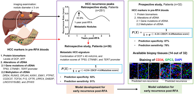
Of 251 HCC patients in the retrospective study, 33.9% experienced recurrence within 1 year post-RFA. The HCC blood signature identified from these patients included des-γ-carboxy-prothrombin ≥40 mAU/mL with cfDNA mutation score, where cfDNA mutations occurred in the genes of TP53, CTNNB1, and TERT promoter. This signature effectively predicted 1-year post-RFA recurrence of HCC with 92% specificity and 91% sensitivity in the retrospective dataset, and with 87% specificity and 76% sensitivity in the prospective dataset (n=32 patients). Among 14 cases in the prospective study with biopsy tissues available, positivity for the HCC blood signature was associated with a higher HCC tissue score and shorter distance between HCC cells and microvasculature.
Conclusions
This study established an HCC blood signature in pre-RFA blood that potentially reflects HCC with metastatic traits and may be valuable for predicting the disease’s early recurrence post-RFA.
Keywords: Liquid biopsy, Liver dysplastic nodules, Radiofrequency ablation, Hepatocellular carcinoma, Recurrence prediction, Metastatic nodules
Graphical abstract
Introduction
Hepatectomy remains the mainstay curative therapy for hepatocellular carcinoma (HCC) cases involving a solitary nodule ≤5 cm in diameter. However, more than half of HCC patients are not candidates for hepatectomy due to liver cirrhosis.1–3 For patients with HCC nodules ≤2–3 cm in diameter, treatment with radiofrequency ablation (RFA) is widely accepted and recommended by oncology experts.1,3 However, previous studies have demonstrated that the rate of tumor recurrence among HCC cases with small nodules is higher following RFA compared to surgery.1,4,5 The major cause of HCC recurrence after RFA therapy is incomplete ablation, which usually occurs in patients with lesions ≥3 cm and/or with multiple nodules.6,7 Even some cases in which complete ablation is achieved still experience recurrence after RFA.8,9 Additional factors known to be associated with an increased risk of HCC recurrence after RFA include intrinsic nodule characteristics (e.g., specific genomic mutations, poor differentiation of tumor cells), tumor location (e.g., periportal HCC), vascular/microvascular invasion, and liver stiffness.10–12 With advancements in systemic therapies for HCC, recent evidence has demonstrated that patients with a high recurrence risk can benefit from adjuvant therapies, such as immune check-point inhibitors, administered during or after RFA or resection.13,14 For appropriate administration of the adjuvant therapies, a means to identify the patients who have a high risk for HCC recurrence post-RFA is necessary.
HCC recurrence prediction is generally established based on the clinicopathological characteristics of tumors among hepatectomy-treated patients. The risk factors contributing to early recurrence of HCC were recognized to be different from those contributing to later recurrence. Early recurrence of HCC generally refers to recurrence within 2 years post-resection and is mainly due to the metastasis of primary tumors, including microscopic vascular invasion. Conversely, late recurrence generally refers to 2–3 years post-resection and is mainly due to de novo hepatocarcinogenesis.1,3,15 For HCC patients with nodules ≤3 cm in diameter treated with RFA, tumor tissue sampling is limited, which in turn, limits the possibility for the comprehensive evaluation of recurrence risk. Imaging-based examinations cannot reliably characterize the pathological characteristics of nodules and discriminate metastatic HCC from dysplastic nodules.1,16,17 Thus, alternative blood markers for pre-RFA analysis are needed. One study reported that some general clinical factors have the ability to predict early recurrence of HCC within 12 months post-RFA, including the presence of multiple tumors and altered pre-RFA blood levels of alpha-fetoprotein (AFP), gamma-glutamyl transferase, and albumin.18 Generally, the levels of circulating tumor cells and the levels of cancer cell products in peripheral blood are known to provide crucial insights into cancer biology and the metastatic process.19 However, studies of the potential value of liquid biopsy assessments for recognizing small HCC nodules with metastatic traits and predicting early recurrence post-RFA were lacking.
Liquid biopsy assays based on the detection of different classes of tumor-related products, including circulating tumor-related DNA (ctDNA), tumor-associated microRNA (miRNA), and proteins, have been translated into clinical practice.19 Comparison with dysplastic nodules in cirrhotic liver, early advanced HCC displays genomic and epigenetic aberrations, and transcriptional deregulation. Integration of data obtained from both preclinical models and human studies has identified that the histopathological features of HCC are closely related to specific molecular alterations at the genomic, epigenomic, and transcriptomic levels.2,20 Previous research found that among the population with a high risk for HCC, the quantification of blood levels of AFP and des-gamma-carboxyprothrombin (DCP), the examination of HCC-related mutations and methylation changes in cell-free DNA (cfDNA), and HCC-related miRNA expression profiling could efficiently detect HCC at the very-early stage.21–25 Additionally, among HCC patients that underwent curative hepatectomy, detection of ctDNA in the plasma post-hepatectomy was able to identify tumors that could not be visualized on imaging.26
In our previous reports, we demonstrated that patients with HCC ≤3 cm in diameter can be identified among individuals with chronic hepatitis B virus (HBV) infection by quantification of blood levels of AFP and DCP along with profiling of mutations and methylation changes in cfDNA in plasma.23,24 In the present study, we aimed to determine if similar markers could provide a pre-RFA blood signature for HCC with metastatic traits in patients with HCC nodules ≤3 cm in diameter to predict early disease recurrence post-RFA, which would support the planning of personalized treatment strategies.
Methods
Patients and study design
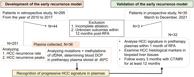
Two cohorts of patients who met the following criteria were included in the retrospective study and enrolled in the prospective study: (1) aged 18–70 years; (2) treatment-naïve HCC related to chronic HBV or hepatitis C virus (HCV) infection; (3) hepatic lesions diagnosed based on contrast-enhanced magnetic resonance imaging (MRI)/computed tomography (CT) examination, without histological confirmation; (4) ≤3 nodules, with each nodule having a diameter ≤3 cm; (5) no signs of tumor vascular invasion or extrahepatic metastases; (6) treatment with RFA within 1 week after imaging-based diagnosis; (7) complete ablation observed on MRI/CT examination at 1 month post-RFA; (8) no signs or history of other malignancies; and (9) no HIV infection. The study design is depicted in Figure 1.
Fig. 1. Study design.
RFA-treated HCC patients met the following criteria were enrolled in the retrospective study and in the prospective study: 1. aged 18–70 years; 2. treatment-naïve HCC related to chronic HBV or HCV infection; 3. diagnosed based on image examination without histological confirmation; 4. ≤3 nodules, each nodule ≤3 cm in diameter; 5. no signs of tumor vascular invasion or extrahepatic metastases; 6. treatment with RFA within 1 week after imaging-based diagnosis; 7. no signs or history of other malignancies; 8. no HIV infection. RFA, radiofrequency ablation; HCC, hepatocellular carcinoma; HBV, hepatitis B virus; HCV, hepatitis C virus; HIV, human immunodeficiency virus; cfDNA, cell-free DNA; DCP, des-γ-carboxy-prothrombin; CT/MRI, computed tomography/magnetic resonance imaging.
In the retrospective study, consecutive HCC patients treated from 2010 to 2017 were included. Clinical laboratory results were obtained 1–3 days before RFA, and treatment outcomes were retrieved from medical records. Disease-free survival (DFS) was defined as the duration from the date of RFA to that of recurrence within the liver or of extrahepatic metastasis based on the modified Response Evaluation Criteria in Solid Tumors (mRECIST). Cases with no outcomes within 12 months post-RFA were excluded according to previously reported results.8,27 One day before RFA performance, some patients who were treated in 2016 and 2017 donated 10 mL EDTA-treated peripheral blood. The pre-RFA plasma was prepared within 2 h after blood draw, snap-frozen, and stored at −80°C.
For the prospective study group, patients were enrolled from March to December 2021 to validate the blood signature of HCC early recurrence established using data from the retrospective patient set. One day before RFA performance, a plasma sample from each participant was prepared from 10 mL EDTA-treated peripheral blood, and HCC-related markers were profiled within 1 month post-RFA. The participants were followed up at 1-month post-RFA and every 3 months thereafter until January 31, 2023 using contrast-enhanced CT/MRI examination. In some patients where biopsy tissue was available, we also validated the HCC blood signature by analyzing the HCC tissue-score,28 and by measuring the distance between HCC cells and tumor vasculature in tissues after multiplex immunohistochemistry staining.
Quantification of blood levels of AFP and DCP
The AFP levels in serum samples, recorded in participants’ medical documents, were determined using reagents from Roche Diagnostic GmbH in Cobas e-602. In plasma samples previously stored at −80°C, the AFP and DCP levels were measured using Abbott reagents with the ARCHITECT i2000SR chemical luminescence immunity analyzer.23
Analysis of cfDNA mutations and methylation changes in stored plasma
HCC-related cfDNA mutations and methylation changes were examined simultaneously in plasma using our previously-described Mutation Capsule Plus (MCP) technology.24 Briefly, cfDNA was extracted from the plasma samples using the Apostle MiniMax cfDNA isolation kit. The cfDNA (5–40 ng) was digested using the methylation-sensitive restriction enzyme Hha I (R0139L, New England BioLabs) and subsequently ligated to customized MCP adapters with random DNA barcodes. The ligated product was amplified using the KAPA HiFi PCR kit (KR0369, Kapa Biosystems) to generate the whole genome library. The library was then amplified using the co-detection panel of mutations and methylations based on the coding regions of TP53 and CTNNB1, and the promoter region of TERT, and the methylation markers of DRD4, RUNX2, OPLAH, AXIN1, LINC01015UBD, EMX1, PTPN7, CCDC37, TCF24, F12, CFTR, LRRC4, DAB2IP, and ZFHX3.24 The product was then sequenced and analyzed on the Illumina NovaSeq 6000 platform (Illumina).
Candidate somatic mutations, including single nucleotide polymorphisms (SNPs) and insertions–deletions (INDELs), were identified by alignment to the hg19 and HBV genomes. Mutations of distinct genes in cfDNA were assigned variation points based on the Region of Interest (ROI) score according to our previous work related to the detection of HCC nodules in the high-risk population.23 The presence of a TP53 mutation was assigned 2.0 points, a CTNNB1 mutation 1.2 points, and a mutation of the TERT promoter or HBV integration at the TERT promoter 1.4 points. The degree of methylation was calculated as the ratio of the number of methylated molecules to the total number of methylated and unmethylated molecules. The sequenced molecules ending with the GCGC sequence were denoted as unmethylated molecules, while those that passed through the GCGC sequence were denoted as methylated molecules.24
Immunochemistry and multiplex immunohistochemistry (mIHC)
Paraffin-embedded biopsy tissues were used for immunochemistry and mIHC analyses. For immunochemistry analysis, sectioned tissues were stained with antibodies against glypican-3 (GPC3), heat-shock protein 70 (HSP70), glutamine synthetase (GS), cytokeratin 7 (CK7), and CD34. All reagents were purchased from ZSGB-Bio (Beijing, China). Sections were scanned and analyzed using Aperio ScanScope software (Aperio Technologies). Three medical doctors independently evaluated levels of HCC markers as described previously.28 GPC3 scoring was slightly modified as follows: 2.0 points for sections that unequivocally overexpressed GPC3 accounting for more than 50% cellularity of the lesion; 1.5 points for 21–50%; 1.0 point for 11–20%; and 0.5 points for 5–10%. Staining of GPC3, HSP70, GS, CK7, and CD34 was scored and summed to generate an HCC tissue-score28 for each individual nodule, with total scores ranging from 0–10.
An Opal Multiplex IHC assay kit (PerkinElmer, USA) was used for mIHC staining of GPC3 and CD34 in the biopsy tissues. The HALO image analysis platform (Indica, USA) was used to measure the distance between GPC3-positive cells and CD34-positive cells as we previously reported.29 When the sectioned samples were negative for GPC3 staining, we defined a value of 80 µm, which was the greatest distance between the two cell types observed in the sections positive for GPC3 staining.
Statistical analysis
GraphPad Prism Version 9.0 was used for statistical analysis. Continuous variables with a normal distribution are presented as mean±standard deviation (SD) and were compared using a t-test. Those with a non-normal distribution are presented as median (interquartile range, IQR) and were compared using the Mann-Whitney U-test. Categorical variables are summarized as frequency (percentage) and were compared using the chi-square test or Fisher’s exact test.
Individual general characteristics and laboratory parameters examined 1–3 days pre- RFA were evaluated by univariable logistic regression analysis to identify risk factors for HCC recurrence within 1 year post-RFA. The predictive value of significant risk factors derived from univariable analysis based on p<0.05 was then evaluated by receiver operating characteristic (ROC) curve analysis, including calculation of the area under the ROC curve (AUC) for predicting 1-year HCC recurrence.
The individual risk factors with p<0.05 were then included in multivariable logistic analysis. Different models were built based on combinations of different risk factors identified from the univariable analysis, and the respective AUC values were calculated. The cut-off value for each factor was set based on the highest Youden index score.30 A formula based on β value (Coefficients) was constructed to generate the risk probability (0–1) for predicting 1-year HCC recurrence post-RFA. A probability ≥0.5 was classified as positive for the HCC blood signature, indicating a risk of HCC recurrence within 1 year post-RFA. For validation of the models, HCC-related biomarkers were examined in pre-RFA plasma samples from each patient in the prospective study. Based on the formula constructed in the retrospective study, the probability (0–1) of HCC recurrence within 1 year was calculated for each case. Cases with a probability ≥0.5 were considered to have an elevated risk of HCC recurrence within 1 year post-RFA.
Results
Clinical characteristics of patients in the retrospective study
For the retrospective data analysis, we included 295 consecutive HCC patients who were diagnosed by imaging examination as having ≤3 nodules each measuring ≤3 cm in diameter and underwent RFA between 2010 and 2017. We excluded 44 cases considered as incomplete ablation by MRI examination 1 month post-RFA or with unknown outcomes over 12 months post-RFA. Finally, 251 eligible patients who underwent sufficient ablation and presented no other lesions in the liver at 1 month post-RFA were included (Fig. 1). The mean DFS and median DFS were 33.9±30.6 months and 23.4 (8.0–49.2) months, respectively.
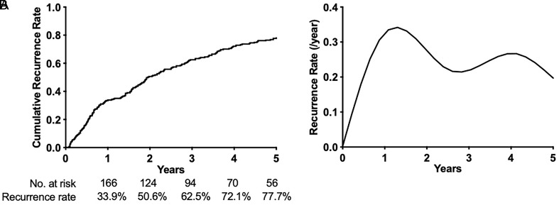
Over the 5-year period post-RFA, the cumulative HCC recurrence rate among the 251 eligible patients was 77.7% (195/251) (Fig. 2A). Two peaks of HCC recurrence were observed. The first peak was observed at approximately 1 year post-RFA, when the recurrence rate was 33.9% (85/251). The second peak was observed approximately 4 years post-RFA, and the recurrence rate beyond 1 year post-RFA was 43.8% (110/251) (Fig. 2B). Based on the results of previous reports,1,3,8,15,27 we classified recurrence within 1 year as early HCC recurrence, and recurrence beyond this time point as late HCC recurrence.
Fig. 2. HCC recurrence rate post-RFA in 251 patients that were radiologically diagnosed as HCC ≤3 cm in diameter and underwent RFA.
(A) Cumulative recurrence rate. (B) Recurrence rate per year post-RFA. RFA, radiofrequency ablation; HCC, hepatocellular carcinoma.
Clinical characteristics associated with early HCC recurrence post-RFA
Generally, HCC late recurrence is associated with the de novo development of a neoplasm in the cirrhotic livers, whereas early recurrence is mainly due to metastasis of primary tumors.1,3,15 Our current analysis excluded the cases considered as incomplete ablation, which was reported as the major cause of early HCC recurrence.6,7 Using data from the eligible 251 patients in the retrospective study, univariable logistic regression analysis indicated that the following clinical factors showed no significant association with early HCC recurrence post-RFA: age, gender, nodule number, nodule size, and types of liver diseases (Table 1).
Table 1. General clinical characters of 251 patients and early HCC recurrence post-RFA.
| Variable, Number (%) | Recurrence |
Non-recurrence |
OR |
p Value |
|---|---|---|---|---|
| N=85 | N=166 | (95% CI) | ||
| Age (years) | ||||
| Mean±SD | 53.5±9.7 | 53.2±8.8 | NA | 0.823# |
| Median (IQR) | 55.0 (47.0–60.0) | 54.5 (48.0–59.0) | ||
| Gender | ||||
| Male | 62 (72.9%) | 128 (77.1%) | 1 | 0.467 |
| Female | 23 (27.1%) | 38 (22.9%) | 1.25 (0.68–2.27) | |
| Tumor numbers | ||||
| 1 | 76 (89.4%) | 143 (86.1%) | 1 | 0.463 |
| >1 | 9 (10.6%) | 23 (13.9%) | 0.74 (0.31–1.62) | |
| Tumor diameter (cm) | ||||
| ≤1 | 9 (10.6%) | 25 (15.1%) | 1 | 0.329 |
| >1 | 76 (89.4%) | 141 (84.9%) | 1.50 (0.68–3.54) | |
| Liver diseases | ||||
| HBV | 79 (92.9%) | 155 (93.4%) | 1 | 0.897 |
| HCV | 6 (7.1%) | 11 (6.6%) | 0.93 (0.34–2.80) | |
#Compared using Unpaired t-test. HCC, hepatocellular carcinoma; RFA, radiofrequency ablation; OR, odds ratio; CI, Confidence Interval; SD, standard deviation; IQR, interquartile range; NA, not available; HBV, Hepatitis B virus; HCV, hepatitis C virus.
Correlation of pre-RFA clinical laboratory results and early HCC recurrence post-RFA in the retrospective study
Among the 251 patients, pre-RFA plasma samples were available for 36 cases treated in 2016–2017. The 1-year HCC recurrence rate among these 36 patients was 30.6% (11/36). No differences in age, gender, type of liver diseases (HBV- or HCV-related), tumor number, tumor size, DFS, or 1-year recurrence rate were observed between the patients with and without blood samples available (Table 2). The DCP levels in the plasma samples of the 36 cases were quantified, and their serum AFP levels and other laboratory results that potentially reflect the severity of liver diseases were retrieved from their medical records. We analyzed the correlation between early HCC recurrence post-RFA and blood HCC biomarker levels, AFP and DCP levels, as well as certain laboratory parameters that could promote HCC development.1,31,32 Cirrhosis-related laboratory variables, including platelet and white blood cell (WBC) counts, albumin and total bilirubin concentrations, and ALBI score, showed negligible impacts on early HCC recurrence (p>0.05). Also, neither AFP serum levels of ≥20 ng/mL or ≥200 ng/mL showed a statistically significant association with early HCC recurrence (p>0.05). However, blood DCP ≥40 mAU/mL was positively associated with early HCC recurrence (p<0.05) (Table 3).
Table 2. Variables of the patients with and without blood donation of the retrospective study.
| Variable, Number (%) | Blood donation |
No blood |
p value |
|---|---|---|---|
| N=36 | N=215 | ||
| Age (years) | |||
| Mean±SD | 51.8±8.5 | 53.6±9.2 | 0.268# |
| Median (IQR) | 52.5 (47.3–59.0) | 55.0 (48.0–59.0) | |
| Gender | |||
| Male | 30 (83.3%) | 160 (74.4%) | 0.248** |
| Female | 6 (16.7) | 55 (25.6%) | |
| Liver diseases | |||
| HBV | 33 (91.7%) | 201 (93.5%) | 0.965* |
| HCV | 3 (8.3%) | 14 (6.5%) | |
| 1-year recurrence | |||
| Yes | 11 (30.6%) | 74 (34.4%) | 0.650** |
| No | 25 (69.4%) | 141 (65.6%) | |
| DFS (Months) | |||
| Mean±SD | 28.0±18.6 | 29.4±22.6 | 0.959## |
| Median (IQR) | 27.0 (9.0–44.8) | 23.0 (7.7–53.0) | |
| Tumor numbers | |||
| 1 | 31 (86.1%) | 188 (87.4%) | 0.961* |
| >1 | 5 (13.9%) | 27 (12.6%) | |
| Tumor size (cm) | |||
| Mean±SD | 1.8±0.5 | 1.7±0.6 | 0.328## |
| Median (IQR) | 1.7 (1.5 - 2.1) | 1.6 (1.2 - 2.1) |
*Compared using Chi-square with Yates’ correction. **Compared using Chi-square test. #Compared using Unpaired t-test. ##Compared using Mann-Whitney test. SD, standard deviation; IQR, interquartile range; HBV, Hepatitis B virus; HCV, hepatitis C virus; DFS, disease free survival.
Table 3. The pre-RFA blood factors related with early HCC recurrence post-RFA in 36 patients who donated blood before RFA treatment.
| Variable, Number (%) | Recurrence |
No recurrence |
Odds ratio |
p value |
|---|---|---|---|---|
| (N=11) | (N=25) | OR (95CI%) | ||
| Age (years) | ||||
| Mean±SD | 50.6±10.8 | 52.2±7.5 | NA | 0.610# |
| Median (IQR) | 53.0 (46.0–59.0) | 52.0 (49.0–59.0) | ||
| Gender | ||||
| Male | 10 (90.9) | 20 (80.0) | 1 | 0.430 |
| Female | 1 (9.1) | 5 (20.0) | 0.40 (0.02–2.95) | |
| Tumor numbers | ||||
| 1 | 10 (90.9) | 21 (84.0) | 1 | 0.586 |
| >1 | 1 (9.1) | 4 (16.0) | 0.53 (0.03–4.15) | |
| Tumor diameter (cm) | ||||
| ≤1 | 2 (18.2) | 2 (8.0) | 1 | 0.383 |
| >1 | 9 (81.8) | 23 (92.0) | 0.39 (0.04–3.66) | |
| Platelet counts, 109/L | ||||
| ≥100 | 4 (36.4) | 9 (36.0) | 1 | 0.983 |
| <100 | 7 (63.6) | 16 (64.0) | 0.98 (0.23–4.62) | |
| WBC counts, 109/L | ||||
| ≥4 | 6 (54.5) | 10 (40.0) | 1 | 0.421 |
| <4 | 5 (45.5) | 15 (60.0) | 0.56 (0.13–2.33) | |
| Albumin, g/L | ||||
| ≥35 | 7 (63.6) | 19 (76.0) | 1 | 0.448 |
| <35 | 4 (36.4) | 6 (24.0) | 1.81 (0.37–8.47) | |
| Total bilirubin, µM | ||||
| <17 | 3 (27.3) | 12 (48.0) | 1 | 0.252 |
| ≥17 | 8 (72.7) | 13 (52.0) | 2.46 (0.56–13.30) | |
| ALBI score | ||||
| 1 (≤−2.60) | 4 (36.4) | 11 (44.0) | 1 | 0.669 |
| ≥2 (>−2.60) | 7 (63.6) | 14 (56.0) | 1.38 (0.33–6.39) | |
| AFP (ng/mL) | ||||
| <20 | 7 (63.6) | 19 (76.0) | 1 | 0.448 |
| ≥20 | 4 (36.4) | 6 (24.0) | 1.81 (0.37–8.47) | |
| AFP (ng/mL) | ||||
| <200 | 8 (72.7) | 23 (92.0) | 1 | 0.144 |
| ≥200 | 3 (27.3) | 2 (8.0) | 4.13 (0.61–37.60) | |
| DCP (mAU/mL) | ||||
| <40 | 2 (18.2) | 17 (68.0) | 1 | 0.011 |
| ≥40 | 9 (81.8) | 8 (32.0) | 9.56 (1.93–73.17) | |
| Methylation score of 14 genes | ||||
| <0.7 | 10 (90.9) | 22 (88.0) | 1 | 0.799 |
| ≥0.7 | 1 (9.1) | 3 (12.0) | 0.73 (0.03–6.58) | |
| CTNNB1 mutation | ||||
| No | 11 (100) | 23 (92.0) | NA | 1 |
| Yes | 0 | 2 (8.0) | ||
| TERT promoter mutation | ||||
| No | 5 (45.5) | 23 (92.0) | 1 | 0.006 |
| Yes | 6 (54.5) | 2 (8.0) | 13.80 (2.42–116.40) | |
| TP53 mutation | ||||
| No | 5 (45.5) | 23 (92.0) | 1 | 0.006 |
| Yes | 6 (54.5) | 2 (8.0) | 13.80 (2.42–116.40) | |
| Mutation score | ||||
| <1.4 | 1 (9.1) | 23 (92.0) | 1 | 0.0002 |
| ≥1.4 | 10 (90.9) | 2 (8.0) | 115.00 (13.45–2,847.00) | |
#Compared using Unpaired t-test. HCC, hepatocellular carcinoma; RFA, radiofrequency ablation; OR, odds ratio; CI, confidence interval; SD, standard deviation; IQR, interquartile range; ALBI, albumin-bilirubin; WBC, white blood cell; AFP, alpha-fetoprotein; DCP, des-γ-carboxy-prothrombin; NA, not available.
Association of genetic and epigenetic alterations in pre-RFA plasma with early HCC recurrence post-RFA
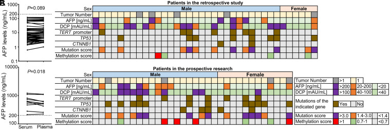
To test the reliability of the frozen plasma samples for detecting the presence of malignancy, we measured the AFP concentrations in 69 frozen plasma samples and compared the results with the corresponding concentrations in matched serum samples. Patients with hepatic lesions and serum AFP levels ≥200 ng/mL are considered to be at high risk of HCC.1 In 59 frozen plasma samples, no significant reduction in AFP concentration was detected compared with that in each matched serum sample with an AFP <200 ng/mL (p=0.089). In 10 frozen plasma samples, the AFP concentrations were slightly lower in frozen plasma samples than those in matched serum samples (p=0.018), but remained above 200 ng/mL (Fig. 3A). These results indicated that the frozen samples could be used for examination of HCC biomarkers.
Fig. 3. HCC-associated biomarkers detected in the pre-RFA blood.
(A) AFP concentrations in freshly-prepared sera retrieved from medical records and in matched frozen plasma samples. Different biomarkers detected in each case enrolled in the retrospective study (B) and in the prospective study (C). AFP, alpha-fetoprotein; DCP, des-γ-carboxy-prothrombin. RFA, radiofrequency ablation; HCC, hepatocellular carcinoma.
In the same blood samples, we analyzed genetic and epigenetic alterations of cfDNA that were previously identified as associated with malignant HCC,23,24 including cfDNA mutations of TP53, CTNNB1, the TERT promoter, and the cfDNA methylation status of the following 14 genes: DRD4, RUNX2, OPLAH, AXIN1, LINC01015UBD, EMX1, PTPN7, CCDC37, TCF24, F12, CFTR, LRRC4, DAB2IP, and ZFHX3. In the frozen plasma samples from patients in the retrospective cohort, genetic and methylation alterations in cfDNA varied, as did the levels of AFP and DCP (Fig. 3B). In the prospectively enrolled patients, distinct patterns of variation in blood AFP and DCP levels as well as cfDNA genetic and methylation alterations were also observed (Fig. 3C), further confirming the reliability of frozen samples for the examination of HCC signatures.
Univariable logistic regression analysis indicated that the CTNNB1 mutation and higher methylation of the 14 targeted genes of cfDNA showed no significant association with early HCC recurrence (p>0.05). However, cfDNA mutations in either TP53 or the TERT promoter were significantly associated with early HCC recurrence post-RFA (p<0.05) (Table 3). In some cases, diagnosed by imaging as having a single nodule, we detected multiple variants of TP53 mutations in cfDNA, indicating that the liver harbored more than one malignant cell clone. Based on our previous finding regarding the detection of HCC nodules in a high-risk population,23 we assigned variated points to different gene mutations based on ROI score to obtain cfDNA mutation scores. A higher cfDNA mutation score was significantly associated with early HCC recurrence post-RFA (p=0.0002) (Table 3).
Pre-RFA HCC blood signature for significant prediction of early HCC recurrence post-RFA
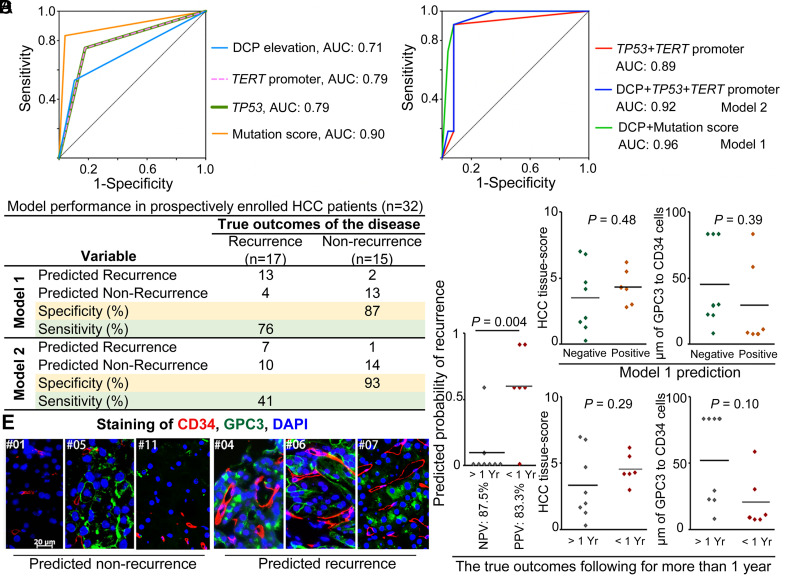
We calculated AUC values to evaluate the individual effectiveness of DCP ≥40 mAU/mL, presence of cfDNA mutations in TP53, presence of cfDNA mutations in the TERT promoter, and cfDNA mutation score for predicting early HCC recurrence post-RFA. The mutation score was identified as the most predictive among the four individual factors (Fig. 4A). We then combined two classes of HCC markers, DCP ≥40 mAU/mL with cfDNA mutations, to examine the effectiveness of this HCC blood signature for predicting early HCC recurrence post-RFA (Fig. 4B, Supplementary Table 1). An algorithm that considered DCP ≥40 mAU/mL combined with cfDNA mutation score (formula of Model 1, ), was better predictive than an algorithm that considered DCP ≥40 mAU/mL and the presence of cfDNA mutations in the TP53 and/or TERT promoter (formula of Model 2, ). The predictive specificity and sensitivity of Model 1 reached 92% and 91%, respectively, whereas those for Model 2 were 92% and 73%, respectively (Table 4).
Fig. 4. The pre-RFA HCC blood signature for prediction of early HCC recurrence post-RFA and validation.
Predictive capability of each single variable (A), combination of variables (B) of different HCC biomarkers, performed in 36 patients of the retrospective study. (C) Validation of the prediction in 32 patients of the prospective study. (D) The HCC tissue-score and cellular distance between GPC3-positive HCC cells and CD34-positve microvascular cells in the patients predicted by Model 1. HCC tissue-score and cellular distance of two cell types are also shown in the patients with and without true early HCC recurrence post-RFA. (E) Representative images show the relation between GPC3-positive HCC cells (green) and CD34-positve microvascular cells (red) of 6 cases as indicated. Nuclei was stained with DAPI (blue). DCP, des-γ-carboxy-prothrombin; AUC, area under curve; HCC, hepatocellular carcinoma; NPV, negative predictive value; PPV, positive predictive value; Yr, year; RFA, radiofrequency ablation.
Table 4. The model coefficients and prediction capability.
| Variable | Coefficients |
|
|---|---|---|
| Model 1 | Model 2 | |
| DCP | 2.00 | 1.89 |
| TP53 mutation | NA | 2.07 |
| TERT promoter mutation | NA | 2.07 |
| Mutation scores | 4.60 | NA |
| Intercept | −4.24 | −3.07 |
| Cutoff | 0.50 | 0.50 |
| Prediction | ||
| Specificity (%) | 92 | 92 |
| Sensitivity (%) | 91 | 73 |
Model 1: ; Model 2: . The value is 1 point for DCP ≥40 mAU/mL, mutation scores ≥1.4, or presence of TP53 mutations, or TERT promoter mutations, respectively. The other combination of individual risk factor derived from Univariable analysis is provided in the Supplementary Table 1. DCP, des-γ-carboxy-prothrombin; NA, not available.
Validation of the HCC blood signature for predicting early HCC recurrence post-RFA
From March to December 2021, we prospectively enrolled 35 patients using the same criteria applied in the retrospective study. In the pre-RFA plasma sample of each patient, AFP and DCP were quantified, and cfDNA mutations of TP53, CTNNB1, and the TERT promoter as well as the cfDNA methylation status of 14 genes were examined (Fig. 3C). After excluding three cases that were considered incomplete ablation on MRI at 1 month post-RFA, a total of 32 cases were followed for at least 12 months post-RFA. Of these, 15 cases were recognized as positive for the HCC blood signature, with a probability ≥0.5 of recurrence calculated using the Model 1 formula. After review of CT/MRI images by two independent radiologists, 17 of the 32 cases were identified as true cases of early HCC recurrence post-RFA. Accordingly, the specificity and sensitivity of the HCC blood signature for predicting early HCC recurrence were 87% and 76%, respectively (Fig. 4C).
Expression of HCC-related markers in biopsy samples from imaging-diagnosed small tumor nodules
Among the 32 enrolled HCC cases diagnosed based on imaging examinations, 14 cases had extra biopsy tissue samples. The follow-up results of CT/MRI examinations showed that 6 cases were true cases of early recurrence, and 8 cases were true cases of non-recurrence post-RFA. Based on the recurrence risk probability calculated using the Model 1 formula, the positive predictive value (PPV) was 83.3% (5/6 cases) and the negative predictive value (NPV) was 87.5% (7/8 cases) (Fig. 4D). We then examined the tissue expression levels of GPC3, HSP70, GS, CD34, and CK7 in biopsy samples, which were previously used to discriminate HCC from dysplasia nodules.28 The tissues of the 14 biopsy samples displayed varying expression levels of HCC-related markers. The staining of the HCC tissue markers in biopsy tissue sections was uneven (Supplementary Fig. 1, Supplementary Table 2). The HCC tissue-score (4.3±0.6) appeared higher in the 6 cases that were positive for the HCC blood signature than in the 8 cases that were not positive for the HCC blood signature (3.5±0.9), but no statistically significant difference was observed (p=0.48). The biopsy samples from the six cases of true early HCC recurrence exhibited the same tendency towards a higher HCC tissue-score (4.6±0.5) compared with the 8 cases without recurrence (3.3±0.9), but again the difference was not statistically significant (p=0.29) (Fig. 4D).
GPC3 expression is predominantly seen in advanced HCC, and vascular invasion is a determinant factor of HCC metastasis.1,3,17,28,33 Thus, we measured the distances between GPC3-positive HCC cells and CD34-positive microvascular cells after mIHC staining. The distance between the two cell types appeared shorter (32.3±13.6 µm) in the 6 cases that were positive for the HCC blood signature than in the 8 cases that were negative (46.9±10.1 µm), but with no statistical significance (p=0.39). Also, the distance between the two cell types appeared shorter in the biopsy samples of the 6 patients that had true early HCC recurrence (25.0±10.0 µm) compared with that in the 8 cases without true recurrence (52.4±10.7 µm), but again the difference was not significant (p=0.10) (Fig. 4D, E). Therefore, the analysis of biopsy tissue from the nodules ≤3 cm in diameter was not effective in predicting prognosis post-RFA.
Discussion
In this study, we retrospectively analyzed data from 251 eligible patients with HCC (≤3 nodules, each nodule ≤3 cm in diameter) diagnosed by contrast-enhanced CT/MRI examination who underwent complete RFA. Over the 5-year follow-up period post-RFA, the cumulative HCC recurrence rate was 77.7%, with two peaks of HCC recurrence. The first peak was at approximately 1 year post-RFA (33.9%), and recurrence during this period was defined as early recurrence of HCC. Consistently, a previous multicenter retrospective study reported that the cut-off point for early vs. late recurrence of HCC after RFA is 12 months.18 Among the 251 cases included in the retrospective study, we obtained pre-RFA blood samples from 36 cases. After analysing HCC-associated markers, including changes in blood levels of AFP and DCP and specified gene mutations and methylations of cfDNA, we developed an HCC blood-signature for predicting early HCC recurrence post-RFA. The HCC blood signature was DCP ≥40 mAU/mL in combination with a cfDNA mutation score that included cfDNA mutations of TP53, CTNNB1, and the TERT promoter. The HCC blood signature was validated in 32 prospectively enrolled patients based on the 1-year follow-up data of these patients. The HCC blood signature achieved a specificity of 87% and sensitivity of 76% for predicting early HCC recurrence post-RFA. These results indicate that the presence of an HCC-specific signature in pre-RFA blood may be used to predict HCC metastasis and/or 1-year recurrence post-RFA.
In the biopsy tissue samples available for patients included in the prospective study, those positive for the HCC blood signature tended to exhibit higher HCC tissue-scores and shorter distances between HCC cells and the microvasculature, as did biopsy samples from patients who experienced HCC recurrence within 1 year. While statistical analyses did not find these differences to be significant, the observed trends, if confirmed in future studies with larger samples, would suggest that the pre-RFA HCC blood signature likely reflects the intrinsic characteristics of liver nodules with metastatic traits. The HCC phenotype is known to be closely related to specific gene mutations. Unique traits in transcriptional, epigenetic, and proteomic changes are associated with vascular invasion, a determinant factor of HCC metastasis.1,3,17,20,28,33,34 Our current analysis did not observe an association of early HCC recurrence with nodule number (>1), serum AFP ≥20 ng/mL, cfDNA mutations in the CTNNB1 gene, or higher methylation levels of the 14 targeted genes. TP53 mutation is more frequent in advanced HCC and HBV-related HCC.2 We frequently detected TP53 mutations in cfDNA. In some cases included in our current study, a single nodule was detected by CT/MRI examination, but multiple variants of TP53 mutations in cfDNA were detected. TERT promoter mutation is recognized as a gatekeeper that drives dysplastic nodules to early HCC.2 CTNNB1 mutation is also recognized to occur in early HCC but not in dysplastic nodules.2 Regarding HCC-related protein biomarkers, abnormal levels of AFP or AFP-L3% did not show a significant association with any clinicopathologic characteristic of the presence of minimal residual disease after curative hepatectomy.26 The algorithm based on ctDNA changes in combination with the DCP levels but not the AFP level and AFP-L3% could identify tumors that had been missed on imaging.23,26 The HCC blood signature developed in our current study was DCP ≥40 mAU/mL in combination with a cfDNA mutation score that included the cfDNA mutations of TP53, CTNNB1, and the TERT promoter.
Contact between the tumor and hepatic vessels has been reported as a risk factor for intrahepatic recurrence in HCC patients.35,36 In the current study, we measured the distance between HCC cells and microvascular cells in cases of early HCC recurrence. Among the 14 cases with available biopsy tissue, we detected a tendency toward a shorter distance between HCC cells and the microvasculature in cases confirmed to experience early HCC recurrence post-RFA. However, the difference was not statistically significant compared to the cases that did not have confirmed early HCC recurrence. This discrepancy between histological and clinical outcomes may reflect the fact that biopsy samples are not ideal for assessing intrinsic tumor characteristics. Nonetheless, it is widely accepted that circulating tumor cells and their products in peripheral blood provide crucial insights into cancer biology and the metastatic process.19 Further research is necessary to optimize liquid biopsy assays for the examination of different classes of tumor-related products to accurately reflect intrinsic nodule traits.
We also found no significant association of HCC early recurrence with several laboratory variables indicating the severity of liver cirrhosis. Some previous studies have analyzed the association of liver matrix stiffness, an important indicator of liver cirrhosis, with HCC recurrence. Their results indicated that the liver stiffness value in RFA-treated patients could not effectively predict early HCC recurrence, but it is an independent predictor for late HCC recurrence after curative resection, transarterial chemoembolization (TACE), or RFA treatment.11,37–40 Substantial evidence has confirmed that the mechanisms of early HCC recurrence differ from those of late recurrence post-surgery.1,3,15 Our current study did not examine the association of alterations in these laboratory parameters with late HCC recurrence, and this topic should be addressed in the future. Another limitation of our study was the small number of patients enrolled in the prospective study, and future studies with more patients are warranted to confirm the results. Further analysis of the predictive value of our HCC blood signature combined with traditional clinical risk factors for HCC recurrence should also be conducted in the future.
In conclusion, the pre-RFA blood HCC signature developed in the present study, which considers the blood levels of DCP (≥40 mAU/mL) and the cfDNA mutation score generated from cfDNA mutations in TP53, CTNNB1 and the TERT promoter, effectively identified small HCC nodules with metastatic traits. The optimal adjuvant therapies for patients at high-risk for early HCC recurrence differ from those for patients at risk for late recurrence. Examination of the pre-RFA HCC blood signature may be used to predict early HCC recurrence post-RFA, potentially informing individualized planning of adjuvant therapy to prevent recurrence of metastatic primary tumors.
Supporting information
The biopsy tissues from the 14 cases were stained with GPC3, HSP70, GS, CD34, and CK7, and representative images from case #02, #04, #06, #07, and #12 are shown. HCC, hepatocellular carcinoma.
Ethical statement
The study was conducted in accordance with the Declaration of Helsinki, and approved by the Ethics Committee of Chinese PLA General Hospital (approval number [S2021-485-01]). The written informed consent was obtained from patients.
Data sharing statement
All data generated or analyzed during this study are included in this article. Further enquiries can be directed to the corresponding author: anonymized dataset of the Study is available to Editors, Reviewers and Readers upon request to the Corresponding Authors.
References
- 1.Omata M, Cheng AL, Kokudo N, Kudo M, Lee JM, Jia J, et al. Asia-Pacific clinical practice guidelines on the management of hepatocellular carcinoma: a 2017 update. Hepatol Int. 2017;11(4):317–370. doi: 10.1007/s12072-017-9799-9. [DOI] [PMC free article] [PubMed] [Google Scholar]
- 2.Rebouissou S, Nault JC. Advances in molecular classification and precision oncology in hepatocellular carcinoma. J Hepatol. 2020;72(2):215–229. doi: 10.1016/j.jhep.2019.08.017. [DOI] [PubMed] [Google Scholar]
- 3.Marrero JA, Kulik LM, Sirlin CB, Zhu AX, Finn RS, Abecassis MM, et al. Diagnosis, Staging, and Management of Hepatocellular Carcinoma: 2018 Practice Guidance by the American Association for the Study of Liver Diseases. Hepatology. 2018;68(2):723–750. doi: 10.1002/hep.29913. [DOI] [PubMed] [Google Scholar]
- 4.Hung HH, Chiou YY, Hsia CY, Su CW, Chou YH, Chiang JH, et al. Survival rates are comparable after radiofrequency ablation or surgery in patients with small hepatocellular carcinomas. Clin Gastroenterol Hepatol. 2011;9(1):79–86. doi: 10.1016/j.cgh.2010.08.018. [DOI] [PubMed] [Google Scholar]
- 5.Lee S, Kang TW, Cha DI, Song KD, Lee MW, Rhim H, et al. Radiofrequency ablation vs. surgery for perivascular hepatocellular carcinoma: Propensity score analyses of long-term outcomes. J Hepatol. 2018;69(1):70–78. doi: 10.1016/j.jhep.2018.02.026. [DOI] [PubMed] [Google Scholar]
- 6.Lam VW, Ng KK, Chok KS, Cheung TT, Yuen J, Tung H, et al. Incomplete ablation after radiofrequency ablation of hepatocellular carcinoma: analysis of risk factors and prognostic factors. Ann Surg Oncol. 2008;15(3):782–790. doi: 10.1245/s10434-007-9733-9. [DOI] [PubMed] [Google Scholar]
- 7.Cho JY, Choi MS, Lee GS, Sohn W, Ahn J, Sinn DH, et al. Clinical significance and predictive factors of early massive recurrence after radiofrequency ablation in patients with a single small hepatocellular carcinoma. Clin Mol Hepatol. 2016;22(4):477–486. doi: 10.3350/cmh.2016.0048. [DOI] [PMC free article] [PubMed] [Google Scholar]
- 8.Lee DH, Lee JM, Lee JY, Kim SH, Yoon JH, Kim YJ, et al. Radiofrequency ablation of hepatocellular carcinoma as first-line treatment: long-term results and prognostic factors in 162 patients with cirrhosis. Radiology. 2014;270(3):900–909. doi: 10.1148/radiol.13130940. [DOI] [PubMed] [Google Scholar]
- 9.Shiina S, Tateishi R, Arano T, Uchino K, Enooku K, Nakagawa H, et al. Radiofrequency ablation for hepatocellular carcinoma: 10-year outcome and prognostic factors. Am J Gastroenterol. 2012;107(4):569–577. doi: 10.1038/ajg.2011.425. [DOI] [PMC free article] [PubMed] [Google Scholar]
- 10.Cao S, Lyu T, Fan Z, Guan H, Song L, Tong X, et al. Long-term outcome of percutaneous radiofrequency ablation for periportal hepatocellular carcinoma: tumor recurrence or progression, survival and clinical significance. Cancer Imaging. 2022;22(1):2. doi: 10.1186/s40644-021-00442-2. [DOI] [PMC free article] [PubMed] [Google Scholar]
- 11.Cho HJ, Kim B, Kim HJ, Huh J, Kim JK, Lee JH, et al. Liver stiffness measured by MR elastography is a predictor of early HCC recurrence after treatment. Eur Radiol. 2020;30(8):4182–4192. doi: 10.1007/s00330-020-06792-y. [DOI] [PubMed] [Google Scholar]
- 12.Poon RT, Lau C, Pang R, Ng KK, Yuen J, Fan ST. High serum vascular endothelial growth factor levels predict poor prognosis after radiofrequency ablation of hepatocellular carcinoma: importance of tumor biomarker in ablative therapies. Ann Surg Oncol. 2007;14(6):1835–1845. doi: 10.1245/s10434-007-9366-z. [DOI] [PubMed] [Google Scholar]
- 13.Li ZX, Zhang QF, Huang JM, Huang SJ, Liang HB, Chen H, et al. Safety and efficacy of postoperative adjuvant therapy with atezolizumab and bevacizumab after radical resection of hepatocellular carcinoma. Clin Res Hepatol Gastroenterol. 2023;47(7):102165. doi: 10.1016/j.clinre.2023.102165. [DOI] [PubMed] [Google Scholar]
- 14.Singal AG, Kudo M, Bruix J. Breakthroughs in Hepatocellular Carcinoma Therapies. Clin Gastroenterol Hepatol. 2023;21(8):2135–2149. doi: 10.1016/j.cgh.2023.01.039. [DOI] [PMC free article] [PubMed] [Google Scholar]
- 15.Wu JC, Huang YH, Chau GY, Su CW, Lai CR, Lee PC, et al. Risk factors for early and late recurrence in hepatitis B-related hepatocellular carcinoma. J Hepatol. 2009;51(5):890–897. doi: 10.1016/j.jhep.2009.07.009. [DOI] [PubMed] [Google Scholar]
- 16.Sato T, Kondo F, Ebara M, Sugiura N, Okabe S, Sunaga M, et al. Natural history of large regenerative nodules and dysplastic nodules in liver cirrhosis: 28-year follow-up study. Hepatol Int. 2015;9(2):330–336. doi: 10.1007/s12072-015-9620-6. [DOI] [PMC free article] [PubMed] [Google Scholar]
- 17.Chen D, Li Z, Zhu W, Cheng Q, Song Q, Qian L, et al. Stromal morphological changes and immunophenotypic features of precancerous lesions and hepatocellular carcinoma. J Clin Pathol. 2019;72(4):295–303. doi: 10.1136/jclinpath-2018-205611. [DOI] [PubMed] [Google Scholar]
- 18.Yang Y, Xin Y, Ye F, Liu N, Zhang X, Wang Y, et al. Early recurrence after radiofrequency ablation for hepatocellular carcinoma: a multicenter retrospective study on definition, patterns and risk factors. Int J Hyperthermia. 2021;38(1):437–446. doi: 10.1080/02656736.2020.1849828. [DOI] [PubMed] [Google Scholar]
- 19.Alix-Panabières C, Pantel K. Liquid Biopsy: From Discovery to Clinical Application. Cancer Discov. 2021;11(4):858–873. doi: 10.1158/2159-8290.CD-20-1311. [DOI] [PubMed] [Google Scholar]
- 20.Calderaro J, Ziol M, Paradis V, Zucman-Rossi J. Molecular and histological correlations in liver cancer. J Hepatol. 2019;71(3):616–630. doi: 10.1016/j.jhep.2019.06.001. [DOI] [PubMed] [Google Scholar]
- 21.Xu RH, Wei W, Krawczyk M, Wang W, Luo H, Flagg K, et al. Circulating tumour DNA methylation markers for diagnosis and prognosis of hepatocellular carcinoma. Nat Mater. 2017;16(11):1155–1161. doi: 10.1038/nmat4997. [DOI] [PubMed] [Google Scholar]
- 22.Cai J, Chen L, Zhang Z, Zhang X, Lu X, Liu W, et al. Genome-wide mapping of 5-hydroxymethylcytosines in circulating cell-free DNA as a non-invasive approach for early detection of hepatocellular carcinoma. Gut. 2019;68(12):2195–2205. doi: 10.1136/gutjnl-2019-318882. [DOI] [PMC free article] [PubMed] [Google Scholar]
- 23.Qu C, Wang Y, Wang P, Chen K, Wang M, Zeng H, et al. Detection of early-stage hepatocellular carcinoma in asymptomatic HBsAg-seropositive individuals by liquid biopsy. Proc Natl Acad Sci U S A. 2019;116(13):6308–6312. doi: 10.1073/pnas.1819799116. [DOI] [PMC free article] [PubMed] [Google Scholar]
- 24.Wang P, Song Q, Ren J, Zhang W, Wang Y, Zhou L, et al. Simultaneous analysis of mutations and methylations in circulating cell-free DNA for hepatocellular carcinoma detection. Sci Transl Med. 2022;14(672):eabp8704. doi: 10.1126/scitranslmed.abp8704. [DOI] [PubMed] [Google Scholar]
- 25.Zhang X, Wang Z, Tang W, Wang X, Liu R, Bao H, et al. Ultrasensitive and affordable assay for early detection of primary liver cancer using plasma cell-free DNA fragmentomics. Hepatology. 2022;76(2):317–329. doi: 10.1002/hep.32308. [DOI] [PubMed] [Google Scholar]
- 26.Cai Z, Chen G, Zeng Y, Dong X, Li Z, Huang Y, et al. Comprehensive Liquid Profiling of Circulating Tumor DNA and Protein Biomarkers in Long-Term Follow-Up Patients with Hepatocellular Carcinoma. Clin Cancer Res. 2019;25(17):5284–5294. doi: 10.1158/1078-0432.CCR-18-3477. [DOI] [PubMed] [Google Scholar]
- 27.N’Kontchou G, Mahamoudi A, Aout M, Ganne-Carrié N, Grando V, Coderc E, et al. Radiofrequency ablation of hepatocellular carcinoma: long-term results and prognostic factors in 235 Western patients with cirrhosis. Hepatology. 2009;50(5):1475–1483. doi: 10.1002/hep.23181. [DOI] [PubMed] [Google Scholar]
- 28.Sciarra A, Di Tommaso L, Nakano M, Destro A, Torzilli G, Donadon M, et al. Morphophenotypic changes in human multistep hepatocarcinogenesis with translational implications. J Hepatol. 2016;64(1):87–93. doi: 10.1016/j.jhep.2015.08.031. [DOI] [PubMed] [Google Scholar]
- 29.Zhang R, Chen K, Gong C, Wu Z, Xu C, Li XN, et al. Abnormal generation of IL-17A represses tumor infiltration of stem-like exhausted CD8(+) T cells to demote the antitumor immunity. BMC Med. 2023;21(1):315. doi: 10.1186/s12916-023-03026-y. [DOI] [PMC free article] [PubMed] [Google Scholar]
- 30.Fluss R, Faraggi D, Reiser B. Estimation of the Youden Index and its associated cutoff point. Biom J. 2005;47(4):458–472. doi: 10.1002/bimj.200410135. [DOI] [PubMed] [Google Scholar]
- 31.Tayob N, Christie I, Richardson P, Feng Z, White DL, Davila J, et al. Validation of the Hepatocellular Carcinoma Early Detection Screening (HES) Algorithm in a Cohort of Veterans With Cirrhosis. Clin Gastroenterol Hepatol. 2019;17(9):1886–1893.e5. doi: 10.1016/j.cgh.2018.12.005. [DOI] [PMC free article] [PubMed] [Google Scholar]
- 32.Matsubara M, Shiraha H, Kataoka J, Iwamuro M, Horiguchi S, Nishina S, et al. Des-γ-carboxyl prothrombin is associated with tumor angiogenesis in hepatocellular carcinoma. J Gastroenterol Hepatol. 2012;27(10):1602–1608. doi: 10.1111/j.1440-1746.2012.07173.x. [DOI] [PubMed] [Google Scholar]
- 33.Wei T, Zhang XF, Bagante F, Ratti F, Marques HP, Silva S, et al. Early Versus Late Recurrence of Hepatocellular Carcinoma After Surgical Resection Based on Post-recurrence Survival: an International Multi-institutional Analysis. J Gastrointest Surg. 2021;25(1):125–133. doi: 10.1007/s11605-020-04553-2. [DOI] [PubMed] [Google Scholar]
- 34.Krishnan MS, Rajan KD A, Park J, Arjunan V, Garcia Marques FJ, Bermudez A, et al. Genomic Analysis of Vascular Invasion in HCC Reveals Molecular Drivers and Predictive Biomarkers. Hepatology. 2021;73(6):2342–2360. doi: 10.1002/hep.31614. [DOI] [PMC free article] [PubMed] [Google Scholar]
- 35.Roayaie S, Blume IN, Thung SN, Guido M, Fiel MI, Hiotis S, et al. A system of classifying microvascular invasion to predict outcome after resection in patients with hepatocellular carcinoma. Gastroenterology. 2009;137(3):850–855. doi: 10.1053/j.gastro.2009.06.003. [DOI] [PMC free article] [PubMed] [Google Scholar]
- 36.Kim YS, Rhim H, Cho OK, Koh BH, Kim Y. Intrahepatic recurrence after percutaneous radiofrequency ablation of hepatocellular carcinoma: analysis of the pattern and risk factors. Eur J Radiol. 2006;59(3):432–441. doi: 10.1016/j.ejrad.2006.03.007. [DOI] [PubMed] [Google Scholar]
- 37.Jung KS, Kim SU, Choi GH, Park JY, Park YN, Kim DY, et al. Prediction of recurrence after curative resection of hepatocellular carcinoma using liver stiffness measurement (FibroScan®) Ann Surg Oncol. 2012;19(13):4278–4286. doi: 10.1245/s10434-012-2422-3. [DOI] [PubMed] [Google Scholar]
- 38.Vestito A, Dajti E, Cortellini F, Montagnani M, Bazzoli F, Zagari RM. Can Liver Ultrasound Elastography Predict the Risk of Hepatocellular Carcinoma Recurrence After Radiofrequency Ablation? A Systematic Review and Meta-Analysis. Ultraschall Med. 2023;44(3):e139–e147. doi: 10.1055/a-1657-8825. [DOI] [PubMed] [Google Scholar]
- 39.Abe H, Shibutani K, Yamazaki S, Kanda T, Moriyama M, Okada M, et al. Tumor stiffness measurement using magnetic resonance elastography can predict recurrence and survival after curative resection of hepatocellular carcinoma. Surgery. 2023;173(2):450–456. doi: 10.1016/j.surg.2022.11.001. [DOI] [PubMed] [Google Scholar]
- 40.Lee YR, Park SY, Kim SU, Jang SY, Tak WY, Kweon YO, et al. Using transient elastography to predict hepatocellular carcinoma recurrence after radiofrequency ablation. J Gastroenterol Hepatol. 2017;32(5):1079–1086. doi: 10.1111/jgh.13644. [DOI] [PubMed] [Google Scholar]
Associated Data
This section collects any data citations, data availability statements, or supplementary materials included in this article.
Supplementary Materials
The biopsy tissues from the 14 cases were stained with GPC3, HSP70, GS, CD34, and CK7, and representative images from case #02, #04, #06, #07, and #12 are shown. HCC, hepatocellular carcinoma.


