

Abstract
Background:
Vasa vasorum (VVs) is a Latin word representing vessels of vessels. VVs are usually found on the adventitia of the parent vessel and infrequently reach the media and intima, depending on the size and type of the parent vessels and physiological and pathological conditions. The VVs include arteries, capillaries, veins, and lymphatic vessels, involving the oxygenation and nourishment of the vessel’s wall to sustain its healthy state. Accumulated studies have revealed that VVs are involved in various intracranial lesions, including atherosclerotic diseases, aneurysms, and shunt diseases. The current review aims to review and integrate past and recent findings and knowledge on VVs and to facilitate our understanding of VVs and intracranial pathology involving VVs.
Methods:
A literature review was carried out with a focus on the role of VVs by searching the Pubmed database.
Results:
We identified 71 articles that discuss the role of VVs. We discussed the anatomical structure, physiological significance, and pathological significance of the VV.
Conclusion:
VV is not only involved in the nutrition and metabolism of the vascular wall but is also deeply involved in the pathogenesis of inflammation, ischemia, and thrombosis of the vascular wall. In addition, in the central nervous system, intracranial vascular wall nutrient particularities and VVs are closely related to the pathogenesis of cerebral aneurysms, subarachnoid hemorrhage, arteriovenous shunt disease, atherosclerotic lesions, and other conditions.
Keywords: Cerebral aneurysms, Intracranial vessels, Vasa vasorum

INTRODUCTION (HISTORY OF VASA VASORUM [VV])
VVs are a Latin word representing vessels of vessels. It is also called vasa nutria [47] due to its role in the nutritional supply of the vessel wall. Thomas Willis first described its existence (1621–1675).[65] He was an English anatomist famous for the intracranial arterial rings that bear his name. The term “Vasa vasorum” was first coined by the German anatomist Christian Gottlieb Ludwig (1709–1773).[41] Since microscopes were unavailable in their time, their observation was conducted with the naked eye. They found tiny vessels on the wall and named them “vasa vasorum.” Later, Gimbert[21] first reported the detailed anatomy and function of VVs. He observed VVs in detail in autopsy cases using microscopes, which developed rapidly in the 19th century. He noted that VVs are distributed in the tunica adventitia of blood vessels, are present in veins and arteries, and are essentially absent in brain blood vessels. Since then, VVs have been commonly recognized as anatomical structures among clinicians and have appeared as an anatomical terminology in textbooks since 1895.[27] The invention of the microradiographic technique in the 1960s facilitated the investigation of VVs, which allowed us to observe the VV microanatomy while maintaining continuity with the surrounding vessels.[9] Then, using corrosion casts enabled observation while preserving the three-dimensional structure.[10,36] In recent years, wall vessel images have made it possible to evaluate the density of VV increments in intracranial vessels.[33] These advancements reveal that VVs are involved in various intracranial lesions, including atherosclerotic diseases, cerebral aneurysms, and arteriovenous shunts (AVSs). The current review aims to review and integrate past and recent findings and knowledge on VVs and to facilitate our understanding of VVs and intracranial pathology involving VVs.
BASIC HISTOLOGICAL ANATOMY OF BLOOD VESSELS
The vessel wall comprises the tunica intima, media, and adventitia[55] [Figure 1]. The tunica intima is divided into vascular endothelial cells and subendothelial connective tissue. The outermost layer of the tunica intima is the internal elastic plate, which borders between the tunica media and tunica intima and provides the vessels with resiliency and resistance to mechanical stress. The tunica media consists of smooth muscle and surrounding collagen and elastic fibers. The tunica media of elastic arteries, for example, the ascending and descending aorta, brachiocephalic artery, common carotid artery, subclavian artery, and common iliac artery, consists of a series of layered structural lamellar units abundant in elastic fibers and smooth muscle [Figure 1]. Indeed, the human aorta is composed of 52 unit layers.[47] In muscular arteries, a layered lamellar unit does not exist, and usually, only the internal/external elastic lamina exists, bordering the intima, media, and adventitia. The external elastic lamina is histologically included in the media. The adventitia, where most VVs exist, comprises sparse connective tissue and fibroblasts with longitudinal elastic fibers and collagen fibers. The structure of the intracranial arteries is almost the same as that of systemic arteries, except for internal and external elastic plates; the internal elastic plate is well developed, while the media and the adventitia show few elastic fibers and lack an external elastic lamina.[38] Interestingly, it has been suggested that the external elastic lamina is once formed but later lost after the age of 2 years.[24]
Figure 1:

The basic structure of a blood vessel (elastic artery) and vasa vasorum (VV). Elastic arteries have a well-developed elastic plate and smooth muscle (lamellar unit). Critical depth is considered to be the limit of oxygen and nutrients from the lumen, and VVs exist on the outer side of the lumen. The critical depth is usually approximately 0.5 mm (30 layers in a lamellar unit). Muscular arteries do not have such a well-developed lamellar unit. In intracranial arteries, the external elastic lamina is absent.
ANATOMICAL STRUCTURE AND PHYSIOLOGICAL SIGNIFICANCE OF THE VV
VVs are usually found on the adventitia of the parent vessel and infrequently reach the media and intima, depending on the size and type of the parent vessels and physiological and pathological conditions. Some articles referred to only VVs extending into the media as true VVs.[39] However, most literature considers the “small vessels that nourish the large vessels” as VVs. We follow the definition and consider small vessels in any wall layer as VVs in this paper. The VVs consist of arteries, capillaries, veins, and even lymphatic vessels[14,15] and constitute the whole vascular network within the parent vessel wall. The VVs conveying the arterial blood are divided into two categories: the VVs directly arising from the arterial lumen are called the “vasa vasorum interna.” The VVs from the remote artery to the adventitia of the different vessels are called the “vasa vasorum externa.” The VVs conveying the venous blood or lymphatic fluid are called the “venous vasa vasorum” or “lymphatic vasa vasorum”[14,15,22] [Figure 2].
Figure 2:

Types of vasa vasorum.
The physiological function of VVs is to supply nutrition and oxygen to the parent vessel walls and to sustain their healthy physiological function, which has been well described in the previous literature. Geiringer investigated the VV distribution in the human aorta and coronary arteries and found that VVs are abundant in the adventitia and outer media. However, he also found that the inner layer of 0.5 mm or 0.35 mm in the aorta or coronary arteries from the lumen is always avascular. He defined this vascular-free layer as the “critical depth,” the limit of oxygen and nutrient diffusion from the lumen inside and outside VVs [Figure 1]. A recent immunohistochemical investigation detected scanty vessels deeper (<0.1 mm) within the classical critical depth.[19] Additional study is necessary to define the true critical depth.
Wolinsky studied various mammalian VVs in the aorta and confirmed that critical depth can be seen in any species. The diffusion has been nicely visualized by Werber and Heistad[62] and Jurrus and Weiss.[29] In a previous study,[62] radioisotope-labeled antipyrine (a substance taken up by the intercellular compartment of the vessel wall) was administered into a dog’s heart. Two hours after injection, the radioisotope concentration was high in the intima and adventitia and low in the media. Then, blockage of VVs of the aorta decreased the concentration of radioisotopes on the adventitia. Jurrus and Weiss[29] directly measured oxygen partial pressure in rabbit aorta walls. Oxygen partial pressure was highest in the innermost and outermost layers and lowest at the medial center. They also showed that the thickened wall in atherosclerotic rabbits obstructed diffusion, and the partial oxygen pressure almost reached zero deep. Such insufficient supply from VVs leads to necrosis in the middle 1/3 of the media, although there is no damage to the tunica intima and inner 1/3 of the tunica media.[63] These studies indicate that the arterial wall is nourished and oxygenized by diffusion from the lumen inside and outside VVs. The middle 1/3 of the tunica media is the most vulnerable watershed layer. The preceding sentences mainly discuss the VVs in the arterial wall. However, does the venous wall also have VVs? If one considers that the oxygen partial pressure in veins is lower than that of the arteries, one may expect that the role of VVs in the veins is more significant than that in arteries. Indeed, the well-developed VV network on the venous wall (vasa venarum) has been reported in the literature.[36,47] O’Nejll[47] studied the canine internal jugular veins and wrote the detailed anatomy of its vasa venarum. Similar to arterial VVs, the vasa venarum consists of arterial, capillary, and venous components. The vasa venarum are arterialized by adjacent arteries, while the venous part empties into the adjacent veins or the parent vein through independent collecting veins on the surface of the venous wall. O’Nejll also investigated the contribution of the vasa venarum to venous wall nourishment by blocking flow either in the vasa venarum or the internal jugular vein. As expected, the flow stasis of the vasa venarum resulted in severe endothelial damage, while that of the jugular vein slightly injured the endothelium. Heistad et al.[26] measured the VV blood flow in various systemic arteries and veins. They found that the VV flow was more significant in the less oxygenized vessels (veins or pulmonary arteries) and more thickened vessels. In summary, the contribution of vasa venarum to sustaining the parent venous wall is generally more significant than that of corresponding arterial VVs.
INTRACRANIAL VV AND INTRACRANIAL VESSEL WALL NUTRITION
The intracranial vessels have been classically considered to lack VVs. Gimbert[22] first reported the absence of VVs in intracranial vessels in his article, which was further confirmed by Clower et al., investigation of intracranial vessels in rats, dogs, and cats.[10] Other researchers investigated human cadaver specimens and reported similar results;[3,8,56] VVs are present only at the proximal segment of the vertebral and internal carotid arteries, and they are absent in arteries 1.5 cm distal to the intradural entry or arteries with a media thickness of 250 µm or less.[3] Interestingly, VVs are thoroughly absent in neonates, and VVs in the proximal intracranial arteries in an adult can be found as an extension of the extradural vessels. Accordingly, they assumed that the VVs in proximal intracranial segments are acquired extensions from the extracranial VVs. As an exception, some authors reported the VVs in distal basilar, anterior cerebral, or middle cerebral arteries,[56] which, however, may be secondarily induced by the atherosclerotic or inflammatory changes in affected arteries (as discussed in a later section). In summary, intracranial arteries are free of VVs under normal physiological conditions except for the proximal segments of the internal carotid and vertebral arteries [Figure 3], which raises the question of how the intracranial arteries sustain the oxygen and nutrition supply without VVs. Some assume that the anatomical features of the intracranial vessels such as thinner medial and adventitial layers, a thicker but fenestrated internal elastic lamina, and the absence of the external elastic lamina, may help greater diffusion from the lumen inside.[52] Others suggest that cerebrospinal fluid (CSF) supplies oxygen and nutrition instead of the VVs. The electron microscopic investigation of the intracranial vessels’ surface revealed the opening of tiny 1–3 µm pores connected through tunnels, making this labyrinthine network structure the “rete vasorum.”[18,39,69] The CSF may deliver oxygen and nutrition through the rete vasorum. Another previous study demonstrated that the leptomeningeal cells surrounding the subarachnoid CSF space do not have a basement membrane,[1] which, in addition to the lack of the external elastic lamina, can be a mechanism to remove the barrier and facilitate substance exchange through the CSF.
Figure 3:
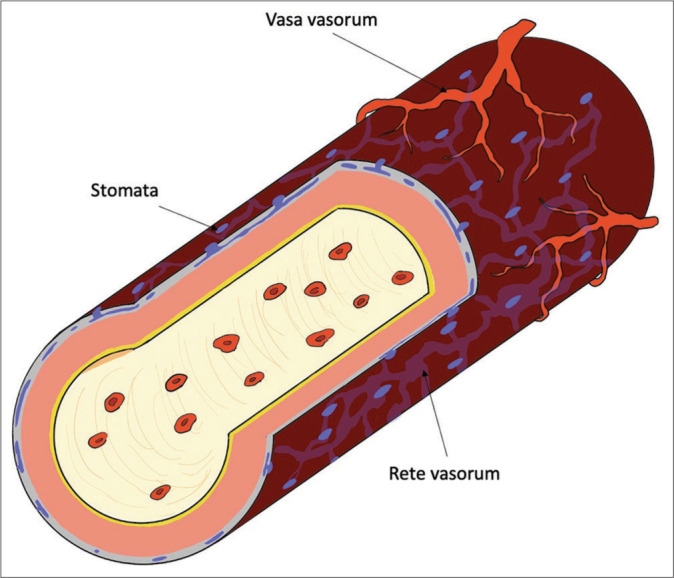
Intracranial vessels and vasa vasorum (VV). In intracranial vessels, the VV is present only in the internal carotid and proximal vertebral arteries. The tunica adventitia of blood vessels in the subarachnoid space has tiny pores called stomata, and there is also a passageway for cerebrospinal fluid called the rete vasorum inside. Oxygen and nutrients are supplied to the tunica media through these channels. If a subarachnoid hemorrhage occurs and the vessel’s surface is covered with blood, this trophic pathway through the rete vasorum may cease functioning.
Pathological VV
Arteriosclerotic lesion
Atherosclerotic lesions are one of the most investigated pathologies involving VVs. In systemic atherosclerotic lesions, hypoxia and inflammation are the fundamental driving forces that trigger VV neovascularization. Atherosclerotic wall thickness directly obstructs diffusion and causes wall hypoxia.[6,29,30] In addition, atherosclerotic lesions increase regional metabolism and aggravate wall ischemia.[5] Then, secondary VV hyperplasia is induced to sustain wall nourishment. Indeed, the authors reported a direct association between VV development or flow and wall thickness.[64] Furthermore, inflammatory cells and cytokines also induce VV neovascularization[67], and VVs usually develop where inflammatory cell infiltration is most prevalently observed.[34]
Induced VV hyperplasia, in turn, leads to the progression of atherosclerosis in many ways. Nakashima and Tashiro investigated lipoprotein deposition at the early stage of atherosclerotic lesions and found that the deposit was seen at the border zone between the intima and media rather than the innermost layer of the intima. The result suggested that the lipoprotein is delivered primarily by the VVs instead of the inner luminal flow.[44] In addition, the impaired integrity of the pathological VV endothelium in atherosclerotic lesions increases its permeability,[54] leading to further inflammatory cell infiltration and lipoprotein deposits, in-plaque hemorrhage, and plaque rupture.[66] Vascular stenting is a major intervention for atherosclerotic lesions. The VVs are also involved with in-stent restenosis after stenting. Neointimal hyperplasia in the restenosis segment is associated with VV development and inflammatory cells[70], while the direct compression of VVs by stent expansion is reported to lead to wall hypoxia, thus promoting medial necrosis, intimal hyperplasia, and restenosis.[13]
The involvement of VVs with intracranial atherosclerotic lesions has been described in only a limited number of articles. The intracranial vessels, as aforementioned, do not have VVs except for their proximal segments. However, VVs can be distally observed in intracranial atherosclerosis[2,56] and can usually be identified as an extension of proximal physiological VVs. Rarely, “isolated” VV internas can also be acquired.[2] After stenting, one case report described well-developed VVs in the intracranial restenotic segment.[20] We know little about the role of VVs in intracranial atherosclerotic lesions, and further study is needed to elucidate it.
THROMBUS RESOLUTION AND SECONDARY AVS FORMATION
The thrombus resolution process is analogous to that of granulation formation in wound formation in wound healing and undergoes inflammation and tissue organization.[43] The initial thrombus mainly consists of red blood cells, platelets, and fibrin. Then, inflammatory cell infiltration initiates the process in a few days, followed by neovessels within the thrombus. The neovessels promote inflammatory cell infiltration, accelerating the resolution process.[60] The neovessels are initially cell-lined small channels but later coalesce by 3–4 weeks and contribute to venous lumen restoration itself.[43] Where do the neovessels come from? The VVs on the affected vascular adventitia actually offer the angiogenesis scaffold for the neovessels, and the neovessels are formed as an extension of VVs.[35] The mechanism of how thrombosis organization induces angiogenesis has not been well elucidated, but inflammation and ischemia can be involved in the process. The administration of the inflammatory chemokine interleukin-8 directly accelerates angiogenesis within the thrombus,[60] possibly by upregulation of the downstream VEGF pathway.[28] The ischemia led by venous hypertension and/or loss of oxygen and nutrient diffusion from the lumen after occlusion of the vessel lumen may remain another debatable hypothesis. In summary, the neovessels within the thrombus, which is a direct extension of the VVs, can be considered as VVs in a broad sense.
Interestingly, some authors reported secondary AVSs on the thrombosed deep venous wall after its resolution.[7,68] The mechanism is still unknown. However, we assume that the pathological misconnection of the neovessels with the original venous lumen during thrombosis resolution can be the origin of these pathological AVSs. We also see a similar pathology in the intracranial lesion; dural AVSs (DAVSs) are the acquired AVSs adjacent to the dura mater and are developed after the pathological opening and growth of preexisting physiological AVSs in dural arteries and veins.[4] No past literature has investigated the involvement of VVs with DAVSs. However, dural veins are extradural channels and certainly have VVs to sustain their healthy wall. Some authors described the physiological AVS in the VV network.[40,47] In addition, the pathological conditions precluding DAVSs include venous thrombosis, venous hypertension, ischemia, infection, and inflammation, which are also strong triggers for VV angiogenesis. Some authors successfully established DAVS animal models induced by venous hypertension and venous thrombosis.[37,57] However, the animals also developed extracranial facial and orbital AVSs in addition to DAVSs.[37,57] The etiology of secondary AVSs, including DAVSs, is common throughout the body. VVs provide angiogenesis scaffolds and play a pivotal role in forming secondary AVSs.
The etiology of secondary AVSs may explain why secondary pial arteriovenous fistula is rarely reported.[51] This is strange, considering that cortical venous thrombosis and dural sinus thrombosis are frequently observed in clinical situations. However, the intradural channels lack VVs. Thus, VVs cannot offer angiogenesis scaffolds for pial lesions. In addition, the vasculature nourished by CSF may not face severe wall ischemia even after luminal occlusion and venous hypertension compared to the extradural vessels. However, secondary pial AVSs can be induced under certain circumstances; the authors described secondary AVS formation after complete occlusion of cerebral and spinal pial macro arteriovenous fistulas (MAVFs).[59] MAVFs are pial lesions with a giant venous ectasia. The thickened wall may be nourished and oxygenized by the arterialize flow inside. However, the complete obliteration of MAVF results in severe wall ischemia and inflammation, which may secondarily induce angiogenesis through VVs and form pathological AVSs. Theologically, the obliteration of any AVSs can potentially recruit secondary AVSs. Among them, MAVF obliteration can cause the severest ischemia and inflammation among any pial AVS lesion, leading to secondary AVSs.
CEREBRAL ANEURYSM AND SUBARACHNOID HEMORRHAGE (SAH)
The association of VVs with unstable intracranial aneurysms inclined to grow or rupture has already been well recognized. As mentioned above, VVs are absent in intracranial vessels except in the proximal segment, but inflammation of the aneurysm wall, wall ischemia associated with decreased diffusion due to a thickened aneurysmal wall, and thrombosis in the aneurysm can trigger secondary VV angiogenesis in the aneurysm wall, as seen in the other pathologies. Induced VVs provide a route for inflammatory cell infiltration and accelerate aneurysm wall inflammation.[31,53] As seen in atherosclerotic lesions, secondary-induced VVs have impaired endothelium integrity,[45,71] increasing permeability and accelerating further inflammatory cell infiltration and causing hemorrhage within or on the surface of the aneurysmal wall.[32,71] Through these mechanisms, VVs are associated with aneurysm wall weakening, growth, and rupture [Figure 4].
Figure 4:

Vasa vasorum (VV) and cerebral aneurysms. The development of the VV is associated with unstable aneurysms.
The aneurysm rupture, SAH, affects the aforementioned peculiar way of nourishing the cerebral vasculature. Studies have reported that SAH induces severe subarachnoid vessel wall injury, including intimal edema and thickening, disruption of the internal elastic lamina, media thickening, smooth muscle cell necrosis, intramural hematoma, and VV recruitment in human cadavers[23] and animal models.[49] Furthermore, in SAH, the elevated intracranial pressure reduces oxygen pressure by diffusion from both the lumen inside and outside VVs, which further aggravates delayed vasospasm. In addition, the blood contents spread into the subarachnoid space, disturbing CSF circulation, which results in oxygen and nutrition supply through the rete vasorum.[49]
Vessel wall imaging (VWI) using enhanced MRI is a modern imaging technique that can detect VV proliferation. In 1991, Atkinson first reported an association between aneurysmal wall enhancement and VVs on the aneurysmal wall.[2] Subsequent studies reported a strong association of aneurysmal wall enhancement with unstable symptomatic aneurysms prone to growth and rupture.[16,42,58] No one still precisely knows what the radiological AWE represents. One may assume that the increased density of VVs reflects vascular wall enhancement. Meanwhile, one study reported that the density of VVs was similar between unruptured and ruptured aneurysms and that VVs with increased permeability were significantly higher in ruptured aneurysms.[45] Accordingly, the “leaky” weakened vessel wall prone to inflammation can be involved in wall enhancement rather than the VV density. VWI is now widely used to identify fragile rupture sites in cerebral aneurysms and other hemorrhagic strokes, including brain arteriovenous malformations[17,48,50] and DAVFs.[12] The representative VWIs in the clinical situation are shown in Figures 5 and 6.
Figure 5:
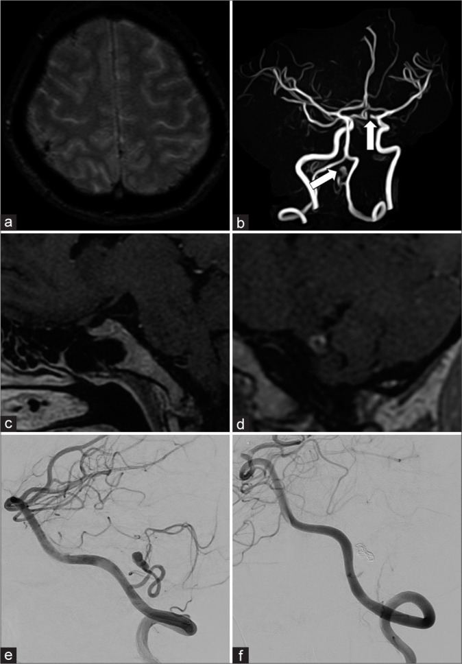
Representative case of vessel wall imaging (VWI) in multiple aneurysms. A 47-year-old female visited our hospital 1 week after headache onset. (a) The initial magnetic resonance (MR) scan revealed cortical subarachnoid hemorrhage. (b) Subsequent MR angiography demonstrated two aneurysms (white arrows): The left internal carotid artery (ICA) aneurysm and the left posterior inferior cerebellar artery (PICA) aneurysm. (c and d) The VWI (enhanced MR) confirmed that the ruptured aneurysm was the left internal PICA aneurysm, and there was no enhancement in the left ICA aneurysm (c shows the left ICA aneurysm, d shows the left PICA aneurysm). (e and f) The PICA aneurysm was subsequently embolized with coils and glue (e: preinterventional radiology [IVR] digital subtraction angiography [DSA], f: post-IVR DSA).
Figure 6:
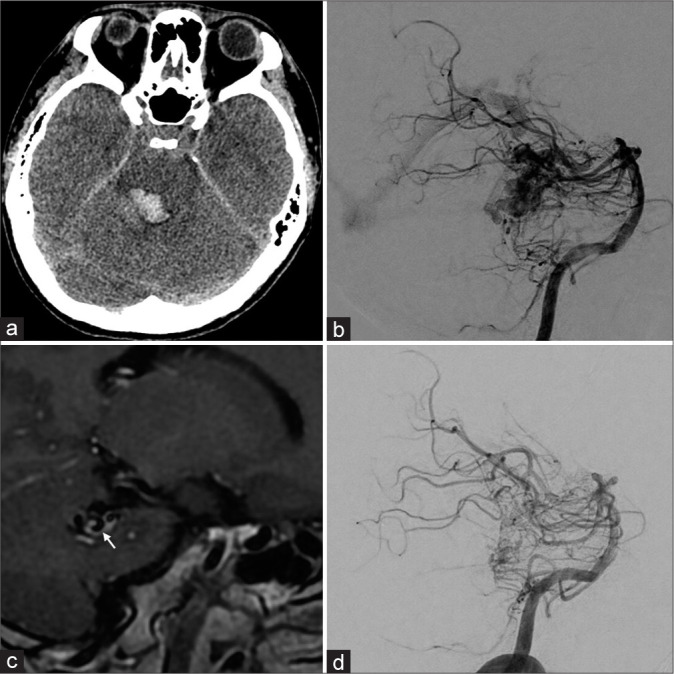
Representative case of vessel wall imaging (VWI) in arteriovenous malformation (AVM). A 27-year-old male visited our hospital with headaches, diplopia, and sensory disturbance. (a) A computed tomography scan demonstrated brainstem hemorrhage. (b) Subsequent digital subtraction angiography (DSA) revealed a brain AVM in the dorsal aspect of the brain stem. (c) In addition, VWI showed vessel wall enhancement in the drainage vein (white arrow). (d) Postinterventional DSA showed a significant decrease in blood flow. Accordingly, reducing the flow burden was a feasible therapeutic option. The patient underwent two transarterial embolization sessions and further stereotactic radiosurgery for the remaining AVM.
The recent advance in endovascular treatment of aneurysms further signifies the association between VVs and aneurysms. Both clipping surgery and endovascular coil embolization block blood influx into the aneurysm. However, they have a fundamental difference as a treatment concerning VVs; in clipping surgery, VVs are also clipped on the aneurysmal neck, while VVs externa remain intact. Accordingly, VVs still supply blood flow to the aneurysmal wall after coil embolization and deliver inflammatory cells, resulting in bleeding from VVs and aneurysmal growth. Indeed, some authors reported a case of a growing aneurysm even after angiographical complete occlusion had been achieved.[45,53] Endovascular treatment cannot cure these lesions, and surgical resection is mandatory. Future treatment may not only target the blood flow inside the aneurysm but also target the background mechanism, including VVs involved in the instability of the aneurysmal wall.
COLLATERAL CHANNELS
On the surface of the vessels, VVs form an abundant network, and the VVs in different territories can anastomose with each other.[9,22,36] Accordingly, when the parent vessel is stenotic or occluded, the VVs connecting the distal and the proximal segments may develop and function as a collateral tract of the parent vessel. A study reported that when porcine coronary arteries were gradually occluded, anastomoses between VVs running in the longitudinal direction develop and constitute fully functional collaterals in a concise timeline, which can be as short as a week.[23] In clinical situations, some authors reported that the collaterals formed by VVs and the occluded internal carotid artery (ICA) in cases with a giant aneurysm in the cavernous sinus segment,[46] with atherosclerotic ICA occlusion adjacent to the carotid bifurcation,[11,26,61] and with atherosclerotic basilar artery occlusion restored its distal flow through VV collaterals. We assume that these VV collaterals usually develop in extracranial segments or proximal parts of the internal carotid or vertebral arteries, depending on their physiological distribution. Since these collaterals are located on the tunica adventitia of the parent artery, carotid stenting or angioplasty may not be recommended due to the high risk of rupture.[26] Radiological features of VV collaterals include the serpiginous and multiple tiny channels running outside the parent vessels’ lumen[11,26,61] (corkscrew sign[26]), which are helpful to distinguish VV collaterals from a mere narrow atherosclerotic narrowing.
A FUTURE TREATMENT TARGETING VVS
This article reviews past and recent articles and discusses the involvement of VVs in various intracranial lesions. In most lesions, VVs aggravate vascular wall inflammation and cause bleeding from or within the vascular wall or plaque, leading to pathological progression. Therefore, the involvement of VVs can be considered as a malignant lesion in one way. At present, no study specifically targeting VVs in intracranial lesions has been reported. However, in the future treatment of intracranial lesions, we need to consider VV involvement or target the VVs themselves. However, at the same time, VVs sustain the normal healthy vascular wall. Thus, complete obliteration of VVs may increase the risk of wall ischemia and medial necrosis. In our opinion, “leaky” pathological VVs should be targeted, and transforming pathological VVs into healthy physiological VVs may change the situation. Further research is needed to elucidate the involvement of VVs in intracranial lesions and facilitate better clinical outcomes.
CONCLUSION
VV is not only involved in the nutrition and metabolism of the vascular wall but is also deeply involved in the pathogenesis of inflammation, ischemia, and thrombosis of the vascular wall. In addition, in the central nervous system, intracranial vascular wall nutrient particularities and VVs are closely related to the pathogenesis of cerebral aneurysms, SAH, AVS disease, atherosclerotic lesions, and other conditions. We believe that the issues discussed in this paper are of great importance to neurosurgeons and endovascular surgeons.
Footnotes
How to cite this article: Yamamoto K, Mizutani K, Akiyama T, Nogawa H, Toda M. Vasa vasorum: The role in intracranial physiology and pathophysiology. Surg Neurol Int. 2024;15:188. 10.25259/SNI_214_2024
Contributor Information
Kosei Yamamoto, Email: y.kousei0509@gmail.com.
Katsuhiro Mizutani, Email: m.coraime@keio.jp.
Takenori Akiyama, Email: akiyamanor@keio.jp.
Hirotsugu Nogawa, Email: hirotsugu0823@gmail.com.
Masahiro Toda, Email: todam@keio.jp.
Ethical approval
The Institutional Review Board approval is not required.
Declaration of patient consent
The authors certify that they have obtained all appropriate patient consent.
Financial support and sponsorship
Nil.
Conflicts of interest
There are no conflicts of interest.
Use of artificial intelligence (AI)-assisted technology for manuscript preparation
The authors confirm that there was no use of artificial intelligence (AI)-assisted technology for assisting in the writing or editing of the manuscript and no images were manipulated using AI.
Disclaimer
The views and opinions expressed in this article are those of the authors and do not necessarily reflect the official policy or position of the Journal or its management. The information contained in this article should not be considered to be medical advice; patients should consult their own physicians for advice as to their specific medical needs.
REFERENCES
- 1.Andres KH. On the fine structure of the arachnoidea and dura mater of mammals. Z Zellforsch Mikrosk Anat. 1967;79:272–95. [PubMed] [Google Scholar]
- 2.Atkinson JL, Okazaki H, Sundt TM, Jr, Nichols DA, Rufenacht DA. Intracranial cerebrovascular vasa vasorum associated with atherosclerosis and large thick-walled aneurysms. Surg Neurol. 1991;36:365–9. doi: 10.1016/0090-3019(91)90025-5. [DOI] [PubMed] [Google Scholar]
- 3.Aydin F. Do human intracranial arteries lack vasa vasorum? A comparative immunohistochemical study of intracranial and systemic arteries. Acta Neuropathol. 1998;96:22–8. doi: 10.1007/s004010050856. [DOI] [PubMed] [Google Scholar]
- 4.Berenstein A, Lasjaunias P, Brugge KG. Surgical neuroangiography. Vol. 2. New York: Springer Science and Business Media; 2012. Clinical and endovascular treatment aspects in adults. [Google Scholar]
- 5.Björnheden T, Bondjers G. Oxygen consumption in aortic tissue from rabbits with diet-induced atherosclerosis. Arteriosclerosis. 1987;7:238–47. doi: 10.1161/01.atv.7.3.238. [DOI] [PubMed] [Google Scholar]
- 6.Björnheden T, Levin M, Evaldsson M, Wiklund O. Evidence of hypoxic areas within the arterial wall in vivo. Arterioscler Thromb Vasc Biol. 1999;19:870–6. doi: 10.1161/01.atv.19.4.870. [DOI] [PubMed] [Google Scholar]
- 7.Brandão GM, Sobreira ML, Malgor RD, Rollo HA. Recanalization rates after acute deep vein thrombosis: A single-center experience using a newly proposed vein diameter variation index. Ann Vasc Surg. 2014;28:1751–60. doi: 10.1016/j.avsg.2014.05.013. [DOI] [PubMed] [Google Scholar]
- 8.Clarke JA. An X-ray microscopic study of the vasa vasorum of the intracranial arteries. Z Anat Entwicklungsgesch. 1965;124:396–400. doi: 10.1007/BF00523521. [DOI] [PubMed] [Google Scholar]
- 9.Clarke JA. An X-ray microscopic study of the vasa vasorum of the normal human ascending aorta. Heart. 1965;27:99–104. doi: 10.1136/hrt.27.1.99. [DOI] [PMC free article] [PubMed] [Google Scholar]
- 10.Clower BR, Sullivan DM, Smith RR. Intracranial vessels lack vasa vasorum. J Neurosurg. 1984;61:44–8. doi: 10.3171/jns.1984.61.1.0044. [DOI] [PubMed] [Google Scholar]
- 11.Colon GP, Deveikis JP, Dickinson LD. Revascularization of occluded internal carotid arteries by hypertrophied vasa vasorum: Report of four cases. Neurosurgery. 1999;45:634–7. doi: 10.1097/00006123-199909000-00040. [DOI] [PubMed] [Google Scholar]
- 12.Cord BJ, Renedo D, Santarosa C, Sujijantarat N, Antonios J, Kim JA, et al. Vessel wall MRI in ruptured cranial dural arteriovenous fistulas. Interv Neuroradiol. 2021;27:553–7. doi: 10.1177/1591019920988205. [DOI] [PMC free article] [PubMed] [Google Scholar]
- 13.Corti A, De Paolis A, Tarbell J, Cardoso L. Stenting-induced vasa vasorum compression and subsequent flow resistance: A finite element study. Biomech Model Mechanobiol. 2021;20:121–33. doi: 10.1007/s10237-020-01372-x. [DOI] [PMC free article] [PubMed] [Google Scholar]
- 14.Drozdz K, Janczak D, Dziegiel P, Podhorska M, Patrzałek D, Ziółkowski P, et al. Adventitial lymphatics of internal carotid artery in healthy and atherosclerotic vessels. Folia Histochem Cytobiol. 2008;46:433–6. doi: 10.2478/v10042-008-0083-7. [DOI] [PubMed] [Google Scholar]
- 15.Drozdz K, Janczak D, Dziegiel P, Podhorska M, Piotrowska A, Patrzalek D, et al. Adventitial lymphatics and atherosclerosis. Lymphology. 2012;45:26–33. [PubMed] [Google Scholar]
- 16.Edjlali M, Gentric JC, Régent-Rodriguez C, Trystram D, Hassen WB, Lion S, et al. Does aneurysmal wall enhancement on vessel wall MRI help to distinguish stable from unstable intracranial aneurysms? Stroke. 2014;45:3704–6. doi: 10.1161/STROKEAHA.114.006626. [DOI] [PubMed] [Google Scholar]
- 17.Eisenmenger LB, Junn JC, Cooke D, Hetts S, Zhu C, Johnson KM, et al. Presence of vessel wall hyperintensity in unruptured arteriovenous malformations on vessel wall magnetic resonance imaging: Pilot study of AVM vessel wall “enhancement. ” Front Neurosci. 2021;15:697432. doi: 10.3389/fnins.2021.697432. [DOI] [PMC free article] [PubMed] [Google Scholar]
- 18.Espinosa F, Weir B, Shnitka T. Electron microscopy of simian cerebral arteries after subarachnoid hemorrhage and after the injection of horseradish peroxidase. Neurosurgery. 1986;19:935–45. doi: 10.1227/00006123-198612000-00007. [DOI] [PubMed] [Google Scholar]
- 19.Federspiel JM, Tschernig T, Laschke MW, Wagenpfeil S, Schnabel P, Schäfers HJ. The vasa vasorum reach deep into the human thoracic aorta. Ann Anat. 2019;225:54–6. doi: 10.1016/j.aanat.2019.06.001. [DOI] [PubMed] [Google Scholar]
- 20.Feng Y, Dmytriw AA, Yang B, Jiao L. Neovascularization in human intracranial atherosclerotic in-stent restenosis. Diagnostics (Basel) 2021;11:322. doi: 10.3390/diagnostics11020322. [DOI] [PMC free article] [PubMed] [Google Scholar]
- 21.Gimbert Memorie sur la structure et sur la texture des artères. J Anatom Physiol Norm Pathol Homme Anim. 1865;2:536–68. [Google Scholar]
- 22.Gössl M, Rosol M, Malyar NM, Fitzpatrick LA, Beighley PE, Zamir M, et al. Functional anatomy and hemodynamic characteristics of vasa vasorum in the walls of porcine coronary arteries. Anat Rec A Discov Mol Cell Evol Biol. 2003;272:526–37. doi: 10.1002/ar.a.10060. [DOI] [PubMed] [Google Scholar]
- 23.Harnoss JM, Krackhardt F, Ritter Z, Granzow S, Felsenberg D, Neumann K, et al. Porcine arteriogenesis based on vasa vasorum in a novel semi-acute occlusion model using high-resolution imaging. Heart Vessels. 2017;32:1400–9. doi: 10.1007/s00380-017-1028-x. [DOI] [PubMed] [Google Scholar]
- 24.Hassler O, Larsson SE. The external elastic layer of the cerebral arteries in different age-groups. Acta Anat (Basel) 1962;48:1–6. doi: 10.1159/000141824. [DOI] [PubMed] [Google Scholar]
- 25.Hawkes C, Durant C, van Adel B. Symptomatic internal carotid artery vasa vasorum treated with surgical occlusion. Can J Neurol Sci. 2022;49:118–9. doi: 10.1017/cjn.2021.49. [DOI] [PubMed] [Google Scholar]
- 26.Heistad DD, Armstrong ML, Amundsen S. Blood flow through vasa vasorum in arteries and veins: effects of luminal PO2. Am J Physiol. 1986;250:H434–42. doi: 10.1152/ajpheart.1986.250.3.H434. [DOI] [PubMed] [Google Scholar]
- 27.His W. Nomina anatomica. Leipzig: Veit; 1895. Die anatomische nomenclatur. [Google Scholar]
- 28.Hou Y, Ryu CH, Jun JA, Kim SM, Jeong CH, Jeun SS. IL-8 enhances the angiogenic potential of human bone marrow mesenchymal stem cells by increasing vascular endothelial growth factor. Cell Biol Int. 2014;38:1050–9. doi: 10.1002/cbin.10294. [DOI] [PubMed] [Google Scholar]
- 29.Jurrus ER, Weiss HS. In vitro tissue oxygen tensions in the rabbit aortic arch. Atherosclerosis. 1977;28:223–32. doi: 10.1016/0021-9150(77)90172-1. [DOI] [PubMed] [Google Scholar]
- 30.Kai H, Kuwahara F, Tokuda K, Shibata R, Kusaba K, Niiyama H, et al. Coexistence of hypercholesterolemia and hypertension impairs adventitial vascularization. Hypertension. 2002;39:455–9. doi: 10.1161/hy0202.103001. [DOI] [PubMed] [Google Scholar]
- 31.Korkmaz E, Kleinloog R, Verweij BH, Allijn IE, Hekking LH, Regli L, et al. Comparative ultrastructural and stereological analyses of unruptured and ruptured saccular intracranial aneurysms. J Neuropathol Exp Neurol. 2017;76:908–16. doi: 10.1093/jnen/nlx075. [DOI] [PubMed] [Google Scholar]
- 32.Krings T, Piske RL, Lasjaunias PL. Intracranial arterial aneurysm vasculopathies: Targeting the outer vessel wall. Neuroradiology. 2005;47:931–7. doi: 10.1007/s00234-005-1438-9. [DOI] [PubMed] [Google Scholar]
- 33.Küker W, Gaertner S, Nagele T, Dopfer C, Schoning M, Fiehler J, et al. Vessel wall contrast enhancement: A diagnostic sign of cerebral vasculitis. Cerebrovasc Dis. 2008;26:23–9. doi: 10.1159/000135649. [DOI] [PMC free article] [PubMed] [Google Scholar]
- 34.Kumamoto M, Nakashima Y, Sueishi K. Intimal neovascularization in human coronary atherosclerosis: Its origin and pathophysiological significance. Hum Pathol. 1995;26:450–6. doi: 10.1016/0046-8177(95)90148-5. [DOI] [PubMed] [Google Scholar]
- 35.Labropoulos N, Bhatti AF, Amaral S, Leon L, Borge M, Rodriguez H, et al. Neovascularization in acute venous thrombosis. J Vasc Surg. 2005;42:515–8. doi: 10.1016/j.jvs.2005.05.036. [DOI] [PubMed] [Google Scholar]
- 36.Lametschwandtner A, Minnich B, Kachlik D, Setina M, Stingl J. Three-dimensional arrangement of the vasa vasorum in explanted segments of the aged human great saphenous vein: Scanning electron microscopy and three-dimensional morphometry of vascular corrosion casts. Anat Rec A Discov Mol Cell Evol Biol. 2004;281:1372–82. doi: 10.1002/ar.a.20098. [DOI] [PubMed] [Google Scholar]
- 37.Lawton MT, Jacobowitz R, Spetzler RF. Redefined role of angiogenesis in the pathogenesis of dural arteriovenous malformations. J Neurosurg. 1997;87:267–74. doi: 10.3171/jns.1997.87.2.0267. [DOI] [PubMed] [Google Scholar]
- 38.Lee RM. Morphology of cerebral arteries. Pharmacol Ther. 1995;66:149–73. doi: 10.1016/0163-7258(94)00071-a. [DOI] [PubMed] [Google Scholar]
- 39.Liszczak TM, Black PM, Varsos VG, Zervas NT. The microcirculation of cerebral arteries: A morphologic and morphometric examination of the major canine cerebral arteries. Am J Anat. 1984;170:223–32. doi: 10.1002/aja.1001700207. [DOI] [PubMed] [Google Scholar]
- 40.Lowenberg RI, Shumacker HB., Jr Experimental studies in vascular repair; morphologic observations of normal vasa vasorum. Yale J Biol Med. 1948;20:395–401. [PMC free article] [PubMed] [Google Scholar]
- 41.Ludwig CG. Lipsiae: Ex Officina Langenhemiana; De arteriarum tunicis; p. 1747. [Google Scholar]
- 42.Matouk CC, Mandell DM, Günel M, Bulsara KR, Malhotra A, Hebert R, et al. Vessel wall magnetic resonance imaging identifies the site of rupture in patients with multiple intracranial aneurysms. Neurosurgery. 2013;72:492–6. doi: 10.1227/NEU.0b013e31827d1012. [DOI] [PubMed] [Google Scholar]
- 43.Modarai B, Burnand KG, Humphries J, Waltham M, Smith A. The role of neovascularisation in the resolution of venous thrombus. Thromb Haemost. 2005;93:801–9. doi: 10.1160/TH04-09-0596. [DOI] [PubMed] [Google Scholar]
- 44.Nakashima T, Tashiro T. Early morphologic stage of human coronary atherosclerosis. Kurume Med J. 1968;15:235–42. doi: 10.2739/kurumemedj.15.235. [DOI] [PubMed] [Google Scholar]
- 45.Noh CY. Vasa vasorum in deep vein thrombus recanalization. J Vasc Ultrasound. 2018;42:33–5. [Google Scholar]
- 46.Numagami Y, Ezura M, Takahashi A, Yoshimoto T. Antegrade recanalization of completely embolized internal carotid artery after treatment of a giant intracavernous aneurysm: A case report. Surg Neurol. 1999;52:611–6. doi: 10.1016/s0090-3019(99)00131-7. [DOI] [PubMed] [Google Scholar]
- 47.O’Nejll JF. The effects on venous endothelium of alterations in blood flow through the vessels in vein walls, and the possible relation to thrombosis. Ann Surg. 1947;126:270–88. [PMC free article] [PubMed] [Google Scholar]
- 48.Omodaka S, Endo H, Fujimura M, Niizuma K, Sato K, Matsumoto Y, et al. High-grade cerebral arteriovenous malformation treated with targeted embolization of a ruptured site: Wall enhancement of an intranidal aneurysm as a sign of ruptured site. Neurol Med Chir. 2015;55:813–7. doi: 10.2176/nmc.cr.2015-0052. [DOI] [PMC free article] [PubMed] [Google Scholar]
- 49.Ozoner B, Cakir T, Kayaci S, Aydin MD, Aydin S, Demirci E. Effect of vasa vasorum on basilar artery vasospasm following subarachnoid hemorrhage. World Neurosurg. 2019;131:e218–25. doi: 10.1016/j.wneu.2019.07.124. [DOI] [PubMed] [Google Scholar]
- 50.Petridis AK, Dibue-Adjei M, Cornelius JF, Suresh MP, Li L, Kamp MA, et al. Contrast enhancement of vascular walls of intracranial high flow malformations in black blood MRI indicates high inflammatory activity. Chin Neurosurg J. 2018;4:13. doi: 10.1186/s41016-018-0120-0. [DOI] [PMC free article] [PubMed] [Google Scholar]
- 51.Phatouros CC, Halbach VV, Dowd CF, Lempert TE, Malek AM, Meyers PM, et al. Acquired pial arteriovenous fistula following cerebral vein thrombosis. Stroke. 1999;30:2487–90. doi: 10.1161/01.str.30.11.2487. [DOI] [PubMed] [Google Scholar]
- 52.Phillippi JA. On vasa vasorum: A history of advances in understanding the vessels of vessels. Sci Adv. 2022;8:eabl6364. doi: 10.1126/sciadv.abl6364. [DOI] [PMC free article] [PubMed] [Google Scholar]
- 53.Quan K, Song J, Zhu W, Wang D, An Q, Huang L, et al. Validation of wall enhancement as a new imaging biomarker of unruptured cerebral aneurysm. Stroke. 2019;50:1570–3. doi: 10.1161/STROKEAHA.118.024195. [DOI] [PubMed] [Google Scholar]
- 54.Sluimer JC, Kolodgie FD, Bijnens AP, Maxfield K, Pacheco E, Kutys B, et al. Thin-walled microvessels in human coronary atherosclerotic plaques show incomplete endothelial junctions relevance of compromised structural integrity for intraplaque microvascular leakage. J Am Coll Cardiol. 2009;53:1517–27. doi: 10.1016/j.jacc.2008.12.056. [DOI] [PMC free article] [PubMed] [Google Scholar]
- 55.Standring S. Netherlands: Elsevier; 2020. Gray’s anatomy: The anatomical basis of clinical practice. [Google Scholar]
- 56.Takaba M, Endo S, Kurimoto M, Kuwayama N, Nishijima M, Takaku A. Vasa vasorum of the intracranial arteries. Acta Neurochir (Wien) 1998;140:411–6. doi: 10.1007/s007010050118. [DOI] [PubMed] [Google Scholar]
- 57.Terada T, Higashida RT, Halbach VV, Dowd CF, Tsuura M, Komai N, et al. Development of acquired arteriovenous fistulas in rats due to venous hypertension. J Neurosurg. 1994;80:884–9. doi: 10.3171/jns.1994.80.5.0884. [DOI] [PubMed] [Google Scholar]
- 58.Texakalidis P, Hilditch CA, Lehman V, Lanzino G, Pereira VM, Brinjikji W. Vessel wall imaging of intracranial aneurysms: Systematic review and meta-analysis. World Neurosurg. 2018;117:453–8.e1. doi: 10.1016/j.wneu.2018.06.008. [DOI] [PubMed] [Google Scholar]
- 59.Tsang CP, Mizutani K, Trenkler J, Holmin S, Rodesch G. De novo arteriovenous shunts after endovascular cure of cerebrospinal macro arteriovenous fistulas. A role for the vasa vasorum? J Neuroradiol. 2021;48:127–31. doi: 10.1016/j.neurad.2020.06.004. [DOI] [PubMed] [Google Scholar]
- 60.Wakefield TW, Linn MJ, Henke PK, Kadell AM, Wilke CA, Wrobleski SK, et al. Neovascularization during venous thrombosis organization: A preliminary study. J Vasc Surg. 1999;30:885–92. doi: 10.1016/s0741-5214(99)70013-3. [DOI] [PubMed] [Google Scholar]
- 61.Wang R, Weng L, Li M. Effect of vasa vasorum in cerebrovascular compensation: 2 case reports. Ann Transl Med. 2020;8:508. doi: 10.21037/atm.2020.03.77. [DOI] [PMC free article] [PubMed] [Google Scholar]
- 62.Werber AH, Heistad DD. Diffusional support of arteries. Am J Physiol. 1985;248:H901–6. doi: 10.1152/ajpheart.1985.248.6.H901. [DOI] [PubMed] [Google Scholar]
- 63.Wilens SL, Malcolm JA, Vazquez JM. Experimental infarction (medial necrosis) of the dog’s aorta. Am J Pathol. 1965;47:695–711. [PMC free article] [PubMed] [Google Scholar]
- 64.Williams JK, Armstrong ML, Heistad DD. Vasa vasorum in atherosclerotic coronary arteries: Responses to vasoactive stimuli and regression of atherosclerosis. Circ Res. 1988;62:515–23. doi: 10.1161/01.res.62.3.515. [DOI] [PubMed] [Google Scholar]
- 65.Willis T. Oxford: E Theatro Sheldoniano; Pharmaceutice rationalis, sive, diatriba de medicamentorum operationibus in humano corpore; p. 1674. [Google Scholar]
- 66.Xu J, Lu X, Shi GP. Vasa vasorum in atherosclerosis and clinical significance. Int J Mol Sci. 2015;16:11574–608. doi: 10.3390/ijms160511574. [DOI] [PMC free article] [PubMed] [Google Scholar]
- 67.Yamashita A, Shoji K, Tsuruda T, Furukoji E, Takahashi M, Nishihira K, et al. Medial and adventitial macrophages are associated with expansive atherosclerotic remodeling in rabbit femoral artery. Histol Histopathol. 2008;23:127–36. doi: 10.14670/HH-23.127. [DOI] [PubMed] [Google Scholar]
- 68.Yuan H, Sun J, Zhou Z, Qi H, Wang M, Dong D, et al. Diagnosis and treatment of acquired arteriovenous fistula after lower extremity deep vein thrombosis. Int Angiol. 2019;38:10–6. doi: 10.23736/S0392-9590.19.04063-X. [DOI] [PubMed] [Google Scholar]
- 69.Zervas NT, Liszczak TM, Mayberg MR, Black PM. Cerebrospinal fluid may nourish cerebral vessels through pathways in the adventitia that may be analogous to systemic vasa vasorum. J Neurosurg. 1982;56:475–81. doi: 10.3171/jns.1982.56.4.0475. [DOI] [PubMed] [Google Scholar]
- 70.Zhang M, Cresswell N, Tavora F, Mont E, Zhao Z, Burke A. Instent restenosis is associated with neointimal angiogenesis and macrophage infiltrates. Pathol Res Pract. 2014;210:1026–30. doi: 10.1016/j.prp.2014.04.004. [DOI] [PubMed] [Google Scholar]
- 71.Zhong W, Su W, Li T, Tan X, Chen C, Wang Q, et al. Aneurysm wall enhancement in unruptured intracranial aneurysms: A histopathological evaluation. J Am Heart Assoc. 2021;10:1026–30. doi: 10.1161/JAHA.120.018633. [DOI] [PMC free article] [PubMed] [Google Scholar]