Abstract
Background Radiation therapy is a mainstay of treatment for brain tumors, but delayed complications include secondary malignancy which may occur months to years after treatment completion.
Methods We reviewed the medical records of a 41-year-old female treated with 60 Gy of radiation for a recurrent astrocytoma, who 6 years later developed a locally advanced sinonasal teratocarcinosarcoma. We searched MEDLINE, Embase, and Web of Science to conduct a scoping review of biopsy-proven sinonasal malignancy in patients who previously received cranial irradiation for a brain tumor.
Results To our knowledge, this is the first report of a patient to present with a sinonasal teratocarcinosarcoma after receiving irradiation for a brain tumor. Our scoping review of 1,907 studies produced 14 similar cases of secondary sinonasal malignancy. Median age of primary cancer diagnosis was 39.5 years old (standard deviation [SD]: 21.9), and median radiation dose was 54 Gy (SD: 20.3). Median latency time between the primary cancer and secondary sinonasal cancer was 9.5 years (SD: 5.8). Olfactory neuroblastoma was the most common sinonasal cancer ( n = 4). Fifty percent of patients died from their sinonasal cancer within 1.5 years.
Conclusion Patients who receive radiation exposure to the sinonasal region for treatment of a primary brain tumor, including low doses or scatter radiation, may be at risk of a secondary sinonasal malignancy later in life. Physicians who monitor at-risk patients must be vigilant of symptoms which may suggest sinonasal malignancy, and surveillance should include radiographic review with careful monitoring for a secondary malignancy throughout the entire irradiated field.
Keywords: sinonasal malignancy, cranial irradiation, radiation-induced malignancy, sinonasal teratocarcinosarcoma, radiation therapy, secondary malignancy, scoping review, case report
Introduction
Radiation therapy (RT) is used as a mainstay of treatment for both primary and metastatic brain tumors, as well as for prophylaxis against brain metastases of extracranial tumors. 1 2 3 By damaging deoxyribonucleic acid (DNA) to the point of irreparability, RT results in cell death and resultant stabilization and/or improvement of neurologic symptoms. 4 Several approaches for delivering RT are currently in use; these include external beam RT as well as brachytherapy. 1 4 While recent advancements in treatment techniques have enhanced RT targeting with the goal of minimizing negative sequelae, cranial irradiation inevitably exposes patients to an amalgam of short- and long-term complications. 4 5 6 In addition to neurocognitive changes and radiation necrosis, delayed complications of RT can include secondary malignancy which may occur months to years after treatment completion. 6 7 Previous studies have demonstrated an increased risk of subsequent central nervous system neoplasms following cranial irradiation, 8 9 and there are several reported instances of sinonasal neoplasm development following delivery of RT adjacent to the brain, such as for retinoblastoma in children. 10 11 12
Tumors of the nasal cavity and paranasal sinuses (sinonasal tumors) are a relatively rare entity, constituting < 5% of all head and neck neoplasms. 13 They have an incidence of < 1 in 100,000 in the United States. 13 Local growth can cause nasal congestion and obstruction, epistaxis, and anosmia, while extension of sinonasal tumors into adjacent structures such as the orbit, oral cavity, nasopharynx, and skull base may result in visual impairments and changes to facial structure. 14 Sinonasal tumors are heterogeneous in both their clinical features and histology, with 5-year overall survival ranging from 52 to 82%. 15 16 17 18 19 20 21 Known risk factors include occupational exposures to wood dust and other industrial compounds, tobacco use, and human papillomavirus infection. 14 However, to date, the association between cranial irradiation for brain tumors and subsequent development of sinonasal malignancy has not been comprehensively studied. Given the proximity of the brain to the nasal cavity and paranasal sinuses, it is conceivable that RT administered for the treatment of a brain tumor may be a contributor to the eventual development of sinonasal malignancy. Several contributing factors may influence such an association, including the approach used to deliver RT, the dose of radiation administered, and the age of the patient receiving RT, but these have also not yet been elucidated.
Herein, we present a case report of a sinonasal teratocarcinosarcoma following cranial irradiation, as well as a scoping review assessing the existing literature on the development of sinonasal tumors in individuals with a history of cranial irradiation. Investigating the association between cranial irradiation and the development of sinonasal malignancy will strengthen our current understanding of the need for enhanced monitoring and/or symptom tracking for at-risk patients, which may lead to earlier diagnosis and treatment. The research question guiding our review was: What is known from the existing literature on the development of sinonasal malignancy in individuals who have previously received cranial irradiation for an intracranial tumor?
Methods
The case report protocol was approved by the Research Ethics Board of Unity Health (REB #24-030) and informed consent was obtained from the patient. Reporting for our scoping review adhered to the Preferred Reporting Items for Systematic Reviews and Meta-Analyses Extension for Scoping Reviews reporting guideline. 22 A protocol was developed by the study team a priori and can be accessed on request from the corresponding author (Y.C.).
Eligibility Criteria
Eligible studies reported on patients with a sinonasal malignancy who previously received cranial irradiation for the treatment of a brain tumor. Patients of any age with any type of brain tumor (i.e., benign, malignant, or metastatic) were eligible for inclusion. To be included, patients must have completed a full course of cranial irradiation prior to their diagnosis of a sinonasal malignancy. Any radiation dosage was permitted. There was no minimum length of time (i.e., latency period) required between completion of cranial irradiation and diagnosis of the sinonasal malignancy. The sinonasal malignancy was required to be proven via biopsy.
All study types and settings were acceptable. Studies were excluded if a sinonasal cancer was diagnosed prior to and/or during a patient's course of cranial irradiation, or if cranial irradiation was administered for the treatment of a nonbrain (e.g., skull base) tumor. Nonoriginal studies such as editorials were excluded. Non-English language studies were also excluded.
Search Strategy and Information Sources
The search was designed and performed by two members of the study team (B.L., M.D.) with consultation from an experienced health sciences librarian (J.M.). Three electronic databases (Ovid MEDLINE, Ovid EMBASE, and Web of Science) were searched from inception to September 18, 2023. Keywords such as “brain neoplasm,” “radiotherapy,” and “sinonasal malignancy” were used with wildcards to account for plurals and spelling variations. The complete search strategy for all databases is available in Supplementary File S1 . The references of included studies were also scanned to ensure all relevant data were captured.
Selection and Extraction of Sources of Evidence
Screening was performed independently by two members of the study team (B.L., M.D.) using Covidence. Titles and abstracts were initially screened for inclusion, and potentially relevant full-text articles were subsequently retrieved and screened. Any disagreements were resolved by consensus or by the corresponding author (Y.C.) if a disagreement persisted. Data were extracted independently by two members of the study team (B.L., M.D.), with disagreements again resolved by consensus or by the corresponding author (Y.C.).
Extracted data included study design, patient demographics (e.g., age, gender, comorbidities), and clinical characteristics such as type of brain and sinonasal tumor, treatment modalities used (i.e., radiotherapy, surgical resection, and/or chemotherapy) with associated details, and latency period for each case. For studies reporting on multiple patients, only data which solely reported on relevant cases were extracted.
Synthesis of Results
Patient cases were synthesized and reported on descriptively. Descriptive nonparametric statistics were computed for all variables where appropriate. Continuous variables were reported as medians with standard deviations (SDs), and categorical variables were reported as unweighted frequencies with percentages.
Case Presentation
Clinical Presentation and Diagnosis
A 41-year-old female patient with a history of astrocytoma was referred to the otolaryngology ambulatory clinic for right nasal obstruction and query chronic rhinosinusitis.
The patient had a history of a grade II, 1p/19q-intact, IDH1-mutated astrocytoma of the right anterior frontal lobe, which was diagnosed incidentally 11 years prior and treated with a surgical resection at that time. She had a recurrence of the astrocytoma 5 years later, for which she underwent a salvage reresection as well as chemoradiotherapy. RT was administered as a dose of 60 Gy in 30 fractions, which was targeted to two sites: (1) the right parasagittal area, which was the location of a remnant from her first resection, and (2) the right frontal cavity, which was the resection site of the recurrence. She was given concurrent temozolomide, as well as adjuvant therapy in the form of 12 additional cycles of temozolomide. Two years later, the patient did have a second recurrence; she was rechallenged with an additional 12 cycles of temozolomide at that time with no further surgery or radiation administered. Following this, she was well clinically and stable radiographically.
The patient presented to the otolaryngology clinic with a 3-month history of right nasal obstruction, local tenderness of the right maxillary sinus, and ongoing yellow/green-colored nasal discharge. This was previously thought to be an acute sinus infection and was treated by a different provider with a 2-week course of cefprozil and an 8-week course of trimethoprim/sulfamethoxazole, as well as various nasal sprays, to no effect. The patient's congestive and obstructive symptoms had gradually worsened over time, resulting in complete right nasal obstruction and anosmia at the time of her referral. On physical examination, the patient had a hyponasal voice. Flexible nasal endoscopy demonstrated a large, red, nonpulsatile mass filling the right nasal cavity with some green mucous, rendering inability to pass into the nasopharynx; on the left side, the nasal cavity was patent except for a red, nonpulsatile mass visualized near the choana which appeared to be originating from the right side. Oral cavity examination was unremarkable. A computed tomography (CT) scan was conducted 1 week prior to the assessment ( Fig. 1 ). While the initial impression from the CT scan was that the mass most likely represented either progression of her primary tumor or metastatic disease, the patient's neurosurgeon believed that this was unlikely to be an extension of her original mass. An incisional biopsy was performed in the clinic on the same day as the patient's assessment. The pathology report favored a poorly differentiated carcinoma.
Fig. 1.
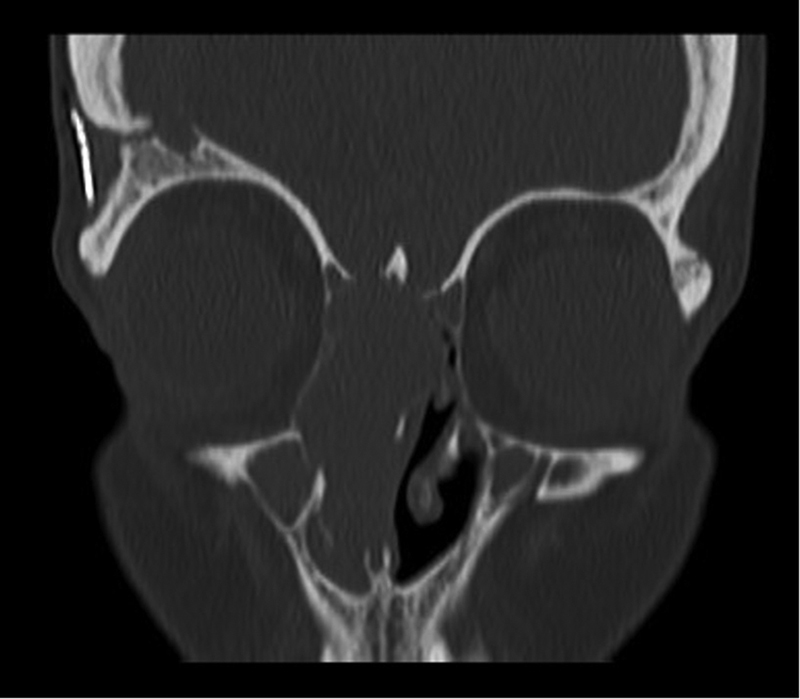
Coronal computed tomography (CT) scan of the sinuses, completed 1 week prior to assessment in clinic.
Magnetic resonance imaging (MRI) of the head and neck was completed 3 days after the patient was assessed in clinic and revealed a large heterogeneous mass likely originating from the right ethmoid region and involving the ethmoid sinuses bilaterally, and the middle and superior nasal cavities bilaterally, with exophytic extension into the nasopharynx ( Fig. 2 ). Intracranial extension was visualized through the right ethmoid roof and right olfactory groove, with progressive superior displacement of the right inferior frontal gyri but no findings to suggest invasion of the right frontal lobe. There was no involvement of the orbits; however, partial involvement of the right nasolacrimal duct was seen.
Fig. 2.
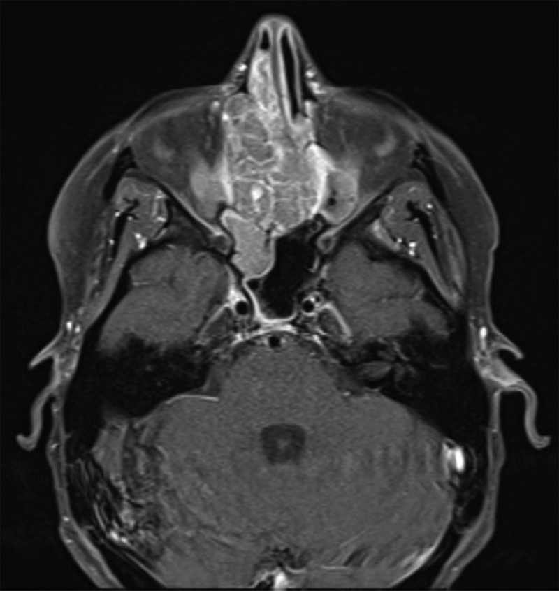
Axial magnetic resonance imaging (MRI) of the head and neck, completed 3 days after assessment in clinic.
Positron emission topography scan was completed the following month and demonstrated a metabolically active lesion, with metabolically active lymph nodes in bilateral neck level IIA and left neck level IIB, suspicious for bilateral lymph node metastasis. However, fine-needle aspiration biopsy was negative for malignancy. There was no convincing evidence of any other metabolically active distant metastatic disease.
Notably, the patient had begun experiencing nasal symptoms before her initial assessment at the otolaryngology clinic. Her medical records show that approximately 1 year prior, she was referred to a respirologist for ongoing fatigue and snoring; a sleep study conducted at that time revealed she had moderate obstructive sleep apnea with an apnea-hypopnea index of > 15. Further review of the patient's serial MRI brain scans, which were conducted as part of her routine neurosurgical follow-up, revealed evidence of a growing sinonasal lesion which was present but not commented on in scans dating as far back as 1 year prior to her assessment ( Fig. 3A–C ).
Fig. 3.
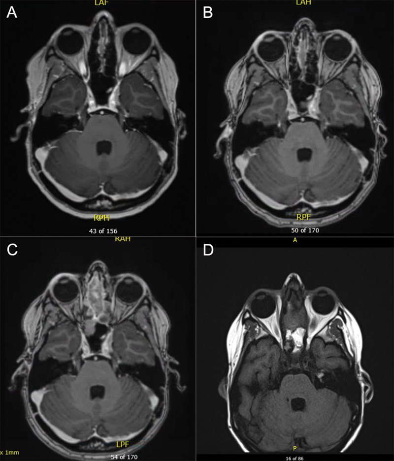
Serial axial magnetic resonance imaging (MRI) of the patient's brain. ( A ) 12 months prior to assessment; ( B ) 8 months prior to assessment; ( C ) 1 month prior to assessment; ( D ) 3 weeks postsurgery.
Management and Surgery
The patient was assessed by the radiation oncology team. Given the patient's history of RT to the right frontal lobe, a significant dose of radiation had already been delivered to several critical structures including the optic nerves bilaterally, optic chiasm, brainstem, and brain. Thus, reirradiation was initially not recommended as it posed an increased risk of permanent blindness, myelopathy, osteoradionecrosis, and brain radiation necrosis.
The patient was subsequently seen by the head and neck oncology and neurosurgery teams, and her sinonasal lesion was deemed to be surgically resectable. Due to the large size of the tumor, neoadjuvant therapy was recommended, with a plan for the patient to undergo up to three cycles prior to definitive surgical management. The patient was started on cisplatin and etoposide for a presumed sinonasal undifferentiated carcinoma (SNUC), with repeat imaging done after the first cycle. The mass appeared stable radiologically at this point, and given the poor response to chemotherapy, a decision was made to proceed directly to surgical resection. Two weeks later, the patient underwent right endoscopic anterior craniofacial resection with orbital dissection and preservation. As a result of dural involvement, a wide dural excision was performed. All surgical margins sent for intraoperative cryosection evaluation were negative for malignancy. The patient tolerated the procedure well with no complications.
Postoperative Management and Outcomes
A MRI scan completed 3 weeks postoperatively revealed no evidence of residual disease ( Fig. 3D ). Intraoperative biopsies from the right maxillary sinus, anterior skull base dura, and right nasal cavity mass were completed. The pathology findings showed a tumor consisting of nests of primitive cells with nuclear atypia and focal cytoplasmic clearing. The stroma showed focal hypercellular spindled areas with no overt differentiation ( Fig. 4 ). There was a focal area of squamous differentiation. The immunohistochemical studies revealed a positive expression of CK5 and LMWCK and a focal CD99 and INSM-1 staining in the tumor cells. The expression of SMARCA4 and SMARCB1 was retained in the tumor. The tumor nests also showed a focal beta catenin (nuclear, cytoplasmic, and membranous) expression. The molecular studies identified a mutation in the APC gene and therefore a teratocarcinosarcoma was favored. 23
Fig. 4.
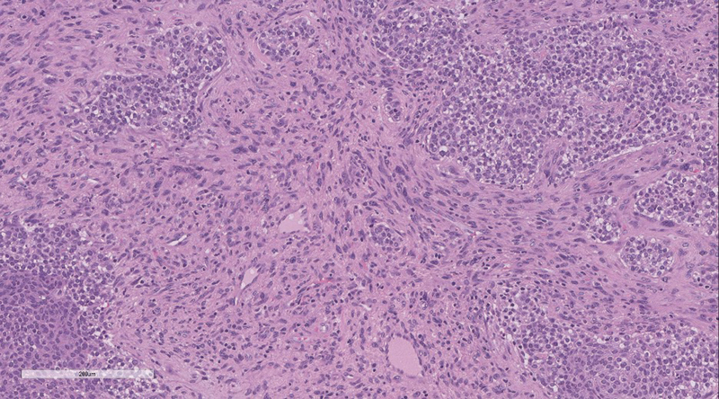
Poorly differentiated malignant neoplasm, teratocarcinosarcoma favored. The tumor showed nests of primitive cells with nuclear atypia, focal cytoplasmic clearing, and occasional mitoses. The stroma showed focal hypercellular spindled areas with no overt differentiation.
At her 6-week postoperative appointment, the patient was doing well clinically, and her nose had healed well. She had no cerebrospinal fluid leakage. Flexible nasal endoscopy revealed a well-healed nasal cavity, nasopharynx, and skull base with no evidence of recurrent disease. Based on the recommendations of the tumor board, the patient is set to undergo adjuvant proton RT.
Results
The literature search retrieved 2,318 studies. Following removal of duplicates, 1,907 articles remained. These articles underwent title and abstract screening, and 15 articles were selected for full-text review. Nine studies (10 patient cases) were immediately included in the review. 24 25 26 27 28 29 30 31 32 A meta-analysis was identified 33 which included two relevant case reports from studies not identified in the initial search; these two case reports were also included. 34 35 One study which described two additional relevant case reports was also identified by screening the references of included studies. 36 Overall, a total of 12 studies (14 patient cases) were included in the review. The full screening process is depicted in Fig. 5 . Details pertaining to each patient case are summarized in Table 1 .
Fig. 5.
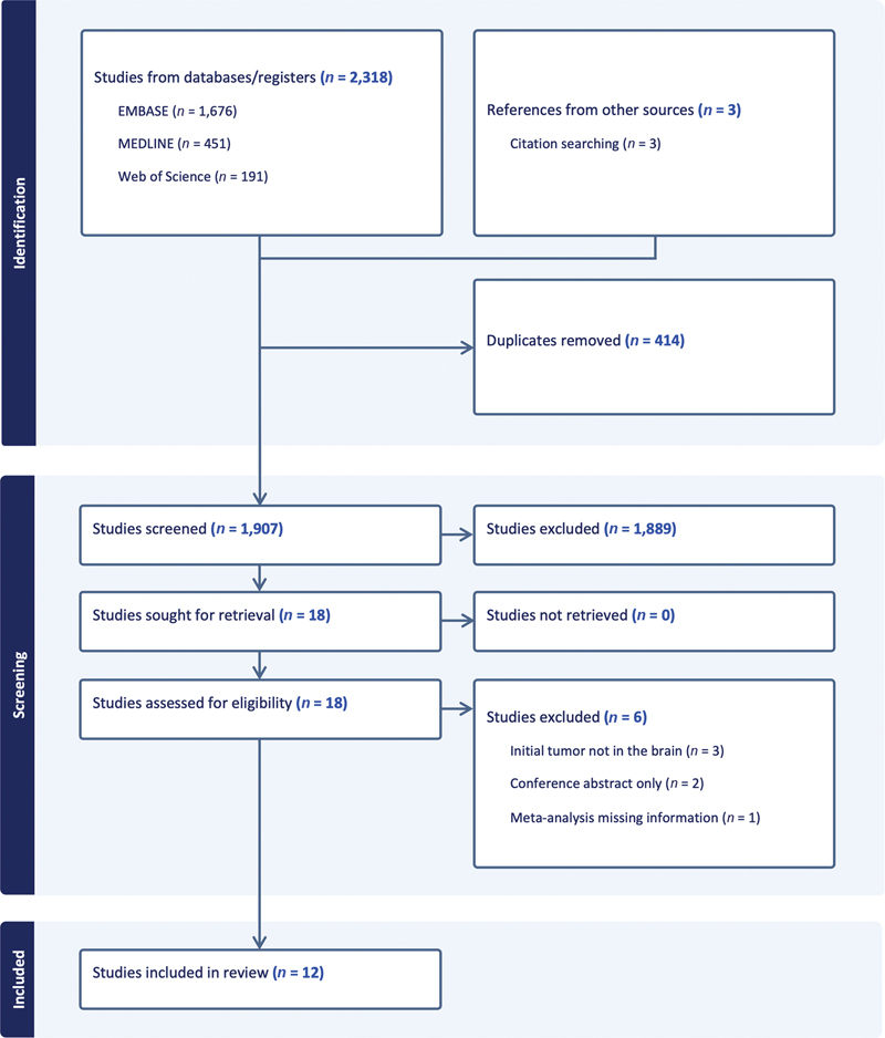
Preferred Reporting Items for Systematic Reviews and Meta-Analyses (PRISMA) flow diagram.
Table 1. Cases of sinonasal malignancy following cranial irradiation for brain tumor ( n = 14) .
| Case # | Author, Year | Age (y) a | Gender | Primary cancer | Primary cancer treatment | Primary cancer radiation dose | Latency period b (y) | Secondary sinonasal cancer | Secondary cancer treatment | Outcome |
|---|---|---|---|---|---|---|---|---|---|---|
| 1 | Goyal et al, 2015 | 17 | M | Glioblastoma multiforme | Radiotherapy, chemotherapy | 50.4 Gy, 28 fractions | 1.5 | Round cell carcinoma (undifferentiated) | Died after four cycles of chemotherapy | Rapid growth; died after 3 months |
| 2 | Ito et al, 2010 | 40 | F | Frontal glioma (low-grade) | Resection, radiotherapy, chemotherapy | 54 Gy, 27 fractions | 16 | Osteosarcoma | Died prior to planned resection | Rapid growth; died |
| 3 | Ito et al, 2010 | 46 | M | Bifrontal anaplastic oligoastrocytoma | Resection, radiotherapy, chemotherapy | 56 Gy, 28 fractions | 12 | Osteosarcoma | Died during radiotherapy treatment | Rapid growth; died |
| 4 | Park et al, 2008 | 39 | F | Pituitary adenoma (prolactinoma) | Resection, radiotherapy | 54 Gy, 30 fractions | 20 | Olfactory neuroblastoma | Resection, chemotherapy | Survived |
| 5 | Perez Garcia et al, 2011 | 43 | F | Bifrontal astrocytoma (low-grade) | Radiotherapy | 58 Gy, 5 fractions per week (holocranial 40 Gy, frontal 18 Gy) | 9 | Olfactory neuroblastoma | Resection, radiotherapy, chemotherapy | Recurrent twice; died after 13 months |
| 6 | Wallin et al, 2007 | 2.5 | M | Medulloblastoma and parietal meningioma | Radiotherapy (medulloblastoma), resection (parietal meningioma) | 45 Gy (23.4 Gy spine, 21.6 Gy posterior fossa) | 16.5 | Neuroendocrine carcinoma (poorly differentiated) | Resection | Survived |
| 7 | Kakkar et al, 2019 | 36 | F | Frontal diffuse astrocytoma | Resection, radiotherapy | 56 Gy, 28 fractions | 5.5 | Papillary adenocarcinoma | Resection | No disease evident at 8 months |
| 8 | Patel et al, 2017 | 54 | M | Anaplastic oligoastrocytoma | Resection, radiotherapy, chemotherapy | NS | 16 | Carcinosarcoma | Resection, Cesium-131 brachytherapy | No disease evident at 9 months |
| 9 | Nery et al, 2022 | 48 | M | Pituitary macroadenoma | Resection, radiotherapy | NS | 12 | Mucoepidermoid carcinoma | Resection, radiotherapy, chemotherapy | Survived |
| 10 | Sahoo et al, 2022 | 12 | M | Medulloblastoma | Resection, radiotherapy, chemotherapy | 56 Gy, 30 fractions | 2 | Olfactory neuroblastoma | Resection | Died after 8 months |
| 11 | Christopherson et al, 2014 | 7.1 c | NS | Medulloblastoma | Resection, radiotherapy, chemotherapy | 54 Gy (craniospinal 28.8 Gy) d | 6.4 | Rhabdomyosarcoma | Radiotherapy, chemotherapy | Treatment successful; last follow-up unknown |
| 12 | Packer et al, 2013 | 6.8 c | NS | Medulloblastoma | Resection, radiotherapy, chemotherapy | 55.8 Gy (23.4 Gy craniospinal, 32.4 Gy posterior fossa) | 4.8 | Spindle cell carcinoma | Unknown | Survived at least 1.67 years |
| 13 | Delank and Ballantyne, 1993 | 68 | M | Pituitary adenoma (prolactinoma) | Resection, radiotherapy, chemotherapy | 1.8 Gy | 6.5 | Malignant melanoma | Resection, radiotherapy | Died after 1.5 years |
| 14 | Delank and Ballantyne, 1993 | 65 | M | Pituitary adenoma | Resection, radiotherapy | 3.0 Gy | 10 | Olfactory neuroblastoma | Radiotherapy, chemotherapy | Died after 9 months |
Abbreviations: F, female; Gy, gray (unit); M, male; NS, not specified.
Age at diagnosis of brain tumor.
Time between irradiation of the brain tumor and diagnosis of the secondary sinonasal malignancy.
Median age of study participants; participant-specific ages not specified.
Median dose administered to study participants; participant-specific doses not specified.
The median age of patients at the time of brain tumor diagnosis was 39.5 years (SD: 21.9, range: 2.5–68). With respect to gender, there were eight males (57%), four females (29%), and two patients with gender not specified (14%). 34 35 All tumors were primary brain tumors; the most common was glioma ( n = 6, 43%), which included low-grade glioma ( n = 1, 7%), 25 glioblastoma ( n = 1, 7%), 24 astrocytoma ( n = 2, 14%), 27 and oligoastrocytoma ( n = 2, 14%). 25 30 Other primary cancers included medulloblastoma ( n = 4, 29%) 28 and pituitary adenoma ( n = 4, 29%). 26 31 36
Every patient received RT for treatment of their primary brain tumor. The median radiation dose was 54 Gy (SD: 20.3, range: 1.8–58); two cases did not report a radiation dose. 30 31 Eight of the 14 cases (57%) 24 25 30 32 34 35 36 used chemotherapy to treat the primary cancer; chemotherapeutic regimens included ACNU, bromocriptine, CCNU, cisplatin, etoposide, and temozolomide, in varying formulations. Twelve of the 14 cases (86%) 25 26 28 29 30 31 32 34 35 36 underwent surgical resection for their primary brain tumor. In one of these cases, a medulloblastoma was treated with radiation and a subsequent parietal meningioma was surgically resected 16 years later. 28
Time between irradiation of the primary tumor and presentation of the secondary sinonasal cancer (i.e., latency period) was a median of 9.5 years (SD: 5.8, range: 1.5–20). Only two cases (14%) had a time interval of less than 4 years between the primary brain tumor and secondary sinonasal cancer, and both of these cases occurred in adolescent patients (i.e., 12 and 17 years old). 24 32 Secondary sinonasal cancers included olfactory neuroblastoma ( n = 4, 29%), 26 27 32 36 osteosarcoma ( n = 2, 14%), 25 neuroendocrine carcinoma ( n = 1, 7%), 28 round cell carcinoma ( n = 1, 7%), 24 papillary adenocarcinoma ( n = 1, 7%), 29 carcinosarcoma ( n = 1, 7%), 30 mucoepidermoid carcinoma ( n = 1, 7%), 31 rhabdomyosarcoma ( n = 1, 7%), 34 spindle cell carcinoma ( n = 1, 7%), 35 and melanoma ( n = 1, 7%). 36
With respect to sinonasal cancer treatment, 9 of the 14 cases (64%) underwent surgical resection, 6 (43%) underwent chemotherapy, and 7 (50%) underwent RT. The treatment for a single patient with secondary sinonasal spindle cell carcinoma was not reported. 35 With respect to outcomes, seven cases (50%) 24 25 27 32 36 died from their secondary sinonasal cancer within 1.5 years, whereas six cases (43%) 26 28 29 30 31 35 were reported to survive or have no evidence of disease up to at least 8 months after treatment. One case did not report on the patient's prognosis following treatment of the secondary cancer. 34
Further data on the radiation fields used, radiotherapy planning, and whether the 14 included cases satisfy the modified Cahan's criteria for diagnosing radiation-induced malignancy 37 38 are outlined in Table 2 .
Table 2. Radiation field, radiotherapy planning, and Cahan's criteria data for included cases ( n = 14) .
| Case # | Author, year | Sinonasal region within irradiated field? | Details of radiation planning for brain tumor provided? a | Does the case satisfy the modified Cahan's criteria? | Authors' most likely described cause of sinonasal tumor |
|---|---|---|---|---|---|
| 1 | Goyal et al, 2015 | No; possible scatter | Yes | No (field b , timing c ) | Metachronous |
| 2 | Ito et al, 2010 | Yes | No | Yes | Radiation-induced |
| 3 | Ito et al, 2010 | Yes | Yes | Yes | Radiation-induced |
| 4 | Park et al, 2008 | Yes | No | Yes | Radiation-induced |
| 5 | Perez Garcia et al, 2011 | Yes | Yes | Yes | Radiation-induced |
| 6 | Wallin et al, 2007 | Not explicitly stated | No | Unknown (field b ) | Radiation-induced |
| 7 | Kakkar et al, 2019 | Yes | No | Yes | Radiation-induced |
| 8 | Patel et al, 2017 | Not explicitly stated | No | Unknown (field b ) | Radiation-induced |
| 9 | Nery et al, 2022 | Not explicitly stated | No | Unknown (field b ) | Radiation-induced |
| 10 | Sahoo et al, 2022 | Not explicitly stated | No | No (timing c ) | Metachronous or genetic |
| 11 | Christopherson et al, 2014 | Yes | No | Yes | Radiation-induced |
| 12 | Packer et al, 2013 | No; scatter only | No | Yes | Radiation-induced |
| 13 | Delank and Ballantyne, 1993 | Not explicitly stated | No | Unknown (field b ) | Metachronous |
| 14 | Delank and Ballantyne, 1993 | Not explicitly stated | No | Unknown (field b ) | Metachronous or radiation-induced |
Defined as inclusion of details beyond radiation dose (i.e., Gy and fractions) and general location, such as provision of a radiation dose distribution map.
Malignancy must arise from an area which has previously been irradiated (criterion 2).
There must be a relatively long, asymptomatic latent period following the initial radiation, which may be 4–5 years in length (criterion 3).
Discussion
While only a small number of cases have been reported in the literature, the results of our scoping review demonstrate that patients who receive radiation exposure to the sinonasal region as part of treatment for a primary brain tumor may be at risk of a secondary sinonasal malignancy later in life. Included cases ( n = 14) were a median of 39.5 years old at the time of diagnosis of their primary brain tumor; there were no reported instances of a primary cancer recurrence. Patients were diagnosed with a secondary sinonasal malignancy at a median of 9.5 years after irradiation of their primary cancer, with most tumors located within or adjacent to the irradiated field. The overall outcome for these patients was relatively poor, as seven patients (50%) succumbed to their secondary sinonasal malignancy within 1.5 years of diagnosis.
Cahan's criteria, first described by Cahan et al. in 1998, have since been modified and continue to be used as the basis for diagnosing radiation-induced cancers. 37 38 39 Four criteria are used in making this diagnosis: (1) there is prior microscopic or radiographic evidence of nonmalignancy in the tissue in which the malignancy is induced, (2) the malignancy arises from an area which has previously been irradiated, (3) there is a relatively long, asymptomatic latent period following the initial irradiation, typically > 4 years in length, and (4) the secondary malignancy is histologically different from the primary malignancy. 37 38 39 Of the 14 cases in our scoping review, 7 (50%) met the modified Cahan's criteria; 6 cases failed criterion 2, and 2 cases failed criterion 3 ( Table 2 ). While several cases did not explicitly state that a patient's sinonasal cancer was within the previously irradiated field, the scatter and/or edge effects of radiation are known to contribute to secondary cancer development. 40 In the case by Goyal et al, the sinonasal cancer was outside of the irradiated field, but the authors do note the possibility that it was induced by the effects of scatter. 24 A meta-analysis of children who received craniospinal irradiation for medulloblastoma conducted by Bavle et al in 2018 found that over a third of secondary neoplasms occurred outside of the irradiated field; the authors posited that the majority of these were due to the effects of exit radiation. 33 This suggests that even if it lies outside of the irradiated field, the sinonasal region may still remain susceptible to radiation-induced secondary malignancy. The importance of defining a specific latency period in the diagnosis of a radiation-induced secondary malignancy has also previously been called into question. 41 For example, in their retrospective study of 20 cases of postradiation sarcoma, Murray et al found no relationship between radiation dose and latency ( r = 0.133), with one case of secondary cancer reported as early as 1 year and 11 months following irradiation. 41 The authors argued that defining an arbitrary cutoff for latency may result in the exclusion of true radiation-induced cases from a report. 41 Our study did not define a specific latency period below which cases were excluded; however, our found median latency of 9.5 years does align with reported averages in the literature, which range between 10 and 20 years. 27 32 34 42
Only three included cases 24 25 27 provided detailed information regarding radiation planning for the primary brain tumor (e.g., radiation dose distribution maps) which could be used to better understand the degree of radiation exposure to the sinonasal cavity. Moreover, no included studies provided detailed dosimetry reports. Most studies which reported on radiation dose in the current review reported a dose of > 45 Gy ( n = 10, 83%), which is considered a high dose. Cantini et al have previously described the relationship between radiation dose and radiation-induced tumors, with benign tumors (e.g., meningioma) tending to occur following radiation doses of < 15 Gy and malignant tumors (e.g., sarcoma) tending to occur at higher doses. 43 The idea that radiation dose can influence the phenotype of the second malignancy is validated across an array of radiation-induced malignancies 44 and is consistent with the outcomes of our review.
The carcinogenic effect of radiation is related to cell injury and genotoxic stress, as radiation exposure can lead to the loss of DNA repair capacity and inactivation of tumor suppressor genes such as TP53 . 42 45 While the majority of primary brain cancers occur in adults, 46 47 our review found five pediatric patients (36%) 24 28 32 34 35 who were under the age of 18 at the time of their primary brain cancer diagnosis. The relatively high prevalence of childhood-onset brain tumors with secondary sinonasal malignancy postradiation detected by our review aligns with the generally accepted premise that children are at higher risk for radiation-induced malignancies, which may be related to the impact of genotoxic injury on stem cells. 8 44 However, a range of other etiologies may also explain the development of a secondary tumor adjacent to the primary tumor, including extension of the primary tumor, metastatic spread, environmental factors, and genetic predisposition. 26 Genetics have been hypothesized to play a role as it is possible for the same genetic anomaly occurring during embryogenesis to be responsible for both a primary and secondary malignancy. 32 For example, Sahoo et al suggested that a common genetic anomaly may be responsible for the development of two malignancies of neuroepithelial origin in the same patient; their patient had a primary medulloblastoma and subsequently developed a secondary esthesioneuroblastoma only 2 years later. 32
Our review found that the most common treatment for secondary sinonasal cancer was surgical resection, which was done in nine cases (64%). Other forms of management included chemotherapy ( n =6, 43%) and reirradiation ( n = 7, 50%). This was an interesting finding, as in our patient case, providers were hesitant to pursue reirradiation given the risk of delivering further radiation to critical structures in the head and neck. Individual cases were also reported where the patient died prior to completion of chemotherapy, surgical resection, and/or RT. 24 25 In the 10 reported cases which received a full course of treatment, 8 were treated surgically (3 ultimately died; 38%), 5 underwent chemotherapy (2 ultimately died; 40%), and 6 underwent radiation (3 ultimately died; 50%) ( Table 1 ). Only two patients received all three modalities of treatment for their secondary cancer; one died 27 and one survived. 31 Given these relatively similar mortality rates, our data suggest that no one treatment modality was superior to another for management of the secondary cancers.
Sinonasal teratocarcinosarcoma is a rare and aggressive entity; a systematic review conducted by Chapurin et al in 2021 found only 127 reported cases in the English literature with a survival rate of 55% and recurrence rate of 38%. 48 As far as we are aware, this is the first case of a sinonasal teratocarcinosarcoma following cranial irradiation to be documented in the literature. The findings of our scoping review demonstrate the importance of carefully monitoring the sinonasal cavity for secondary malignancy in patients who have received RT for a primary brain tumor, as cranial irradiation may increase the risk for a subsequent sinonasal malignancy. Given the high mortality and substantial impact on quality of life associated with all sinonasal tumors, 49 50 physicians monitoring these patients must be vigilant of potential symptoms which may be indicative of sinonasal malignancy, including anosmia, epistaxis, facial/sinus tenderness, obstructive sleep apnea, and/or persistent nasal discharge. Surveillance should include radiographic assessment as well since patients with a history of high-dose irradiation for primary brain tumor may undergo serial MRI brain scans to monitor for recurrence, as was seen in our reported case. In these instances, it is important to carefully evaluate not only the location of the primary tumor, but the entire irradiated field and periphery for signs of a secondary malignancy. While the rarity of secondary sinonasal malignancy relative to other long-term complications of radiotherapy makes it impractical to perform serial brain MRIs solely to screen for secondary sinonasal disease, these scans should be comprehensively evaluated if performed.
Genetics play an increasing role in the diagnosis, evaluation, and management of sinonasal malignancies. 51 52 Sinonasal malignancies may be characterized by gene mutations in SMARCA4 and SMARCB1 , which play a central role in tumor suppression and along with mutations in IDH2 may serve as driver genetic events in SNUC. 52 In our described case, the patient did not have a deficiency in either SMARCA4 or SMARCB1 , which may be seen as a positive prognostic indicator. There remains limited data on the specific impact(s) of radiation exposure and role of germline versus induced somatic mutations of SMARCA4 or SMARCB1 in the pathogenesis of SNUC. However, our case report did identify a mutation in the patient's APC gene, the abnormal expression of which in vivo animal studies have shown is induced by irradiation and results in tumor development (although this evidence is limited to intestinal tumors). 53 54 While the exact causative mechanism for the presentation of the secondary sinonasal malignancy in our study remains unknown, genetic damage due to cranial irradiation is a plausible explanation. As modern tumor classification and management continues to increasingly rely on tumor genotype, future studies reporting on radiation-induced malignancy should prioritize inclusion of genotype details for the primary brain tumor and secondary sinonasal cancer.
Our study is supported by several strengths, including a comprehensive literature search strategy designed in collaboration with a health sciences librarian (J.M.), and broad inclusion criteria which captured all patient ages and primary brain tumor types with no minimum latency period. We also acknowledge several limitations. Given the rarity of sinonasal cancers, only a small number of cases met the criteria for inclusion in our review. Case studies were the only included study type as studies with more rigorous designs did not meet the criteria for inclusion; this was expected given the low frequency of these secondary sinonasal malignancies after cranial irradiation and the difficultly of conducting such studies over a long period. The need for relevant studies to be conducted over an extended duration may explain the unavailability of information about specific cases, including details such as radiation doses and follow-up periods after secondary cancer treatment. Moreover, older included studies may reflect antiquated classifications, diagnostic criteria, and/or treatment algorithms for both brain tumors and sinonasal cancers, which limit the homogeneity of included cases and generalizability of our conclusions. The absence of genetic information offered by the studies also limited our ability to comment on trends within genotypic data. Finally, information on radiation planning for the initial treatment of the primary brain tumor was rarely provided, which may be in part due to the difficulty of retrieving relevant patient information after many years. This made it challenging to draw any conclusions on the impact of specific variables within the radiation plan (e.g., dosage, location) on the secondary sinonasal malignancy and/or patient outcomes.
Conclusion
To our knowledge, this is the first case of a sinonasal teratocarcinosarcoma following cranial irradiation to be documented in the literature. Findings from our scoping review demonstrate that patients who receive radiation exposure to the sinonasal region as part of treatment for a primary brain tumor, including low doses or scatter radiation, may be at risk of a secondary sinonasal malignancy later in life. Given the paucity of reported cases, it is difficult to draw conclusions on the impact of the radiation plan for the primary brain tumor on secondary sinonasal cancer development, and there is currently no definite evidence of a causal relationship. Physicians who monitor at-risk patients must be vigilant of symptoms which may be indicative of a sinonasal malignancy, and surveillance should extend to radiographic review with careful monitoring for secondary malignancy throughout the entire irradiated field.
Acknowledgment
The authors thank Ms. Julia Martyniuk for her assistance with developing the literature search strategy.
Footnotes
Conflict of Interest None declared.
Supplementary Material
References
- 1.Perkins A, Liu G. Primary brain tumors in adults: diagnosis and treatment. Am Fam Physician. 2016;93(03):211–217. [PubMed] [Google Scholar]
- 2.Owonikoko T K, Arbiser J, Zelnak A et al. Current approaches to the treatment of metastatic brain tumours. Nat Rev Clin Oncol. 2014;11(04):203–222. doi: 10.1038/nrclinonc.2014.25. [DOI] [PMC free article] [PubMed] [Google Scholar]
- 3.McTyre E, Scott J, Chinnaiyan P.Whole brain radiotherapy for brain metastasis Surg Neurol Int 20134(5, Suppl 4):S236–S244. [DOI] [PMC free article] [PubMed] [Google Scholar]
- 4.Rahman R, Sulman E, Haas-Kogan D, Cagney D N. Update on radiation therapy for central nervous system tumors. Hematol Oncol Clin North Am. 2022;36(01):77–93. doi: 10.1016/j.hoc.2021.08.006. [DOI] [PubMed] [Google Scholar]
- 5.for NRG Oncology . Brown P D, Gondi V, Pugh S et al. Hippocampal avoidance during whole-brain radiotherapy plus memantine for patients with brain metastases: phase III trial NRG Oncology CC001. J Clin Oncol. 2020;38(10):1019–1029. doi: 10.1200/JCO.19.02767. [DOI] [PMC free article] [PubMed] [Google Scholar]
- 6.Rahman R, Alexander B M, Wen P Y. Neurologic complications of cranial radiation therapy and strategies to prevent or reduce radiation toxicity. Curr Neurol Neurosci Rep. 2020;20(08):34. doi: 10.1007/s11910-020-01051-5. [DOI] [PubMed] [Google Scholar]
- 7.Makale M T, McDonald C R, Hattangadi-Gluth J A, Kesari S. Mechanisms of radiotherapy-associated cognitive disability in patients with brain tumours. Nat Rev Neurol. 2017;13(01):52–64. doi: 10.1038/nrneurol.2016.185. [DOI] [PMC free article] [PubMed] [Google Scholar]
- 8.Neglia J P, Robison L L, Stovall M et al. New primary neoplasms of the central nervous system in survivors of childhood cancer: a report from the Childhood Cancer Survivor Study. J Natl Cancer Inst. 2006;98(21):1528–1537. doi: 10.1093/jnci/djj411. [DOI] [PubMed] [Google Scholar]
- 9.Bowers D C, Nathan P C, Constine L et al. Subsequent neoplasms of the CNS among survivors of childhood cancer: a systematic review. Lancet Oncol. 2013;14(08):e321–e328. doi: 10.1016/S1470-2045(13)70107-4. [DOI] [PMC free article] [PubMed] [Google Scholar]
- 10.Bhagia P, Colanta A B, Abramson D H et al. Sinonasal adenocarcinoma: a rare second malignancy in long term retinoblastoma survivors. Pediatr Blood Cancer. 2011;57(04):693–695. doi: 10.1002/pbc.23161. [DOI] [PMC free article] [PubMed] [Google Scholar]
- 11.Franchi A, Sardi I, Cetica V et al. Pediatric sinonasal neuroendocrine carcinoma after treatment of retinoblastoma. Hum Pathol. 2009;40(05):750–755. doi: 10.1016/j.humpath.2008.09.018. [DOI] [PubMed] [Google Scholar]
- 12.Saw D, Chan J KC, Jagirdar J, Greco M A, Lee M. Sinonasal small cell neoplasm developing after radiation therapy for retinoblastoma: an immunohistologic, ultrastructural, and cytogenetic study. Hum Pathol. 1992;23(08):896–899. doi: 10.1016/0046-8177(92)90401-n. [DOI] [PubMed] [Google Scholar]
- 13.Thawani R, Kim M S, Arastu A et al. The contemporary management of cancers of the sinonasal tract in adults. CA Cancer J Clin. 2023;73(01):72–112. doi: 10.3322/caac.21752. [DOI] [PMC free article] [PubMed] [Google Scholar]
- 14.Bracigliano A, Tatangelo F, Perri F et al. Malignant sinonasal tumors: update on histological and clinical management. Curr Oncol. 2021;28(04):2420–2438. doi: 10.3390/curroncol28040222. [DOI] [PMC free article] [PubMed] [Google Scholar]
- 15.Hoppe B S, Stegman L D, Zelefsky M J et al. Treatment of nasal cavity and paranasal sinus cancer with modern radiotherapy techniques in the postoperative setting–the MSKCC experience. Int J Radiat Oncol Biol Phys. 2007;67(03):691–702. doi: 10.1016/j.ijrobp.2006.09.023. [DOI] [PubMed] [Google Scholar]
- 16.Chen A M, Daly M E, Bucci M K et al. Carcinomas of the paranasal sinuses and nasal cavity treated with radiotherapy at a single institution over five decades: are we making improvement? Int J Radiat Oncol Biol Phys. 2007;69(01):141–147. doi: 10.1016/j.ijrobp.2007.02.031. [DOI] [PubMed] [Google Scholar]
- 17.Allen M W, Schwartz D L, Rana V et al. Long-term radiotherapy outcomes for nasal cavity and septal cancers. Int J Radiat Oncol Biol Phys. 2008;71(02):401–406. doi: 10.1016/j.ijrobp.2007.10.031. [DOI] [PMC free article] [PubMed] [Google Scholar]
- 18.Mendenhall W M, Amdur R J, Morris C G et al. Carcinoma of the nasal cavity and paranasal sinuses. Laryngoscope. 2009;119(05):899–906. doi: 10.1002/lary.20196. [DOI] [PubMed] [Google Scholar]
- 19.Duprez F, Madani I, Morbée L et al. IMRT for sinonasal tumors minimizes severe late ocular toxicity and preserves disease control and survival. Int J Radiat Oncol Biol Phys. 2012;83(01):252–259. doi: 10.1016/j.ijrobp.2011.06.1977. [DOI] [PubMed] [Google Scholar]
- 20.Dulguerov P, Jacobsen M S, Allal A S, Lehmann W, Calcaterra T. Nasal and paranasal sinus carcinoma: are we making progress? A series of 220 patients and a systematic review. Cancer. 2001;92(12):3012–3029. doi: 10.1002/1097-0142(20011215)92:12<3012::aid-cncr10131>3.0.co;2-e. [DOI] [PubMed] [Google Scholar]
- 21.Dagan R, Bryant C, Li Z et al. Outcomes of sinonasal cancer treated with proton therapy. Int J Radiat Oncol Biol Phys. 2016;95(01):377–385. doi: 10.1016/j.ijrobp.2016.02.019. [DOI] [PubMed] [Google Scholar]
- 22.Tricco A C, Lillie E, Zarin W et al. PRISMA Extension for Scoping Reviews (PRISMA-ScR): checklist and explanation. Ann Intern Med. 2018;169(07):467–473. doi: 10.7326/M18-0850. [DOI] [PubMed] [Google Scholar]
- 23.Rooper L M, Agaimy A, Gagan J et al. Comprehensive molecular profiling of sinonasal teratocarcinosarcoma highlights recurrent SMARCA4 inactivation and CTNNB1 mutations. Am J Surg Pathol. 2023;47(02):224–233. doi: 10.1097/PAS.0000000000001976. [DOI] [PubMed] [Google Scholar]
- 24.Goyal S, Singh R R, Balukrishna S, Bindra M, Backianathan S. An early and rare second malignancy in a treated glioblastoma multiforme: is it radiation or temozolomide? J Clin Diagn Res. 2015;9(04):TD05–TD07. doi: 10.7860/JCDR/2015/13588.5833. [DOI] [PMC free article] [PubMed] [Google Scholar]
- 25.Ito T, Ozaki Y, Sato K et al. Radiation-induced osteosarcomas after treatment for frontal gliomas: a report of two cases. Brain Tumor Pathol. 2010;27(02):103–109. doi: 10.1007/s10014-010-0267-7. [DOI] [PubMed] [Google Scholar]
- 26.Park K-J, Kang S H, Lee H-G, Chung Y-G. Olfactory neuroblastoma following treatment for pituitary adenoma. J Neurooncol. 2008;90(02):237–241. doi: 10.1007/s11060-008-9657-7. [DOI] [PubMed] [Google Scholar]
- 27.Perez Garcia V, Martinez Izquierdo MdeL. Radiation-induced olfactory neuroblastoma: a new etiology is possible. Oral Maxillofac Surg. 2011;15(02):71–77. doi: 10.1007/s10006-010-0234-9. [DOI] [PubMed] [Google Scholar]
- 28.Wallin J L, Tanna N, Misra S, Puri P K, Sadeghi N. Sinonasal carcinoma after irradiation for medulloblastoma in nevoid basal cell carcinoma syndrome. Am J Otolaryngol. 2007;28(05):360–362. doi: 10.1016/j.amjoto.2006.10.006. [DOI] [PubMed] [Google Scholar]
- 29.Kakkar A, Sakthivel P, Mahajan S, Thakar A. Nasopharyngeal papillary adenocarcinoma as a second head and neck malignancy. Head Neck Pathol. 2019;13(04):699–704. doi: 10.1007/s12105-018-0944-0. [DOI] [PMC free article] [PubMed] [Google Scholar]
- 30.Patel K R, Talei B, Hsu A K, Parashar B, Scognamiglio T et al. Cesium-131 brachytherapy seeds for the treatment of sinonasal carcinosarcoma: a case report and literature review. J Otolaryngol Head Neck Surg. 2017;3:7. [Google Scholar]
- 31.Nery B, Costa V RX, Pinto G D et al. Nasal mucoepidermoid carcinoma after radiotherapy: case report. Braz Neurosurg. 2022;41(01):E85–E89. [Google Scholar]
- 32.Sahoo S K, Madan R, Chatterjee D, Parkhi M, Kumar A. Medulloblastoma and esthesioneuroblastoma in a pediatric patient: a co-incidence or result of common genetic anomaly. Childs Nerv Syst. 2022;38(05):1023–1027. doi: 10.1007/s00381-021-05346-x. [DOI] [PubMed] [Google Scholar]
- 33.Bavle A, Tewari S, Sisson A, Chintagumpala M, Anderson M, Paulino A C. Meta-analysis of the incidence and patterns of second neoplasms after photon craniospinal irradiation in children with medulloblastoma. Pediatr Blood Cancer. 2018;65(08):e27095. doi: 10.1002/pbc.27095. [DOI] [PubMed] [Google Scholar]
- 34.Christopherson K M, Rotondo R L, Bradley J A et al. Late toxicity following craniospinal radiation for early-stage medulloblastoma. Acta Oncol. 2014;53(04):471–480. doi: 10.3109/0284186X.2013.862596. [DOI] [PubMed] [Google Scholar]
- 35.Packer R J, Zhou T, Holmes E, Vezina G, Gajjar A. Survival and secondary tumors in children with medulloblastoma receiving radiotherapy and adjuvant chemotherapy: results of Children's Oncology Group trial A9961. Neuro-oncol. 2013;15(01):97–103. doi: 10.1093/neuonc/nos267. [DOI] [PMC free article] [PubMed] [Google Scholar]
- 36.Delank K-W, Ballantyne A J. Tumors of the nasal cavity occurring after hypophysectomy. Neurochirurgia (Stuttg) 1993;36(06):203–206. doi: 10.1055/s-2008-1053828. [DOI] [PubMed] [Google Scholar]
- 37.Cahan W G, Woodard H Q, Higinbotham N L, Stewart F W, Coley B L. Sarcoma arising in irradiated bone: report of eleven cases. 1948. Cancer. 1998;82(01):8–34. doi: 10.1002/(sici)1097-0142(19980101)82:1<8::aid-cncr3>3.0.co;2-w. [DOI] [PubMed] [Google Scholar]
- 38.Cahan W G. Radiation-induced sarcoma–50 years later. Cancer. 1998;82(01):6–7. doi: 10.1002/(sici)1097-0142(19980101)82:1<6::aid-cncr2>3.0.co;2-t. [DOI] [PubMed] [Google Scholar]
- 39.Chen W-Y, Lu S-H, Wang Y-M et al. Post-irradiation sarcoma after definitive radiation therapy for nasopharyngeal carcinoma. Radiother Oncol. 2023;178:109423. doi: 10.1016/j.radonc.2022.11.012. [DOI] [PubMed] [Google Scholar]
- 40.Diallo I, Haddy N, Adjadj E et al. Frequency distribution of second solid cancer locations in relation to the irradiated volume among 115 patients treated for childhood cancer. Int J Radiat Oncol Biol Phys. 2009;74(03):876–883. doi: 10.1016/j.ijrobp.2009.01.040. [DOI] [PubMed] [Google Scholar]
- 41.Murray E M, Werner D, Greeff E A, Taylor D A. Postradiation sarcomas: 20 cases and a literature review. Int J Radiat Oncol Biol Phys. 1999;45(04):951–961. doi: 10.1016/s0360-3016(99)00279-5. [DOI] [PubMed] [Google Scholar]
- 42.Halperin E C, Constine L S, Tarbell N J, Kun L E. Lippincott, Philadelphia, PA; 2004. Pediatric Radiation Oncology. [Google Scholar]
- 43.Cantini R, Burchianti M, Fabrini M G, Valleriani A M, Simi U. Postirradiation meningioma. Childs Nerv Syst. 1987;3(06):382–384. doi: 10.1007/BF00270714. [DOI] [PubMed] [Google Scholar]
- 44.Singh G K, Yadav V, Singh P, Bhowmik K T. Radiation-induced malignancies making radiotherapy a “two-edged sword”: a review of literature. World J Oncol. 2017;8(01):1–6. doi: 10.14740/wjon996w. [DOI] [PMC free article] [PubMed] [Google Scholar]
- 45.Stone H B, Coleman C N, Anscher M S, McBride W H. Effects of radiation on normal tissue: consequences and mechanisms. Lancet Oncol. 2003;4(09):529–536. doi: 10.1016/s1470-2045(03)01191-4. [DOI] [PubMed] [Google Scholar]
- 46.Arora R S, Alston R D, Eden T OB, Estlin E J, Moran A, Birch J M. Age-incidence patterns of primary CNS tumors in children, adolescents, and adults in England. Neuro-oncol. 2009;11(04):403–413. doi: 10.1215/15228517-2008-097. [DOI] [PMC free article] [PubMed] [Google Scholar]
- 47.Porter K R, McCarthy B J, Freels S, Kim Y, Davis F G. Prevalence estimates for primary brain tumors in the United States by age, gender, behavior, and histology. Neuro-oncol. 2010;12(06):520–527. doi: 10.1093/neuonc/nop066. [DOI] [PMC free article] [PubMed] [Google Scholar]
- 48.Chapurin N, Totten D J, Morse J C et al. Treatment of sinonasal teratocarcinosarcoma: a systematic review and survival analysis. Am J Rhinol Allergy. 2021;35(01):132–141. doi: 10.1177/1945892420959585. [DOI] [PMC free article] [PubMed] [Google Scholar]
- 49.American Society of Clinical Oncology (ASCO) . Nasal Cavity and Paranasal Sinus Cancer: Statistics;2019
- 50.Canadian Cancer Statistics Advisory Committee . Canadian Cancer Statistics 2021;2021
- 51.Heft Neal M E, Birkeland A C, Bhangale A D et al. Genetic analysis of sinonasal undifferentiated carcinoma discovers recurrent SWI/SNF alterations and a novel PGAP3-SRPK1 fusion gene. BMC Cancer. 2021;21(01):636. doi: 10.1186/s12885-021-08370-x. [DOI] [PMC free article] [PubMed] [Google Scholar]
- 52.Taverna C, Agaimy A, Franchi A. Towards a molecular classification of sinonasal carcinomas: clinical implications and opportunities. Cancers (Basel) 2022;14(06):1463. doi: 10.3390/cancers14061463. [DOI] [PMC free article] [PubMed] [Google Scholar]
- 53.Iizuka D, Sasatani M, Ishikawa A, Daino K, Hirouchi T, Kamiya K. Newly discovered genomic mutation patterns in radiation-induced small intestinal tumors of ApcMin/+ mice. PLoS One. 2023;18(10):e0292643. doi: 10.1371/journal.pone.0292643. [DOI] [PMC free article] [PubMed] [Google Scholar]
- 54.Yanagihara H, Morioka T, Yamazaki S et al. Interstitial deletion of the Apc locus in β-catenin-overexpressing cells is a signature of radiation-induced intestinal tumors in C3B6F1 ApcMin/+ mice. J Radiat Res (Tokyo) 2023;64(03):622–631. doi: 10.1093/jrr/rrad021. [DOI] [PMC free article] [PubMed] [Google Scholar]
Associated Data
This section collects any data citations, data availability statements, or supplementary materials included in this article.


