Abstract
Objective
Although early-detected cervical cancer is associated with good survival, the prognosis for late-stage disease is poor and treatment options are sparse. Mismatch repair deficiency (MMR-D) has surfaced as a predictor of prognosis and response to immune checkpoint inhibitor(s) in several cancer types, but its value in cervical cancer remains unclear. This study aimed to define the prevalence of MMR-D in cervical cancer and assess the prognostic value of MMR protein expression.
Methods
Expression of the MMR proteins MLH-1, PMS-2, MSH-2, and MSH-6 was investigated by immunohistochemical staining in a prospectively collected cervical cancer cohort (n=508) with corresponding clinicopathological and follow-up data. Sections were scored as either loss or intact expression to define MMR-D, and by a staining index, based on staining intensity and area, evaluating the prognostic potential. RNA and whole exome sequencing data were available for 72 and 75 of the patients and were used for gene set enrichment and mutational analyses, respectively.
Results
Five (1%) tumors were MMR-deficient, three of which were of neuroendocrine histology. MMR status did not predict survival (HR 1.93, p=0.17). MSH-2 low (n=48) was associated with poor survival (HR 1.94, p=0.02), also when adjusting for tumor stage, tumor type, and patient age (HR 2.06, p=0.013). MSH-2 low tumors had higher tumor mutational burden (p=0.003) and higher frequency of (frameshift) mutations in the double-strand break repair gene RAD50 (p<0.01).
Conclusion
MMR-D is rare in cervical cancer, yet low MSH-2 expression is an independent predictor of poor survival.
Keywords: Cervical Cancer, Pathology
WHAT IS ALREADY KNOWN ON THIS TOPIC
Mismatch repair deficiency (MMR-D) as well as individual MMR protein expression is prognostic in multiple cancer types. Recently, MMR-D has emerged as a marker for immune checkpoint inhibitor response. In cervical cancer the role of MMR proteins remains unclear.
WHAT THIS STUDY ADDS
MMR-D is rare in cervical cancer (1%), although it presents in 30% of neuroendocrine tumors. Moreover, this study reveals low MSH-2 as an independent marker for poor prognosis in cervical cancer. Tumors with low MSH-2 expression associate with higher mutational burden and immune activation as well as p53 abnormalities and RAD50 frameshift mutations.
HOW THIS STUDY MIGHT AFFECT RESEARCH, PRACTICE OR POLICY
MSH-2 may aid in providing a more complete risk profile within cervical cancer. Additionally, cervical cancer patients with MSH-2 low tumors may be candidates for immune checkpoint inhibitor treatment.
Introduction
Cervical cancer was the fourth most common cancer in women worldwide in 2022, with 661 021 new cases reported and 348 189 deaths.1 Standard treatment is surgery for early-stage disease and concomitant pelvic chemoradiotherapy with brachytherapy in locally advanced disease.2 However, 30–40% of patients with advanced disease respond poorly to treatment and few alternative treatment options are available.3 Recently, the immune checkpoint inhibitor pembrolizumab was approved by the U.S. Food and Drug Administration (FDA) for use in combination with chemotherapy as first-line treatment in programmed death-ligand 1 (PD-L1)-positive cervical cancers,4 and as monotherapy in unselected patients with metastatic and recurrent disease.5 Although PD-L1 positivity has shown promise as an immune checkpoint inhibitor response marker,6 additional markers are needed to stratify patients more accurately and improve overall response rates.
Patient selection for immune checkpoint inhibitor based on mutational burden or microsatellite instability (MSI) assessment is suggested and often used clinically.6 The latter is preferrable due to faster testing time and lower costs. MSI is an accumulation of single-nucleotide mutations and indels due to defects in the DNA mismatch repair (MMR) system, of which four proteins play major roles; MutS homolog 6 (MSH-6), MutS homolog 2 (MSH-2), PMS2 homolog 2 (PMS-2), and MutL homolog 1 (MLH-1). MSH-6 interacts with MSH-2 to form the heterodimer MutSα, whereas PMS-2 dimerizes with MLH-1 to form MutLα. Both complexes are involved in recognizing, excising, and resynthesizing single-base mismatches and indel mispairings. Loss of MMR protein expression (MMR deficiency; MMR-D) leads to high mutational burden and generation of neoantigens, which trigger recruitment of immune cells to the site. These “immune-hot” tumors are more likely to respond to immune checkpoint inhibitor treatment.6 MSI is detected clinically either using polymerase chain reaction (PCR) on specific microsatellite markers or by immunohistochemical (IHC) staining of two or four of the MMR proteins MSH-6, MSH-2, PMS-2, and MLH-1. Both methods are easily available, are low cost, and are widely used, facilitating clinical implementation globally.7 Response rates to immune checkpoint inhibitors are significantly better in patients with MMR-D tumors, and IHC MMR detection is thus already recommended for selecting patients for immune checkpoint inhibitor treatment for these cancer types.8
MMR-D is also suggested as a prognostic marker in colorectal cancer, where MMR-D predicts favorable disease outcome.9 In endometrial cancer, no significant difference in survival for MMR-D and MMR-proficient (MMR-P) tumors is reported,10 but high levels of MSH-6 independently predict poor survival.11 For MSH-2, high expression is related to poor outcome in oral squamous carcinoma.12 The prognostic impact of differential MMR expression has, to our knowledge, not previously been explored in cervical cancer. We aimed to determine the prevalence of MMR-D in cervical carcinomas and to explore the predictive power of individual MMR protein expression in a large and prospectively collected cervical cancer cohort.
Methods
Patients
A population-based cervical cancer cohort was prospectively collected at Haukeland University Hospital (Bergen, Norway) between 2001 and 2020 as previously described.13 Clinical data including age at diagnosis, disease stage, body mass index (BMI), lymph node metastasis, treatment, and follow-up were retrospectively extracted from patient records until March 2021.14 Patients were staged according to the International Federation of Gynecology and Obstetrics (FIGO) 2018 guidelines.15 Histological type and grade, depth of invasion, inflammation reaction, and vascular space invasion was reviewed by an expert pathologist (BIB) as previously described.13
Immunohistochemistry
The tissue microarrays were constructed from formalin-fixed, paraffin-embedded tissue as previously described13 (see online supplemental appendix A and table 1 for staining details).
ijgc-2024-005377supp001.pdf (392.1KB, pdf)
Scoring of tumor tissue was performed (MCB) blinded to clinicopathological features. To define MMR status, protein expression was scored either as intact or lost nuclear expression. Tumors with loss of one or more MMR proteins were defined as MMR-D. Tumors with lack of internal positive control (positive stromal/immune cells) were excluded from the study. All negative tumors with available tissue (n=7) were further evaluated through staining of full sections to confirm their negative protein status on a larger tumor surface.
To evaluate the prognostic value of individual MMR proteins, sections were also scored using the semi-quantitative staining index method, yielding a score between 0 and 9 (detailed in online supplemental appendix A). In statistical analyses, expression levels were dichotomized based on the best cut-off for prediction of disease-specific survival by the Youden index of the staining index of the specific proteins; cut-off values are shown in online supplemental table 2. To investigate other MMR-D cut-offs described in the cervical cancer literature,16 slides were also scored for containing ≤10% or >10% stained cells.
Transcriptome Analyses
RNA sequencing data for 72 patients was available from our previous study.17 All 72 cases were MMR-P. Gene expression analyses of MSH-2 low versus high tumors (n=68) were performed using the J-Express software (Molmine, Bergen, Norway).18 High and low expression groups were based on staining index and defined as described in online supplemental table 2. Gene set enrichment analyses were used to identify differentially expressed gene sets between MSH-2 low (SI 0–4) and MSH-2 high (SI 6–9) tumors. Gene set collections of the Molecular Signatures Database v4.0 (MSigDB; Broad Institute, Cambridge, MA, USA), namely Ontology gene sets (C5)19 and Hallmark (H)20 gene sets, were used to compute overlaps of differentially expressed genes.
Mutational Analyses
Whole exome sequencing was performed on 75 of the cervical carcinomas. For details regarding DNA extraction, library set up, sequence alignment, and variant calling, see online supplemental appendix A. Total number of sequenced reads, unique reads, covered bases, and coverage per base are summarized in online supplemental table 3. The R/Bioconductor package Maftools (version 2.12.0) was applied to display oncoplots, coBarplot, and lollipopplot2, and the mafCompare and tmb functions were applied to identify differentially mutated genes and mutational burden.21 A prespecified list of significantly mutated genes in cervical cancer was applied in the oncoplots.22 Mutational burden and gene expression levels were illustrated by boxplots using Statistical Package for the Social Sciences (SPSS) version 26 (SPSS Inc., Chicago, IL, USA).
Statistical Analysis
Chi-square and Fisher’s exact tests were used to evaluate associations between categorical variables as appropriate. The Mann–Whitney U test was used for comparison between groups of continuous variables. Interobserver reliability was analyzed with the (weighted) Cohen’s kappa. Survival was estimated by log-rank (Mantel–Cox) test for group differences and illustrated by Kaplan–Meier curves. Multivariate survival analyses were performed using the Cox’s proportional regression hazard model. Entry date for disease-specific survival analyses was defined as time of primary treatment, and end date was defined as last day of follow-up or death from disease. Statistical significance was defined as p<0.05, and all p-values were two-sided. All statistical analyses were performed in SPSS version 26.
Results
Study Population
Sufficient tumor tissue for MMR status assessment was available for 508 patients. Apart from higher FIGO stage, the clinicopathological characteristics of the study cohort represent the full population-based cohort (n=865) (Table 1). Median follow-up time was 62 months. Maximum magnetic resonance imaging (MRI)-assessed tumor diameter was available for 243 patients by reassessment of pelvic MRIs performed as part of the primary workup.14
Table 1.
Clinicopathological characteristics of the study cohorts
| Variable | Population-based* (n=865) n (%) |
TMAs (n=508) n (%) |
P-value† | RNAseq (n=72) n (%) |
P-value† | WES (n=75) n (%) |
P-value† |
| Median age (years) | 0.410 | 0.254 | 0.036 | ||||
| <44 | 432 (50) | 242 (48) | 41 (57) | 28 (37) | |||
| ≥44 | 433 (50) | 266 (52) | 31 (43) | 47 (63) | |||
| FIGO-18 stage‡ | <0.001 | <0.001 | <0.001 | ||||
| I-IB1 | 409 (47) | 180 (35) | 15 (21) | 7 (9) | |||
| IB2-IV | 456 (53) | 328 (64) | 57 (79) | 68 (91) | |||
| Histological type§ | 0.194 | 0.023 | 0.16 | ||||
| SCC | 616 (72) | 372 (73) | 45 (63) | 58 (77) | |||
| AC | 199 (23) | 101 (20) | 18 (25) | 11 (15) | |||
| Other | 43 (5) | 34 (7) | 9 (12) | 6 (8) | |||
| Grade¶ | 0.324 | 0.212 | 0.55 | ||||
| 1/2 | 590 (84) | 430 (85) | 56 (78) | 63 (86) | |||
| 3 | 116 (16) | 72 (14) | 16 (22) | 10 (14) | |||
| Primary treatment | 0.194 | <0.001 | <0.001 | ||||
| (Chemo)radiation | 240 (27) | 153 (30) | 7 (10) | 43 (57) | |||
| Surgery | 581 (67) | 331 (65) | 64 (89) | 27 (36) | |||
| Other | 44 (5) | 24 (5) | 1 (1) | 5 (7) |
Other histological type: adenosquamous carcinoma, neuroendocrine carcinoma, and undifferentiated tumor. Other primary treatment: palliative treatment without chemotherapy, paclitaxel/carboplatin, 5-fluorouracil/cisplatin, and cisplatin/paclitaxel.
*Prospective cohort collected at Haukeland University Hospital.
†Chi-square in comparison to the population-based cohort.
‡Data are missing from n=4 in the population-based cohort.
§Data are missing from n=7 in the population-based cohort and n=1 in the immunohistochemistry cohort.
¶Data are missing from n=159 in the population-based cohort, n=5 in the immunohistochemistry cohort, n=2 in the RNA sequencing cohort, and n=2 in the WES cohort.
AC, adenocarcinoma; FIGO, International Federation of Gynecology and Obstetrics; IHC, immunohistochemistry; RNAseq, RNA sequencing cohort; SCC, squamous cell carcinoma; TMA, tissue microarray; WES, whole exome sequencing.
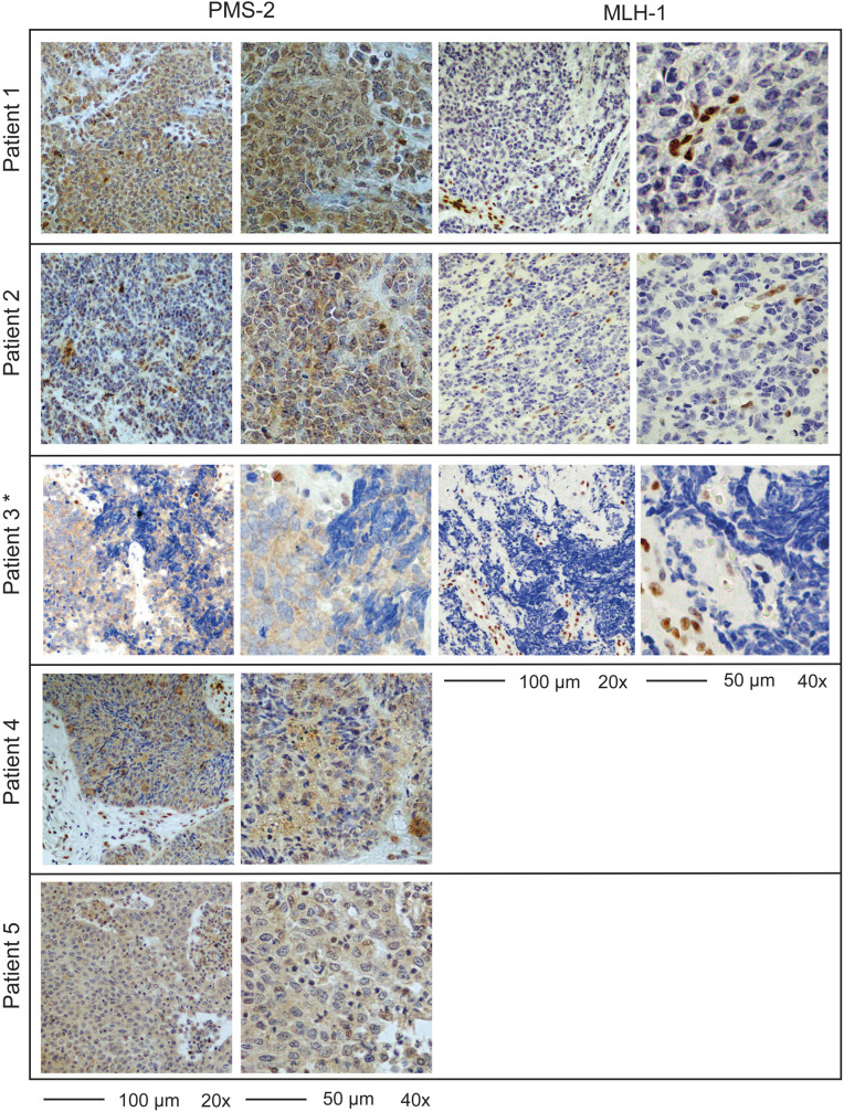
MMR-D was initially detected in 8 of 508 cervical tumors on tissue microarrays (1.6%). To reduce the risk of falsely classifying the tissue microarray tumor sections as MMR-D, full sections of these tumors were re-stained resulting in three tumors being reclassified as MMR-P. Within the remaining five MMR-D tumors, all had loss of PMS-2, and three had combined loss of MLH-1 and PMS-2 (Figure 1, online supplemental table 4). No significant association between MMR-D and disease-specific survival was found (p=0.17; online supplemental figure 1A). Rescoring using <10% as cut-off for MMR-D was also not prognostic (online supplemental figure 1B). The prevalence of MMR-D was highest within the rare and very aggressive neuroendocrine carcinomas. Three of ten (30%) neuroendocrine carcinomas were MMR-D, representing 60% of all MMR-D cases (online supplemental table 5). Proficient and deficient neuroendocrine carcinomas had similar 5-year disease-specific survival (online supplemental figure 2).
Figure 1.
Full section staining of PMS2 and MLH1 validates mismatch repair deficiency (MMR-D) in five patients. Representative images of full sections stained for MMR proteins with negative nuclear staining of tumor cells and positive staining of stroma or immune cells. Although some tumor cells exhibited cytoplasmic staining, no nuclear staining was found. This was confirmed by an expert pathologist. *Tissue block missing, photographs of tissue microarray (TMA).
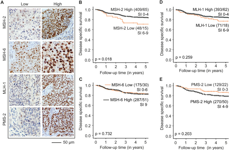
To evaluate the prognostic value of individual MMR proteins, differential expression of MSH-6, MSH-2, PMS-2, and MLH-1 was scored from tissue microarrays (n=508) using the staining index method (Figure 2A). Weighted kappa scores were calculated from independently scoring (MCB and MKH) of a subset of cases demonstrating overall good concordance: k=0.826 for MLH-1 (n=65), k=0.839 for MSH-2 (n=63), k=0.771 for MSH-6 (n=66), and k=0.703 for PMS-2 (n=60).
Figure 2.
Differential expression of MSH-2, but not MSH-6, MLH-1, and PMS-2, associates with disease-specific survival in cervical cancer. (A): Tumors were defined as ‘low’ or ‘high’ for each of the mismatch repair (MMR) proteins. Low and high MMR protein expression was defined based on staining index (SI) 0–9. (B–E) Disease-specific survival relative to MSH-2 (B), MSH-6 (C), MLH-1 (D), and PMS-2 (E) protein levels. Best cut-off for predicting disease-specific survival (Youden index) was applied (MSH-2: ‘low’ SI 0–4 and ‘high’ SI 6–9, MLH-1: ‘low’ SI 0–6 and ‘high’ SI 9, MSH-6: ‘low’ SI 0–6 and ‘high’ SI 9, PMS-2: ‘low’ SI 0–3 and ‘high’ SI 4–9). P-values are given by log-rank (Mantel–Cox) test. Numbers in parentheses indicate total number of patients/events.
High and low expression were defined from the Youden index as described in the Methods section. MSH-2 low (staining index 0–4) associated with poor disease-specific survival (p=0.018) (Figure 2B), whereas differential expression of MSH-6, MLH-1, or PMS-2 did not associate with survival (all p>0.05) (Figure 2C–E). MSH-2 protein level did not associate with any clinicopathological variables except for p53 status (online supplemental table 6). However, MSH-2 low independently predicted poor disease-specific survival after adjusting for age, FIGO stage, and histological type (HR 1.77, 95% CI 1.00 to 3.17, p=0.049) (Table 2). To account for possible interactions between age, FIGO stage, and histological type, interaction terms age*FIGO stage, FIGO stage*histological type, and histological type*age were explored. None influenced the effect. To evaluate the reproducibility and applicability of low MSH-2 tumor expression as a potential biomarker in future clinical settings, interobserver agreement for scoring MSH-2 low versus MSH-2 high was analyzed. Kappa agreement for categorizing MSH-2 in high or low was almost perfect (k=0.924).
Table 2.
Multivariate survival analysis of patients (n=457*) with high MSH-2 versus low MSH-2 staining index according to Cox’s proportional hazard regression method.
| Variable | Unadjusted | Adjusted | ||||
| HR | 95% CI | P-value | HR | 95% CI | P-value | |
| MSH-2 ≤4 | 1.94 | 1.11 to 3.41 | 0.020 | 1.77 | 1.00 to 3.13 | 0.049 |
| Age | 1.04 | 1.03 to 1.05 | <0.001 | 1.03 | 1.02 to 1.05 | <0.001 |
| FIGO-18 ≥IB2 | 7.60 | 4.60 to 12.57 | <0.001 | 12.82 | 4.03 to 40.80 | <0.001 |
| Histological type | ||||||
| Squamous cell carcinoma | <0.001 | |||||
| Adenocarcinoma | 0.71 | 0.38 to 1.33 | 0.286 | 1.41 | 0.70 to 2.85 | 0.34 |
| Other | 3.55 | 2.07 to 6.100 | <0.001 | 3.71 | 2.08 to 6.59 | <0.001 |
Other histological type: adenosquamous carcinoma, neuroendocrine carcinoma, and undifferentiated carcinoma.
*Only cases with available data for all variables in the multivariate analyses were included in the univariate analyses.
CI, confidence interval; FIGO, International Federation of Gynecology and Obstetrics; HR, hazard ratio.
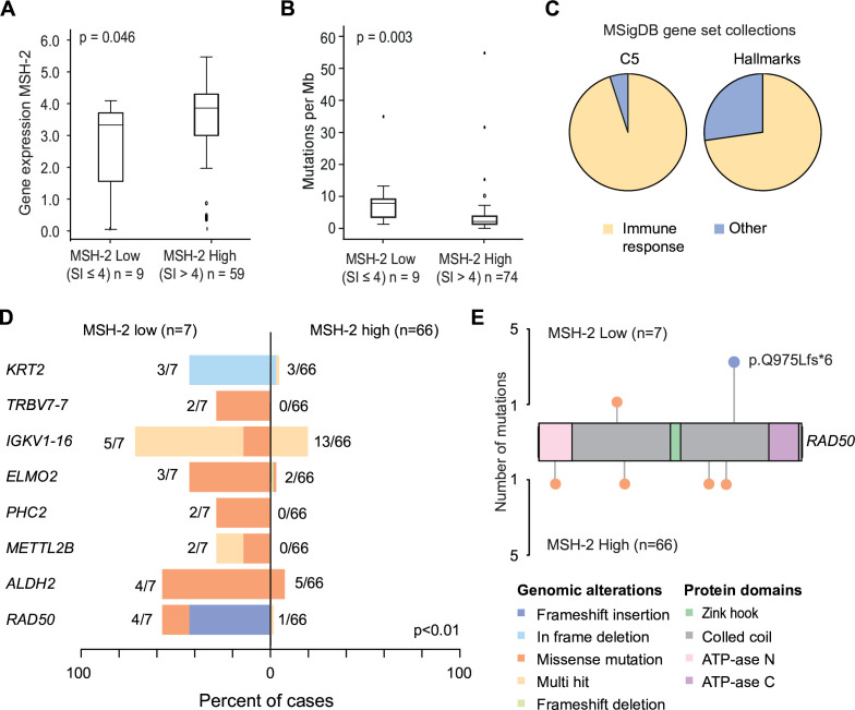
RNA sequencing and whole exome sequencing data were available for 72 and 75 patients, respectively. MSH-2 mRNA levels were significantly correlated with MSH-2 protein expression in overlapping samples (p=0.046, n=68) (Figure 3A). MSH-2 low tumors (n=9) had a higher mutational burden compared with MSH-2 high tumors (p=0.003, n=74) (Figure 3B). In gene set enrichment analyses, gene sets related to immune activation were enriched in MSH-2 low tumors. Among the 20 top-ranked gene sets in MSH-2 low tumors, 90% in the Ontology gene sets (C5) and 70% of Hallmark gene sets were related to immune response (GO ‘adaptive immune response’, ‘T-cell activation’ and Hallmark ‘interferon gamma response’, ‘inflammatory response’ (Figure 3C, online supplemental table 7). In MSH-2 high tumors, none of the enriched gene sets were related to immune response.
Figure 3.
Transcriptomic and genomic characterization reveal high mutational burden and immune cell signaling in MSH-2 low tumors. (A/B) Immunohistochemical protein expression (MSH-2: SI 0–4 and SI 6–9) in relation to corresponding MSH-2 mRNA expression (A) and mutational load (B). (C) Gene set enrichment analysis showing distribution of top 20 ranked enriched gene sets within the C5 and Hallmark gene set collections (MSigDB) for tumors with low MSH-2 expression. Other Hallmark gene sets: ‘coagulation’, ‘KRAS signaling’, ‘p53 pathway’. Other C5 gene sets: ‘cornification’. (D) Top eight differentially mutated genes of MSH-2 low tumors compared with MSH-2 high tumors. Multiple mutations per case per gene is indicated as ‘multi-hit’. Numbers indicate frequencies of patients with mutations and percentages are indicated on the bar below. (E) RAD50 co-lollipop plot illustrating type and location of mutations. Three of four RAD50 mutations in MSH-2 low tumors were frameshift mutations at location p.Q975Lfs*6. In MSH-2 high all four missense mutations were detected within the same patient tumor. P-values are given by Mann–Whitney U test. SI, staining index.
Mutational analyses grouped for MSH-2 low (n=7) versus MSH-2 high (n=66) revealed a significantly higher mutational frequency in MSH-2 low tumors (p<0.001) (Figure 3C). Eight genes (KRT2, TRBV7-7, IGKV1-16, ELMO2, PHC2, METTL2B, ALDH2, and RAD50) had significantly higher mutation frequency in MSH-2 low tumors (p>0.01) (Figure 3D). RAD50 mutations were previously correlated to survival in other cancer types23 24 and were therefore further explored, revealing a recurrent (n=4) frameshift insertion mutation in MSH-2 low tumors (Figure 3E).
Discussion
Summary of Main Results
Loss of MMR proteins is a common event in cancer and has been proposed as a marker for response to immune checkpoint inhibitors for several cancer types.8 In cervical cancer, the frequency and role of MMR loss is not clearly determined. We herein demonstrate that MMR loss is extremely rare in cervical cancer. Only 1% (n=5) of patients showed complete loss of one or more MMR proteins. When examining levels of MMR proteins individually, we found that MSH-2 low independently predicted poor outcome in cervical cancer and that MSH-2 low associated with higher mutational burden, RAD50 frameshift mutations, and an immune reactive transcriptome.
Results in the Context of Published Literature
Three of the five (60%) MMR-deficient tumors were of neuroendocrine histology which accounts for 33% (3/10) of all cervical neuroendocrine carcinomas included in this study. Other studies have found similar levels of MMR-D in cervical neuroendocrine carcinomas (30%, n=2025 and 33%, n=926). Neuroendocrine carcinomas are aggressive tumors that are challenging to characterize due to their rareness and, to date, no effective treatment regimens exist. As MMR-D is a successful biomarker for immune checkpoint inhibitor response,8 the present and previous studies could indicate that immune checkpoint inhibitors could be a valid treatment option for up to a third of these neuroendocrine carcinoma patients.
Regarding MMR-D levels, our findings contrast with previous studies where substantially higher MMR-D frequencies were found, generally ranging from 11% to 22% (n=102–186) of cervical cancers.27–29 This may be partly due to different sample size as well as the different detection and scoring methods used. Noh et al27 used both IHC and PCR to identify MMR-D (observing 11.3% MMR-D and/or MSI, without further specification) and Nijhuis et al29 used full sections with a lower cut-off (1%) for MMR loss (observing 22% MMR-D). However, none of these studies reported positive stroma cells as an internal control for MMR expression. Consistent with our findings, Bonneville et al analyzed whole exome sequencing data from The Cancer Genome Atlas (TCGA) cervical cancer cohort (n=305) and identified MSI in 2.6% of the tumors.30 We did not detect any significant differences in survival between patients with MMR-deficient and MMR-proficient tumors, which is in line with previous reports.27 30
This study reveals low MSH-2 as an independent marker for poor prognosis in cervical cancer. Furthermore, our study reveals that 80% of the MSH-2 low tumors had aberrant p53 staining. We have previously shown that aberrant p53 associates with poor survival.13 p53 is a major apoptotic regulator in cancer.31 MMR proteins, and specifically MSH-2, have also been found to influence apoptotic signaling by recognizing damaged DNA independently of their repair function.32 Thus, a combination of impaired MSH-2 and aberrant p53 may lead to increased mutagenesis and disrupted apoptotic signaling. We did indeed identify a significantly higher mutational burden in MSH-2 low versus high tumors, suggesting that a low level of MSH-2 may impair DNA MMR thereby inducing a higher mutational load, regardless of expression levels of the other MMR proteins. This is supported by a previous study demonstrating that a reduction of MSH-2 mRNA expression by 25% or more significantly decreases MMR efficacies,33 yet for MLH-1 a 50% decrease is necessary to find similar effects.33
A recurrent RAD50 p.Q975Lfs*6 frameshift insertion was detected in four of seven MSH-2 low tumors with available mutational data. Pan-cancer analysis has revealed that RAD50 is the most frequently altered gene in MSI tumors.34 Also, mutations in and/or low expression of RAD50 have been associated with poor survival in multiple cancer types.23 24 RAD50 is part of the DNA damage repair Mre11-Rad50-Nbs1 (MRN)-complex and depletion links to double-strand break accumulation and increased apoptosis independent of p53.35 Loss of RAD50 function may thus play a role in the tumorigenesis of these tumors.
Strengths and Weaknesses
In our study all negative TMA tumor specimens were re-stained using full sections and verified by a trained pathologist to ensure true negative lesions. This is to date the largest cervical cancer study of MMR-D protein levels and their relationship to patient outcomes. Still, despite the comprehensive and population-based nature of this study, the genomic and clinicopathological analyses were hampered by a limited number of MMR-D and MSH-2 low patients.
Implications for Practice and Future Research
To our knowledge, low MSH-2 has not previously been identified as an independent prognostic marker for poor survival. Whether MSH-2 could aid in providing a more complete risk profile within cervical cancer subgroups warrants further investigation. Mutational burden is one hallmark of immune checkpoint inhibitor response alongside immune activation,6 and in our genomic analyses we found higher mutational burden and enriched immune signaling in the MSH-2 low tumors. If our findings are confirmed in follow-up studies, cervical cancer patients with MSH-2 low tumors may be candidates for immune checkpoint inhibitor treatment. Furthermore, this is the first discovery of the RAD50 p.Q975Lfs*6 frameshift mutation in cervical cancer. A possible link between RAD50 and the MMR system should be further investigated in cervical cancer.
Conclusions
This comprehensive characterization of MMR protein expression confirms that MMR-D is rare in cervical cancer, yet in neuroendocrine tumors MMR-D is abundant. Low expression of the MMR protein MSH-2 is discovered as an independent poor prognosis marker and associates with high mutational burden, immune activation, and RAD50 frameshift mutations. Together these findings indicate that MSH-2 low and not MMR-D could be considered as an immune checkpoint inhibitor response marker in cervical cancer. As our results are impaired by a limited number of MSH-2 low tumors (n=48), the clinical applicability of MSH-2 as a prognostic and predictive marker warrants further validation in multi-institutional studies.
Acknowledgments
We sincerely thank Olivera Bozickovic, Kadri Madissoo, Jenny Margrethe Dugstad, Helene Midtun Flatekvål, and Bendik Nordanger for their excellent technical assistance. The Genomics Core Facility (GCF) at the University of Bergen, which is a part of the NorSeq consortium, provided services on whole exome sequencing. The GCF is supported in part by major grants from the Research Council of Norway (grant number 245979/F50) and Mohn Foundation (grant numbers BFS2017TMT04 and BFS2017TMT08).
Footnotes
Presented at: This work was presented as a poster at the European Society of Gynaecological Oncology (ESGO) Conference held in Berlin, Germany, October 27–30, 2022.
GContributors: Conception and design: CK, MKH. Development of methodology: MCB, CK, MKH. Acquisition of radiological data: ISH. Acquisition of pathological data: MCB, HE, BIB, MKH. Resources: TS, AIO, CK. Formal analysis and interpretation of data: MCB, HFB, TS, EAH, KW, ISH, JT, CK, MKH. Data access and verification: MCB, CK, MKH. Writing, review and revision of the article: all authors. Study supervision: JT, CK, MKH. Guarantors: MCB, MKH. The work reported in this article has been performed by the authors, unless clearly specified otherwise in the text.
Funding: This study was supported by the Trond Mohn Foundation (809119 and BFS2018TMT06); University of Bergen; the Research Council of Norway (311350, 326348, and 273280); Norwegian Cancer Society (190202 and 223283); and Western Norway Regional Health Authority (Helse Vest) (HV912263 and F-12542). None of the funders was involved in the study design, data collection, data analysis, interpretation, or writing of the manuscript.
Competing interests: None declared.
Provenance and peer review: Not commissioned; externally peer reviewed.
Supplemental material: This content has been supplied by the author(s). It has not been vetted by BMJ Publishing Group Limited (BMJ) and may not have been peer-reviewed. Any opinions or recommendations discussed are solely those of the author(s) and are not endorsed by BMJ. BMJ disclaims all liability and responsibility arising from any reliance placed on the content. Where the content includes any translated material, BMJ does not warrant the accuracy and reliability of the translations (including but not limited to local regulations, clinical guidelines, terminology, drug names and drug dosages), and is not responsible for any error and/or omissions arising from translation and adaptation or otherwise.
Data availability statement
Data are available upon reasonable request. In accordance with the journal’s guidelines, we will provide our data for independent analysis by a selected team by the Editorial Team for the purposes of additional data analysis or for the reproducibility of this study in other centers if such is requested.
Ethics statements
Patient consent for publication
Not applicable.
Ethics approval
This retrospective study involves human participants and was performed under Institutional Review Board (IRB)-approved protocols (2015/2333 and 2018/591 - Regional Committee for Medical Research Ethics, Western Norway (REK Vest)). Participants gave informed consent to participate in the study before taking part.
References
- 1. Bray F, Laversanne M, Sung H, et al. Global cancer statistics 2022: GLOBOCAN estimates of incidence and mortality worldwide for 36 cancers in 185 countries. CA Cancer J Clin 2024;74:229–63. 10.3322/caac.21834 [DOI] [PubMed] [Google Scholar]
- 2. Eskander RN, Tewari KS. Chemotherapy in the treatment of metastatic, persistent, and recurrent cervical cancer. Curr Opin Obstet Gynecol 2014;26:314–21. 10.1097/GCO.0000000000000042 [DOI] [PubMed] [Google Scholar]
- 3. Kumar L, Gupta S. Integrating chemotherapy in the management of cervical cancer: a critical appraisal. Oncology 2016;91 Suppl 1:8–17. 10.1159/000447576 [DOI] [PubMed] [Google Scholar]
- 4. Colombo N, Dubot C, Lorusso D, et al. Pembrolizumab for persistent, recurrent, or metastatic cervical cancer. N Engl J Med 2021;385:1856–67. 10.1056/NEJMoa2112435 [DOI] [PubMed] [Google Scholar]
- 5. Chung HC, Ros W, Delord J-P, et al. Efficacy and safety of pembrolizumab in previously treated advanced cervical cancer: results from the phase II KEYNOTE-158 study. J Clin Oncol 2019;37:1470–8. 10.1200/JCO.18.01265 [DOI] [PubMed] [Google Scholar]
- 6. Bai R, Lv Z, Xu D, et al. Predictive biomarkers for cancer immunotherapy with immune checkpoint inhibitors. Biomark Res 2020;8:34. 10.1186/s40364-020-00209-0 [DOI] [PMC free article] [PubMed] [Google Scholar]
- 7. Vikas P, Messersmith H, Compton C, et al. Mismatch repair and microsatellite instability testing for immune checkpoint inhibitor therapy: ASCO endorsement of College of American Pathologists guideline. J Clin Oncol 2023;41:1943–8. 10.1200/JCO.22.02462 [DOI] [PubMed] [Google Scholar]
- 8. Le DT, Durham JN, Smith KN, et al. Mismatch repair deficiency predicts response of solid tumors to PD-1 blockade. Science 2017;357:409–13. 10.1126/science.aan6733 [DOI] [PMC free article] [PubMed] [Google Scholar]
- 9. Vilar E, Gruber SB. Microsatellite instability in colorectal cancer—the stable evidence. Nat Rev Clin Oncol 2010;7:153–62. 10.1038/nrclinonc.2009.237 [DOI] [PMC free article] [PubMed] [Google Scholar]
- 10. Kandoth C, Schultz N, Cherniack AD, et al. Integrated genomic characterization of endometrial carcinoma. Nature 2013;497:67–73. 10.1038/nature12113 [DOI] [PMC free article] [PubMed] [Google Scholar]
- 11. Berg HF, Engerud H, Myrvold M, et al. Mismatch repair markers in preoperative and operative endometrial cancer samples; expression concordance and prognostic value. Br J Cancer 2023;128:647–55. 10.1038/s41416-022-02063-3 [DOI] [PMC free article] [PubMed] [Google Scholar]
- 12. Pereira CS, Oliveira M de, Barros LO, et al. Low expression of MSH2 DNA repair protein is associated with poor prognosis in head and neck squamous cell carcinoma. J Appl Oral Sci 2013;21:416–21. 10.1590/1679-775720130206 [DOI] [PMC free article] [PubMed] [Google Scholar]
- 13. Halle MK, Ojesina AI, Engerud H, et al. Clinicopathologic and molecular markers in cervical carcinoma: a prospective cohort study. Am J Obstet Gynecol 2017;217:432. 10.1016/j.ajog.2017.05.068 [DOI] [PubMed] [Google Scholar]
- 14. Halle MK, Sødal M, Forsse D, et al. A 10-gene prognostic signature points to LIMCH1 and HLA-DQB1 as important players in aggressive cervical cancer disease. Br J Cancer 2021;124:1690–8. 10.1038/s41416-021-01305-0 [DOI] [PMC free article] [PubMed] [Google Scholar]
- 15. Bhatla N, Aoki D, Sharma DN, et al. Cancer of the cervix uteri: 2021 update. Int J Gynaecol Obstet 2021;155 Suppl 1:28–44. 10.1002/ijgo.13865 [DOI] [PMC free article] [PubMed] [Google Scholar]
- 16. Zhang Y, Shu YM, Wang SF, et al. Stabilization of mismatch repair gene PMS2 by glycogen synthase kinase 3Β is implicated in the treatment of cervical carcinoma. BMC Cancer 2010;10:58. 10.1186/1471-2407-10-58 [DOI] [PMC free article] [PubMed] [Google Scholar]
- 17. Ojesina AI, Lichtenstein L, Freeman SS, et al. Landscape of genomic alterations in cervical carcinomas. Nature 2014;506:371–5. 10.1038/nature12881 [DOI] [PMC free article] [PubMed] [Google Scholar]
- 18. Dysvik B, Jonassen I. J-Express: exploring gene expression data using Java. Bioinformatics 2001;17:369–70. 10.1093/bioinformatics/17.4.369 [DOI] [PubMed] [Google Scholar]
- 19. Ashburner M, Ball CA, Blake JA, et al. Gene ontology: tool for the unification of biology. Nat Genet 2000;25:25–9. 10.1038/75556 [DOI] [PMC free article] [PubMed] [Google Scholar]
- 20. Liberzon A, Birger C, Thorvaldsdóttir H, et al. The Molecular Signatures Database (MSigDB) hallmark gene set collection. Cell Syst 2015;1:417–25. 10.1016/j.cels.2015.12.004 [DOI] [PMC free article] [PubMed] [Google Scholar]
- 21. Mayakonda A, Lin DC, Assenov Y, et al. Maftools: efficient and comprehensive analysis of somatic variants in cancer. Genome Res 2018;28:1747–56. 10.1101/gr.239244.118 [DOI] [PMC free article] [PubMed] [Google Scholar]
- 22. Halle MK, Sundaresan A, Zhang J, et al. Genomic alterations associated with mutational signatures, DNA damage repair and chromatin remodeling pathways in cervical carcinoma. NPJ Genom Med 2021;6:82. 10.1038/s41525-021-00244-2 [DOI] [PMC free article] [PubMed] [Google Scholar]
- 23. Fan C, Zhang J, Ouyang T, et al. RAD50 germline mutations are associated with poor survival in BRCA1/2-negative breast cancer patients. Int J Cancer 2018;143:1935–42. 10.1002/ijc.31579 [DOI] [PubMed] [Google Scholar]
- 24. Ho V, Chung L, Singh A, et al. Early postoperative low expression of RAD50 in rectal cancer patients associates with disease-free survival. Cancers (Basel) 2017;9:163. 10.3390/cancers9120163 [DOI] [PMC free article] [PubMed] [Google Scholar]
- 25. Ji X, Sui L, Song K, et al. PD‐L1, PARP1, and MMRs as potential therapeutic biomarkers for neuroendocrine cervical cancer. Cancer Med 2021;10:4743–51. 10.1002/cam4.4034 [DOI] [PMC free article] [PubMed] [Google Scholar]
- 26. Morgan S, Slodkowska E, Parra‐Herran C, et al. PD ‐L1, RB 1 and mismatch repair protein immunohistochemical expression in neuroendocrine carcinoma, small cell type, of the uterine cervix. Histopathology 2019;74:997–1004. 10.1111/his.13825 [DOI] [PubMed] [Google Scholar]
- 27. Noh JJ, Kim MK, Choi MC, et al. Frequency of mismatch repair deficiency/high microsatellite instability and its role as a predictive biomarker of response to immune checkpoint inhibitors in gynecologic cancers. Cancer Res Treat 2022;54:1200–8. 10.4143/crt.2021.828 [DOI] [PMC free article] [PubMed] [Google Scholar]
- 28. Köster F, Schröer A, Fischer D, et al. Correlation of DNA mismatch repair protein hMSS2 immunohistochemistry with P53 and apoptosis in cervical carcinoma. Anticancer Res 2007;27:63–8. [PubMed] [Google Scholar]
- 29. Nijhuis ER, Nijman HW, Oien KA, et al. Loss of MSH2 protein expression is a risk factor in early stage cervical cancer. J Clin Pathol 2007;60:824–30. 10.1136/jcp.2005.036038 [DOI] [PMC free article] [PubMed] [Google Scholar]
- 30. Bonneville R, Krook MA, Kautto EA, et al. Landscape of microsatellite instability across 39 cancer types. JCO Precis Oncol 2017;2017. 10.1200/PO.17.00073 [DOI] [PMC free article] [PubMed] [Google Scholar]
- 31. Aubrey BJ, Kelly GL, Janic A, et al. How does P53 induce apoptosis and how does this relate to P53-mediated tumour suppression? Cell Death Differ 2018;25:104–13. 10.1038/cdd.2017.169 [DOI] [PMC free article] [PubMed] [Google Scholar]
- 32. Salsbury FR, Clodfelter JE, Gentry MB, et al. The molecular mechanism of DNA damage recognition by MutS homologs and its consequences for cell death response. Nucleic Acids Res 2006;34:2173–85. 10.1093/nar/gkl238 [DOI] [PMC free article] [PubMed] [Google Scholar]
- 33. Kansikas M, Kasela M, Kantelinen J, et al. Assessing how reduced expression levels of the mismatch repair genes MLH1, MSH2, and MSH6 affect repair efficiency. Hum Mutat 2014;35:1123–7. 10.1002/humu.22605 [DOI] [PubMed] [Google Scholar]
- 34. Shi P, Scott MA, Ghosh B, et al. Synapse microarray identification of small molecules that enhance synaptogenesis. Nat Commun 2011;2:510. 10.1038/ncomms1518 [DOI] [PMC free article] [PubMed] [Google Scholar]
- 35. Fagan-Solis KD, Simpson DA, Kumar RJ, et al. A P53-independent DNA damage response suppresses oncogenic proliferation and genome instability. Cell Rep 2020;30:1385–99. 10.1016/j.celrep.2020.01.020 [DOI] [PMC free article] [PubMed] [Google Scholar]
Associated Data
This section collects any data citations, data availability statements, or supplementary materials included in this article.
Supplementary Materials
ijgc-2024-005377supp001.pdf (392.1KB, pdf)
Data Availability Statement
Data are available upon reasonable request. In accordance with the journal’s guidelines, we will provide our data for independent analysis by a selected team by the Editorial Team for the purposes of additional data analysis or for the reproducibility of this study in other centers if such is requested.


