Abstract
Vesicular stomatitis virus (VSV) is the prototype of the Rhabdoviridae and contains nonsegmented negative-sense RNA as its genome. The 11-kb genome encodes five genes in the order 3′-N-P-M-G-L-5′, and transcription is obligatorily sequential from the single 3′ promoter. As a result, genes at promoter-proximal positions are transcribed at higher levels than those at promoter-distal positions. Previous work demonstrated that moving the gene encoding the nucleocapsid protein N to successively more promoter-distal positions resulted in stepwise attenuation of replication and lethality for mice. In the present study we investigated whether moving the gene for the attachment glycoprotein G, which encodes the major neutralizing epitopes, from its fourth position up to first in the gene order would increase G protein expression in cells and alter the immune response in inoculated animals. In addition to moving the G gene alone, we also constructed viruses having both the G and N genes rearranged. This produced three variant viruses having the orders 3′-G-N-P-M-L-5′ (G1N2), 3′-P-M-G-N-L-5′ (G3N4), and 3′-G-P-M-N-L-5′ (G1N4), respectively. These viruses differed from one another and from wild-type virus in their levels of gene expression and replication in cell culture. The viruses also differed in their pathogenesis, immunogenicity, and level of protection of mice against challenge with wild-type VSV. Translocation of the G gene altered the kinetics and level of the antibody response in mice, and simultaneous reduction of N protein expression reduced replication and lethality for animals. These studies demonstrate that gene rearrangement can be exploited to design nonsegmented negative-sense RNA viruses that have characteristics desirable in candidates for live attenuated vaccines.
The order Mononegavirales is composed of four families, Rhabdoviridae, Paramyxoviridae, Filoviridae, and Bornaviridae. The viruses in these families contain a single strand of nonsegmented negative-sense RNA and are responsible for a wide range of significant diseases in fish, plants, and animals (31). Viral gene expression is controlled at the level of transcription by the order of the genes on the genome relative to the single 3′ promoter. Gene order throughout the Mononegavirales is highly conserved; genes encoding products required in stoichiometric amounts for replication are always at or near the 3′ end of the genome, while those whose products are needed in catalytic amounts are more promoter distal.
Vesicular stomatitis virus (VSV) is the prototypic virus of the Rhabdoviridae. Its 11-kb genome has five genes which encode the five structural proteins of the virus: the nucleocapsid protein, N, which is required in stoichiometric amounts for encapsidation of the replicated RNA; the phosphoprotein, P, which is a cofactor of the RNA-dependent RNA polymerase, L; the matrix protein, M; and the attachment glycoprotein, G. The order of genes in the genome is 3′-N-P-M-G-L-5′, and previous work has shown that expression is obligatorily sequential from a single 3′ promoter (1, 3). Due to attenuation at each gene junction (15), the 3′-most genes are transcribed more abundantly than those which are more promoter distal (15, 41). In nature, VSV infects a wide range of animals, of which horses, cattle, and domestic swine are the most economically important. Infection results in the appearance of lesions around the mouth, hooves, and udder teats; while seldom fatal, it leads to a loss in meat and milk production along with the expense of quarantine and vaccination (13). There are two main VSV serotypes, Indiana and New Jersey; while these viruses are endemic in Central and South American countries, outbreaks also occur within the United States. The most recent outbreak in the United States occurred in 1997 in horses (40) and was of the Indiana serotype, while previous cases identified in 1995 (6) and 1982 to 1983 (43) were of the New Jersey serotype. The ease with which these viruses are transmitted, and the similarity of their symptoms to those caused by foot-and-mouth disease virus in cattle and domestic swine, makes VSV a pathogen of concern to sections of the agriculture industry.
In recent work we rearranged the three internal genes of VSV, P, M, and G, to all combinations and recovered viruses having the six possible gene orders (4). In every case, the relative levels of protein expression in cells were in concordance with the distance of the corresponding genes from the 3′ promoter. These studies demonstrated that viruses with rearranged genes were viable and that expression of a gene could be modulated in a predictable manner according to its positioning relative to the 3′ promoter.
VSV genome RNA replication is proportional to the amount of N protein synthesized (2, 27), and so in a second study we examined the effects of moving the N gene from its promoter-proximal site to successively promoter-distal positions. N transcription was reduced in a systematic manner (45), and this resulted in correspondingly reduced N protein synthesis, a stepwise reduction in the ability of these viruses to replicate in cell culture, and an attenuation of their lethality for mice. Mice that survived inoculation with the rearranged variants were protected from challenge with a lethal dose of wild-type (wt) virus. Therefore, despite attenuation of replication in cell culture and lethality in vivo, these viruses stimulated a protective immune response. These findings show that despite the highly conserved gene order of VSV, gene rearrangement was not lethal to the virus and could be used to generate viruses with predictable degrees of attenuation. Translocation of an essential gene of a nonsegmented negative-strand virus provides a new approach for engineering viruses with different degrees of replication ability by design rather than traditional empirical methods.
In the present study we examined whether we could increase the expression of a promoter-distal gene, the gene that encodes the attachment glycoprotein G, by moving it to a promoter-proximal site. The attachment glycoprotein contains the neutralizing site (17, 21, 35, 36), along with helper T-cell (7) and cytotoxic T-cell epitopes (19, 37), of the virus. It has been shown that stimulation of neutralizing antibodies is an important feature in the protection of mice (12, 20) and the natural host against infection with VSV (22). To determine if an increase in the production of G protein in cells during infection could elicit a greater protective immune response, we engineered changes into an infectious cDNA clone of the VSV genome and recovered two novel viruses in which the glycoprotein gene was moved from its normal fourth position to the first position in the gene order. One virus, referred to as G1N2, had the gene order 3′-G-N-P-M-L-5′; the gene order of the second, G1N4, was 3′-G-P-M-N-L-5′. The in vitro and in vivo characteristics of these viruses were assessed and compared to those of viruses having the gene orders 3′-P-M-G-N-L-5′ (G3N4) and 3′-N-P-M-G-L-5′ (N1G4), the latter being the wt gene order. Differences were observed in the replication of these viruses in cell culture, their lethality in mice, the kinetics and levels of antibody production after inoculation, and their ability to protect mice against challenge with a lethal dose of wt VSV.
MATERIALS AND METHODS
Viruses and cells.
The San Juan isolate of the Indiana serotype of VSV provided the template for all of the cDNA clone of the VSV genome except the G protein gene, which was derived from the Orsay isolate of VSV-Indiana (46). All viruses were recovered from cDNAs in baby hamster kidney (BHK-21) cells. BHK-21 cells were also used for single-step growth assays and radioisotopic labeling of viral RNAs and proteins. Plaque assays were performed on the African green monkey kidney cell line Vero-76.
Plasmid construction and recovery of infectious virus.
The construction of a full-length cDNA clone of the VSV genome and its use for the recovery of infectious virus have been described elsewhere (46). This infectious clone was manipulated using methods which allowed the genome to be assembled with the genes in different orders (4). No other changes were made in the genome except for a single nucleotide in the intergenic region downstream of the P gene. This change, from 3′-CA-5′ to 3′-GA-5′, has little effect on transcription (5).
To recover infectious viruses from the rearranged cDNA clones, BHK-21 cells were infected with a recombinant vaccinia virus expressing the T7 RNA polymerase (vTF7-3) (11). One hour later, the cells were transfected with the rearranged VSV cDNA along with three plasmids, which expressed the N, P, and L proteins required for encapsidation and replication of the antigenomic RNA (4, 45, 46). Infectious viruses were harvested from the supernatant medium and amplified in BHK-21 cells at a low multiplicity of infection (MOI) to avoid formation of defective interfering particles and in the presence of cytosine arabinoside (25 μg/ml) to suppress the replication of vaccinia virus. Supernatant medium was filtered through 0.2-μm-pore-size filters, and the virus was banded on 15 to 45% sucrose velocity gradients to separate it from any remaining vTF7-3. The gene orders of the recovered viruses were confirmed by amplifying the rearranged portions of the genomes using reverse transcription and PCR followed by restriction enzyme analysis.
Analysis of viral protein synthesis.
Viral protein synthesis directed by each of the variant viruses was measured in BHK-21 cells infected at an MOI of 50, with actinomycin D (5 μg/ml) added at 3 h postinfection. At 5 h postinfection, the cells were washed and incubated in methionine-free medium for 30 min. Cells were exposed to [35S]methionine (30 μCi/ml; specific activity, 10.2 mCi/ml) for 1 h. Cell monolayers were harvested directly into gel loading buffer; after normalizing for equal counts per minute, the viral proteins were separated on 10% polyacrylamide gels using a low bisacrylamide-to-acrylamide ratio to separate the P and N proteins. Viral proteins were quantitated using a phosphorimager, and their molar ratios were calculated.
Analysis of virion proteins.
To assess the quantity of each of the proteins in the mature virions, BHK-21 cells were infected at an MOI of 5. After 2 h, the cells were washed and incubated in methionine-free medium for 30 min. Cells were labeled with [35S]methionine (50 μCi/ml; specific activity, 10.2 mCi/ml) overnight, with unlabeled methionine added to 10% of normal medium level. Supernatant fluid was collected, cell debris was removed by centrifugation, and virus was collected by centrifugation through 10% sucrose. After normalizing the counts per minute, the viral pellet was resuspended in gel loading buffer and virion proteins were separated on a 10% polyacrylamide gel. Virion proteins were quantitated using a phosphorimager, and the molar ratios were determined.
Single-cycle virus replication.
BHK-21 cells were infected at an MOI of 3. After 1 h of adsorption, the inoculum was removed and the monolayer was washed twice. Fresh medium was added, and the cells were incubated at 37°C. Supernatant fluids were harvested at indicated intervals over a 30-h period, and viral yields were determined by plaque assay on Vero-76 cells.
Lethality in mice.
Male Swiss Webster mice, 3 to 4 weeks old, were purchased from Taconic Farms, Germantown, N.Y., and housed under BL2 containment conditions. Groups of six mice were lightly anesthetized with ketamine-xylazine and inoculated intranasally with 10-μl aliquots of serial 10-fold viral dilutions of the individual viruses in Dulbecco's modified Eagle medium. Control animals were given a similar volume of DMEM. Animals were observed, and each group was weighed daily. The 50% lethal dose (LD50) for each virus was calculated by the method of Reed and Muench (33).
Determination of serum antibody levels and neutralization titers.
After virus inoculation, blood was collected at weekly intervals from groups of two to four animals. Serum samples were pooled and heated to 57°C for 40 min to inactivate complement. Cell monolayers infected with wt VSV (N1G4) and uninfected BHK-21 cells were lysed in detergent buffer (1% NP-40, 0.4% sodium deoxycholate, 66 mM EDTA, 10 mM Tris-HCl [pH 7.4]) and used as antigen in a direct enzyme-linked immunosorbent assay (ELISA). Samples were serially diluted and detected using goat anti-mouse immunoglobulin conjugated to horseradish peroxidase. The optical density (OD) was read at 450 nm, and the antibody titers were calculated by linear regression analysis of a plot of OD versus serum dilution. The endpoint titers (log10) were deduced at an OD 1.5 times that of the preimmune samples. Serum neutralizing antibody titers on day of challenge were determined by a standard plaque reduction assay on Vero-76 cells, and the titer was expressed as the reciprocal of the dilution giving 50% neutralization.
Protection of mice from wt challenge.
Mice were immunized intranasally with doses of each virus ranging from 1 to 10,000 PFU in DMEM. Twenty-one days postinoculation, groups of mice that received nonlethal doses of each of the variant viruses were challenged intranasally with 5.4 × 10⁶ PFU of N1G4 (wt) virus. Challenged animals and controls were monitored for a further 21 days. At weekly intervals, blood was collected by tail bleeds for serum antibody titrations.
RESULTS
Generation and recovery of rearranged viruses.
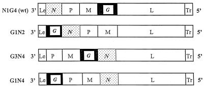
In previous work we generated a full-length cDNA clone of the genome of VSV from which infectious virus was recovered (46). We subsequently developed a way to rearrange the order of the genes at the cDNA level without introducing other changes into the genome by using remote cutting restriction enzymes to cleave within the conserved sequences at the 3′ and 5′ ends of each gene (4). This strategy allowed us to recover viruses from rearranged cDNAs in which the N gene had been moved to successively promoter-distal positions in order to downregulate its expression (45). In the present work, we used this approach to generate cDNA clones in which the G gene was moved from its normal position of fourth in the gene order to the first, most promoter-proximal position to determine if doing so would increase its expression. Two new gene rearrangements were generated: one in which the G gene was moved to first in the gene order and the remaining four genes were left undisturbed to generate the order 3′-G-N-P-M-L-5′ (G1N2), and the second in which the positions of the G and N genes were exchanged to generate the order 3′-G-P-M-N-L-5′ (G1N4) (Fig. 1). These cDNAs were transfected into cells as described previously (45), and virus was recovered in both cases. The recovered viruses were designated G1N2 and G1N4, respectively, according to the positions of the N and G genes in the rearranged gene order. The properties of these viruses were examined in comparison to a virus derived from a cDNA clone created using the same gene rearrangement process to regenerate the wt gene order (N1G4) and a virus with the gene order 3′-P-M-G-N-L-5′ (G3N4) (45).
FIG. 1.
Gene orders of N1G4 (wt), G1N2, G3N4, and G1N4. Le, leader; Tr, trailer.
Effect of gene rearrangement on viral protein expression.
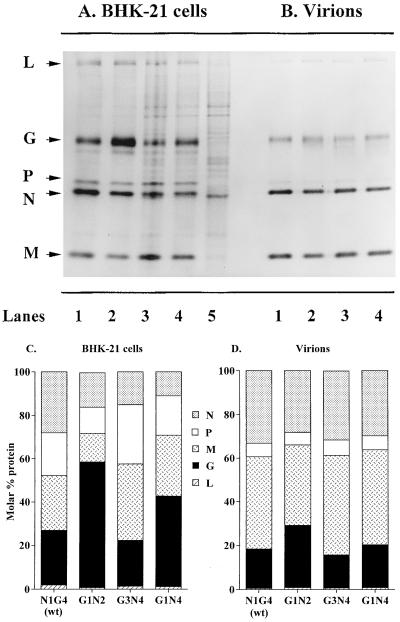
BHK-21 cells were infected with viruses with rearranged genomes, and the relative levels of viral protein synthesis were examined by labeling for 1 h with [35S]methionine at 5 h postinfection. Total cellular proteins were resolved by sodium dodecyl sulfate-polyacrylamide gel electrophoresis (SDS-PAGE) and visualized by autoradiography. A typical gel is shown in Fig. 2A. Infection with wt VSV and the rearranged variants resulted in rapid inhibition of host protein synthesis which allowed the viral N, P, M, G, and L proteins to be detected directly. Synthesis of G protein was significantly increased relative to the other viral proteins in cells infected with G1N2 and G1N4 viruses (Fig. 2A, lanes 2 and 4) compared to wt (N1G4)-infected cells (Fig. 2A, lane 1).
FIG. 2.
Synthesis of viral proteins in BHK-21 cells infected with the variant viruses. (A) BHK-21 cells were infected at an MOI of 50 and incubated at 37°C for 5 h in the presence of actinomycin D (5 μg/ml) for the final 2 h. Infected cells were then starved for methionine for 30 min and exposed to medium containing [35S]methionine (30 μCi/ml) for 1 h. Total infected cell proteins were analyzed by SDS-PAGE. (B) Virions were isolated from supernatant fluids of BHK-21 cells infected at an MOI of 5 and exposed to [35S]methionine (50 μCi/ml) from 2.5 to 12 h postinfection. Virus particles were purified by centrifugation through 10% sucrose, and their protein contents were determined by SDS-PAGE. Viral proteins shown in panels A and B were quantitated by phosphorimaging and expressed as molar percentages of each viral protein in infected BHK-21 cells (C) or purified virions (D). Data shown are averages from two independent experiments. Lanes: 1, N1G4 (wt); 2, G1N2; 3, G3N4; 4, G1N4; 5, uninfected cells.
Proteins were quantitated by phosphorimaging. The molar percentage of G protein synthesized during a 1-h labeling period was 2.3-fold higher in G1N2-infected cells and 1.7-fold higher in G1N4-infected cells than in cells infected with wt virus. Similarly, translocation of the N gene from its promoter-proximal position to a more distal position in viruses G1N2, G3N4, and G1N4 decreased the rate of N protein synthesis (Fig. 2C).
The protein contents of purified virus particles were also examined to determine if changes in protein synthesis in cells affected protein assembly into virions. BHK-21 cells were infected with each of the viruses and labeled with [35S]methionine overnight, and virions were harvested from supernatant fluids and separated from cell debris by centrifugation through 10% sucrose. Analysis of the virion proteins by SDS-PAGE (Fig. 2B) showed no gross differences in the relative protein contents. Phosphorimager quantitation confirmed that despite the altered relative levels of protein synthesis in infected cells, the amounts of proteins in virions were similar to that of wild-type virus except for virus G1N2, in which the level of G was 1.6-fold higher than in the wt virus or other rearranged viruses (Fig. 2D). This observation is being investigated further.
Virus replication in cell culture.
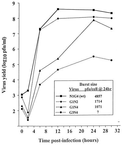
Replication of the rearranged viruses under single-step growth conditions was examined in cultured BHK-21 cells infected at an MOI of 3 followed by incubation at 37°C. Supernatant fluids were harvested at various times, and the virus yields were measured by plaque assay. As described previously (45), translocation of the N gene away from the promoter-proximal position resulted in stepwise reduction of replication as the gene was moved further from the first position. Movement of N to the second position (G1N2) decreased replication 3-fold, whereas moving N to the fourth position (G3N4) reduced replication as much as 1,000-fold (Fig. 3). However, the two viruses with N in the fourth position (G3N4 and G1N4) replicated to different levels under single-step growth conditions. We attribute this to the fact that the molar ratio of N to P, which is critical for optimal replication, was less perturbed in G1N4 than G3N4. Measurement of the intracellular rates of protein synthesis 5 h after infection showed molar ratios of N to P of 1:1.6 in cells infected with G1N4 and 1:1.8 in G3N4-infected cells (Fig. 2C). An N:P molar ratio of between 1:0.5 and 1:1 is optimal for replication (14, 29, 45), as shown by the N:P ratios of 1:0.7 in wt-infected cells and 1:0.8 for cells infected with G1N2. Both the wt virus and G1N2 have N directly followed by P in the gene order (Fig. 1). Too much or too little P relative to N decreases replication significantly (14, 26, 28, 29); thus, in cells infected with G3N4, not only is N limiting, but also the molar ratio of N:P is more than twice the optimal value. The kinetics of replication of G3N4 and G1N4 were delayed in comparison to wt and G1N2. Single-step growth of G3N4 and G1N4 was not complete until 24 h postinfection, compared to 12 h for N1G4 and G1N2. It is unlikely that the overabundance of G in the infected cell was responsible for this delay in replication since G1N2 showed no delay in replication relative to wild-type virus.
FIG. 3.
Single-step growth analysis. Viruses were assayed for the ability to replicate by single-step growth in BHK-21 cells at 37°C. Cells were infected at an MOI of 3, and samples of the supernatant medium were harvested at the indicated time points. Samples were titrated in duplicate by plaque assay on Vero-76 cells. Average virus yields per cell were determined at 24 h postinfection (inset).
Lethality in mice.
Young mice provide a sensitive animal model for the study of neuropathology caused by VSV and its mutants, (24, 38, 42), and inoculation of mice with wt VSV via the intranasal route results in fatal encephalitis. We compared the pathogenesis of the rearranged variant viruses to that of wt (N1G4) virus after intranasal inoculation in 3 to 4 week-old Swiss-Webster mice. The doses that constituted LD50s were 100, 50, >100,000, and 19,000 PFU of N1G4, G1N2, G3N4, and G1N4, respectively.
All of the viruses were lethal for mice if given in sufficiently high doses, although the doses of G3N4 administered in these experiments did not reach the LD50 seen previously (45). In general, the position of the N gene, the N:P ratio, and the resulting level of virus replication were major determinants of lethality. Viruses in which the N gene was moved away from the promoter required greatly increased doses to constitute an LD50. These results confirmed our previous observations with viruses N1 to N4 in which the N gene was moved sequentially (45). However, the results presented here show that for viruses with N in the fourth position (G3N4 and G1N4), both the replication ability and the LD50 also were affected by the position of the G gene.
The LD50s reported here are expressed in terms of the viral titers on Vero-76 cells. These titers are about 10-fold higher than titers on BSC-40 cells, as reported in our previous publications (4, 45). We changed cell lines because we discovered that rearranging the gene order of VSV could affect the interactions of the variant viruses with the interferon system (reference 23 and unpublished results). BSC-40 cells are competent to produce interferon after infection, while Vero cells are not (10). Therefore, changing to Vero cells circumvented possible differences in interferon induction or sensitivity. Studies of the interactions of the rearranged viruses with the interferon system are in progress.
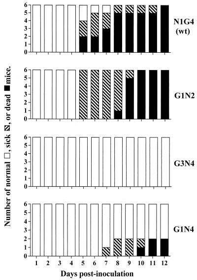
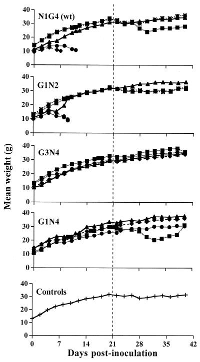
The first symptoms of sickness (a haunched posture and hind-limb paralysis) appeared 5 days after inoculation with both N1G4 and G1N2 viruses, although the first deaths occurred earlier in animals inoculated with N1G4 (Fig. 4). The viruses with N in the fourth position induced symptoms more slowly; at a dose of 1,000 PFU per mouse, G3N4 caused neither morbidity nor mortality, as observed before (45). In an attempt to detect subclinical signs of sickness, the groups of mice were weighed daily throughout the study period (Fig. 5). However, whereas the mice that showed symptoms invariably lost weight and died, those that showed no symptoms showed no weight differences from uninoculated control animals (Fig. 5). Similar results were observed after challenge of the inoculated mice with wt virus; all animals that developed symptoms subsequently died, and those that did not develop symptoms also showed no weight loss.
FIG. 4.
Pathogenesis in mice. The viruses shown were administered intranasally to groups of six mice at a dose of 1,000 PFU per mouse, and the animals were monitored daily for signs of morbidity and mortality. No further changes occurred after day 12.
FIG. 5.
Average weights of mice inoculated with the rearranged viruses. Groups of six mice were inoculated intranasally with serial 10-fold dilutions of N1G4 (wt), G1N2, G3N4, or G1N4 ranging from 10,000 to 1 PFU/animal. Control mice received inoculation medium alone. The vertical dotted line indicates day of challenge with 5.4 × 10⁶ PFU of wt virus per mouse. For each group, all living animals were weighed together and the average weight was determined. ⧫, 10,000 PFU; ●, 1,000 PFU; ▴, 100 PFU; ■, 10 PFU; ∗, 1 PFU; +, medium.
Serum antibody.
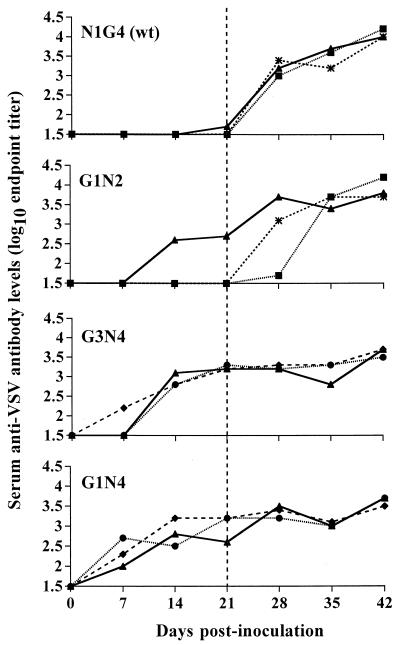
To assess the effect of inoculation of viruses with rearranged G genes on the humoral immune response, mice were inoculated intranasally with serial 10-fold dilutions of each of the variant viruses. Blood was collected at weekly intervals by tail bleed, and the level of serum antibody was determined by ELISA. Since survival of the inoculation was a prerequisite for this experiment, only doses at or below the LD50 were used. Translocation of the G gene changed the kinetics and magnitude of the antibody response (Fig. 6). Mice inoculated with wt virus made barely detectable levels of antibody within 21 days, whereas animals that received 100 PFU of G1N2 and G1N4, respectively, had significant antibody titers by 14 and 7 days postinoculation. This accelerated and enhanced response can be seen most clearly by comparing the mice that received 100 PFU (Fig. 6). The results demonstrate that translocation of the G gene from the fourth to the first position enhanced the humoral immune response to VSV. Mice given G1N4 synthesized antibody earlier and at higher levels than those given G3N4. This further confirmed the observation that putting the G gene first in the gene order increased the immunogenicity of VSV.
FIG. 6.
Kinetics of antibody production in response to inoculation with the rearranged and wt viruses. Groups of six mice were inoculated intranasally with serial 10-fold dilutions of N1G4 (wt), G1N2, G3N4, or G1N4 ranging from 10,000 to 1 PFU/animal. Control mice received inoculation medium only. The vertical dotted line indicates the day of challenge with 5.4 × 10⁶ PFU of wt virus per mouse. Serum was collected by tail bleeds from two to four animals at weekly intervals, the serum samples were pooled, and the level of antibody raised against VSV was determined by titration on detergent-lysed VSV-infected cell antigen in an ELISA. Antibody levels are expressed as log10 titers. ⧫, 10,000 PFU; ●, 1,000 PFU; ▴, 100 PFU; ■, 10 PFU; ∗, 1 PFU.
Twenty-one days postinoculation, the mice were challenged with 5.4 × 10⁶ PFU of wt VSV. A rapid increase in antibody titer was observed in animals given either N1G4 or G1N2, although there was no further rise in the already high titers that had been achieved prior to challenge in mice inoculated with G3N4 or G1N4.
Neutralizing antibody titer after inoculation.
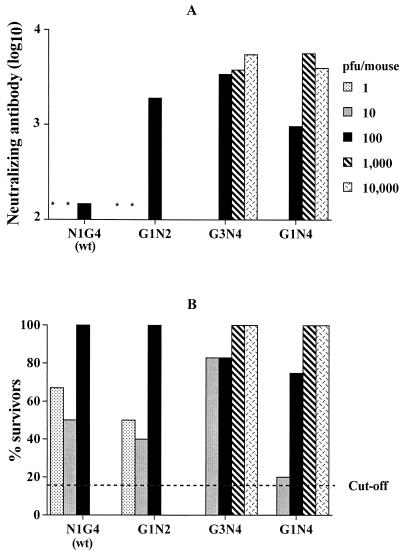
The level of neutralizing antibody in the serum at the time of challenge was measured. In mice and cattle, neutralizing antibodies are an important element in protection against VSV infection (12, 20, 22). On the day of challenge, mice were bled and serum samples were assayed for the ability to neutralize wt VSV in a standard plaque reduction assay on Vero-76 cells. The reciprocal of the highest dilution that gave a 50% reduction of plaque numbers was calculated to determine the neutralizing titers of the sera.
All of the viruses with rearranged genomes elicited serum neutralizing antibody in mice (Fig. 7A). Neutralizing antibody was not detected at a dose of 1 or 10 PFU/mouse of either N1G4 or G1N2, but both viruses elicited detectable titers at doses of 100 PFU, the response to G1N2 being 10-fold higher than that to wt virus. Thus, for N1G4 and G1N2, the level of neutralizing antibody did not correlate with virus replication in cell culture, where the wt virus replicated two- to threefold more abundantly than G1N2 (Fig. 3). This conclusion was reinforced by the response to G3N4 and G1N4, which elicited approximately 10-fold-higher titers than the wt virus despite greatly reduced replication potential.
FIG. 7.
Groups of six mice were inoculated intranasally with serial 10-fold dilutions of N1G4 (wt), G1N2, G3N4, or G1N4 ranging from 10,000 to 1 PFU/animal. Control mice received inoculation medium only. (A) Neutralizing antibody levels as measured in serum samples on the day of challenge by plaque reduction assay. Neutralizing antibody levels are expressed as the reciprocal of the highest dilution giving a 50% reduction in wt virus plaques on Vero-76 cells. Asterisks denote that sera from animals given 1 or 10 PFU of N1G4 or G1N2 virus had background levels of neutralizing antibody. (B) Ability to survive intranasal challenge by 5.4 × 10⁶ PFU of N1G4 virus. The dotted line shows the lethality of this dose (83%) in unvaccinated, age-matched, control animals 21 days after challenge.
In summary, viruses which overexpressed G and underexpressed N in infected cells yielded increased levels of neutralizing antibody compared to wt virus (N1G4) following intranasal inoculation. The combination of overexpressing G and underexpressing N combined this enhanced immunogenicity with virus attenuation, which allowed the administration of higher doses that elicited correspondingly higher titers of neutralizing antibodies. Moreover, because of the lower lethality of these viruses, 100 times more virus could be administered without detriment, and under these conditions they elicited up to 100-fold more neutralizing antibody than could be attained in response to wt virus.
Protection of mice from challenge.
These results establish that nonpathogenic doses of the viruses that overexpressed G protein could elicit significant humoral immune responses in mice. To see whether immunization with the rearranged viruses could confer protection against VSV disease, animals that survived inoculation with each of the rearranged viruses were challenged after 21 days with 5.4 × 10⁶ PFU of wt virus. This dose was sufficient to kill 83% of the uninoculated, age-matched, control group of animals.
All of the viruses with rearranged genomes conferred protection, the level of which varied with the dose of inoculum (Fig. 7B). The levels of protection elicited by N1G4 and G1N2 were alike, reflecting the comparable levels of replication and lethality of these viruses described previously (Fig. 3; see also “Lethality in mice,” above). Similarly, the protection conferred by G1N4 resembled that of G3N4. By 21 days postinoculation, both viruses elicited solid immunity at doses of 1,000 PFU per mouse. Importantly, these fully protective doses were 20- to 100-fold less than the corresponding LD50s. This emphasizes the conclusion that gene rearrangement is an effective method to systematically change the phenotype of VSV to optimize the properties required of a live attenuated vaccine.
DISCUSSION
The recovery of infectious viruses from cDNA clones of the Mononegavirales permits experimental manipulation of the viral genome (8, 9). Gene expression in these viruses is controlled at the transcriptional level by the order of the genes relative to the single promoter at the 3′ end of the viral genome (15, 41). We developed a method to rearrange the order of the genes without introducing other changes into the genome (4, 45). Gene rearrangement altered the relative levels of synthesis of the viral proteins, as expected, and produced infectious viruses having a variety of different phenotypes. In previous work we showed that moving the N gene from its promoter-proximal position to more distal positions resulted in a stepwise decrease in N protein synthesis, viral RNA replication, infectious virus production, and lethality of the variant viruses for mice (45). The present studies examined the consequences of moving the G protein gene, which encodes the major neutralizing epitopes of the virus, from its promoter-distal position to first in the gene order. As predicted by our previous work, expression of G protein in infected cells was significantly increased when its gene was moved from the fourth to the first position. However, the protein content of the purified virus particles was largely unaffected by changes in the viral gene order. Any differences that may exist were at the limits of the quantitation methods used in this study and will require the application of more precise techniques.
The overexpression of G protein by these viruses allowed us to explore whether they elicited an altered humoral immune response in animals. The data in Fig. 6 show that at an inoculum dose of 100 PFU, antibody was produced more quickly and at higher levels in animals infected with the viruses with G moved to a promoter-proximal position compared to the wt virus. Doses higher than 100 PFU could not be assayed with the N1G4 (wt) and G1N2 viruses because of their lethality. At the dose of 100 PFU, viruses G1N2, G3N4, and G1N4 all elicited higher antibody titers more rapidly than N1G4. The reduced lethality of the G1N4 and G3N4 viruses allowed higher doses to be administered, and in these cases antibody levels increased more rapidly than at lower doses.
The observation that all three viruses which had G moved closer to the promoter elicited an enhanced humoral immune response in mice has implications for our understanding of protective immunity in this system. Although we do not know the relative levels of replication of the variant inocula in the cells that are most relevant for induction of the immune response, it seems likely that they mirrored, at least qualitatively, the relative levels of replication seen in cell culture. If this is the case, G1N2, G3N4, and G1N4 expressed higher levels of G protein per inoculated mouse only during the first round of replication. After that, the more robust replication of the wt virus should have more than compensated for its weaker G protein synthesis. Yet at the same inoculated dose of 100 PFU per mouse, the variant viruses elicited an enhanced and accelerated humoral immune response compared to the wt-inoculated animals.
The results suggest that the kinetics and magnitude of the humoral immune response become established very early in infection. Either there is a short temporal window during which the scale of the immune response becomes established irrevocably or the immune response to VSV infection is somehow determined by the level of G protein synthesis per infected cell rather than by the aggregate immunogenic load. A similar conclusion is suggested by the efficacy of vaccines using recombinant canarypox vectors under conditions where they are unable to replicate (25). Robust synthesis of antigen by a highly attenuated vector appears to be an effective vaccine strategy that warrants further exploration.
Studies outside the scope of this report are under way to investigate the types and subtypes of antibodies produced by the variant viruses and to investigate T-cell responses and localization of the viruses following infection. It is difficult to compare our present work with that of others on the pathogenesis of VSV in mice and the complexities of the immune response because of significant differences in the ages and strains of the experimental animals as well as in the routes of inoculation and challenge (12, 20, 36). In the work presented here, we used the Swiss-Webster mouse model of VSV established by Sabin and Olitsky (38) and Wagner (42), and all experiments were controlled relative to the action of the wt virus.
In agreement with previous findings, we observed that the position of the N gene and the level of N protein expression correlated with efficiency of replication because the N protein is required in stoichiometric amounts for genomic RNA replication. The wt virus N1G4 replicated to the highest titers, followed by G1N2 virus and finally G1N4 and G3N4, which replicated least well. G1N4, however, replicated significantly better than G3N4, although they both had the N gene in the fourth position. Both of these viruses showed delayed replication kinetics, as might be expected if the formation of progeny virus was limited by the supply of N protein.
It is known that the relative levels of N and P proteins, in addition to the absolute amount of N protein, are critical for efficient replication (14, 29). One function of P protein is to maintain N in a soluble state such that it is able to support encapsidation of newly replicated RNA (14, 44). Consistent with this, G1N2 virus, while having reduced N protein expression (Fig. 2A and C), had the N and P genes in the same relative order as the N1G4 (wt) (Fig. 1). Accordingly, G1N2 expressed the N and P proteins at about the same relative rates as wt virus, 1:0.8 and 1:0.7, respectively. In agreement with this, G1N2 replicated only slightly less than N1G4. Further to this point, although G1N4 and G3N4 both had N in the fourth position, G1N4 replicated substantially better than G3N4 (Fig. 3). The ratio between the rates of synthesis of N and P proteins was disparate from the wt value in both of these viruses. However, G3N4, which had P in the first position, had an N:P ratio in infected cells of 1:1.8, whereas the N:P ratio in cells infected with G1N4, where P was in the second position, was 1:1.6, closer to that of wt virus. There was also a difference between these two viruses in the rates of G protein expression, and it is possible that increased levels of G protein provided an advantage for replication of G1N4.
The reduced lethality of the viruses with gene rearrangements was also consistent with our previous work showing that attenuation of lethality in mice correlated with reduced replication capacity. Reduced replication, in turn, was related to the overall expression levels of N protein and the N:P ratio as discussed above. Obviously any gene rearrangement which brings the G gene to the first position will displace the N gene from its wt position and therefore decrease N protein expression. It will also alter the molar ratios of proteins whose gene positions relative to one another are changed by the rearrangement in question. Both types of change would be expected to alter replication efficiency and lethality. As noted in Results, the viruses which replicated best, wt and G1N2, required only 50 to 100 PFU to constitute an LD50, whereas 200 and 1,000 times more G1N4 and G3N4, respectively, were required for an LD50. The data presented here show that rearrangement of genes allowed the manipulation of two important aspects of the viral phenotype, lethality and the stimulation of neutralizing antibody. By reducing N protein expression and altering the N:P ratio, it was possible to decrease replication potential and lethality for animals; by increasing G protein expression, it was possible to alter the kinetics and level of antibody synthesis.
These results expand on our earlier demonstration that gene rearrangement can be used to generate viruses with novel, beneficial phenotypes. This approach provides the ability to alter the phenotype in a stepwise manner to achieve a desired level of attenuation or to alter the expression of a particular gene. It allows the level of attenuation and immunogenicity to be modulated independently and systematically, exactly what is needed to generate and manipulate live attenuated vaccine candidates. This approach should be applicable to other members of the Mononegavirales, all of which have a common mechanism for the control of gene expression via obligatorily sequential transcription originating from a single 3′ promoter. Furthermore, viruses of the Mononegavirales have not been found to undergo homologous recombination; therefore, changes made to the gene order should be irreversible by natural processes (30, 32). Several foreign genes have been expressed from VSV (16, 18, 34, 39), and in one study mice were protected against the corresponding pathogen (34). These properties make this virus an excellent candidate in which to generate future vaccines directed against VSV itself or against other pathogens. Studies designed to evaluate the pathogenesis and immunogenicity of the G1N2, G3N4, and G1N4 viruses in a natural host are under way.
ACKNOWLEDGMENTS
This work was supported by Public Health Service grants R37 AI 12464 to G.W.W. and R37 AI 18270 to L.A.B.
We thank Claire Hankin for experimental assistance and the members of the Wertz and Ball laboratories for constructive comments.
REFERENCES
- 1.Abraham G, Banerjee A K. Sequential transcription of the genes of vesicular stomatitis virus. Proc Natl Acad Sci USA. 1976;73:1504–1508. doi: 10.1073/pnas.73.5.1504. [DOI] [PMC free article] [PubMed] [Google Scholar]
- 2.Arnheiter H, Davis N L, Wertz G, Schubert M, Lazzarini R A. Role of the nucleocapsid protein in regulating vesicular stomatitis virus RNA synthesis. Cell. 1985;41:259–267. doi: 10.1016/0092-8674(85)90079-0. [DOI] [PubMed] [Google Scholar]
- 3.Ball L A, White C N. Order of transcription of genes of vesicular stomatitis virus. Proc Natl Acad Sci USA. 1976;73:442–446. doi: 10.1073/pnas.73.2.442. [DOI] [PMC free article] [PubMed] [Google Scholar]
- 4.Ball L A, Pringle C P, Flanagan B, Perepelitsa V P, Wertz G W. Phenotypic consequences of rearranging the P, M, and G genes of vesicular stomatitis virus. J Virol. 1999;73:4705–4712. doi: 10.1128/jvi.73.6.4705-4712.1999. [DOI] [PMC free article] [PubMed] [Google Scholar]
- 5.Barr J N, Whelan S P J, Wertz G W. Role of the intergenic dinucleotide in vesicular stomatitis virus RNA transcription. J Virol. 1997;71:1794–1801. doi: 10.1128/jvi.71.3.1794-1801.1997. [DOI] [PMC free article] [PubMed] [Google Scholar]
- 6.Bridges V E, McCluskey B J, Salman M D, Hurd H S, Dick J. Review of the 1995 vesicular stomatitis outbreak in the western United States. J Am Vet Med Assoc. 1997;211:556–560. [PubMed] [Google Scholar]
- 7.Burkhart C, Freer G, Castro R, Adorini L, Wiesmüller K-H, Zinkernagel R M, Hengartner H. Characterization of T-helper epitopes of the glycoprotein of vesicular stomatitis virus. J Virol. 1994;68:1573–1580. doi: 10.1128/jvi.68.3.1573-1580.1994. [DOI] [PMC free article] [PubMed] [Google Scholar]
- 8.Conzelmann K-K. Genetic manipulation of non-segmented negative-strand RNA viruses. J Gen Virol. 1996;77:381–389. doi: 10.1099/0022-1317-77-3-381. [DOI] [PubMed] [Google Scholar]
- 9.Conzelmann K-K. Nonsegmented negative-strand RNA viruses: genetics and manipulation of viral genomes. Annu Rev Genet. 1998;32:123–162. doi: 10.1146/annurev.genet.32.1.123. [DOI] [PubMed] [Google Scholar]
- 10.Desmyter J, Melnick J L, Rawls W E. Defectiveness of interferon production and of rubella virus interference in a line of African green monkey kidney cells (Vero) J Virol. 1968;2:955–961. doi: 10.1128/jvi.2.10.955-961.1968. [DOI] [PMC free article] [PubMed] [Google Scholar]
- 11.Fuerst T R, Niles E G, Studier F W, Moss B. Eukaryotic transient-expression system based on recombinant vaccinia virus that synthesizes bacteriophage T7 RNA polymerase. Proc Natl Acad Sci USA. 1986;83:8122–8126. doi: 10.1073/pnas.83.21.8122. [DOI] [PMC free article] [PubMed] [Google Scholar]
- 12.Gobet R, Cerny A, Rüedi E, Hengartner H, Zinkernagel R M. The role of antibodies in natural and acquired resistance of mice to vesicular stomatitis virus. Exp Cell Biol. 1988;56:175–180. doi: 10.1159/000163477. [DOI] [PubMed] [Google Scholar]
- 13.Hayek A M, McCluskey B J, Chavez G T, Salman M D. Financial impact of the 1995 outbreak of vesicular stomatitis on 16 beef ranches in Colorado. J Am Vet Med Assoc. 1998;212:820–823. [PubMed] [Google Scholar]
- 14.Howard M, Wertz G. Vesicular stomatitis virus RNA replication: a role for the NS protein. J Gen Virol. 1989;70:2683–2694. doi: 10.1099/0022-1317-70-10-2683. [DOI] [PubMed] [Google Scholar]
- 15.Iverson L E, Rose J K. Localization attenuation and discontinuous synthesis during vesicular stomatitis virus transcription. Cell. 1981;23:477–484. doi: 10.1016/0092-8674(81)90143-4. [DOI] [PubMed] [Google Scholar]
- 16.Kahn J S, Schnell M J, Buonocore L, Rose J K. Recombinant vesicular stomatitis virus expressing the respiratory syncytial virus (RSV) glycoproteins: RSV fusion protein can mediate infection and cell fusion. Virology. 1999;254:81–91. doi: 10.1006/viro.1998.9535. [DOI] [PubMed] [Google Scholar]
- 17.Kelley J R, Emerson S U, Wagner R R. The glycoprotein of vesicular stomatitis virus is the antigen that gives rise to and reacts with neutralizing antibody. J Virol. 1972;10:1231–1235. doi: 10.1128/jvi.10.6.1231-1235.1972. [DOI] [PMC free article] [PubMed] [Google Scholar]
- 18.Kretzschmur E, Buonocore L, Schnell M J, Rose J K. High-efficiency incorporation of functional influenza virus glycoproteins into recombinant vesicular stomatitis viruses. J Virol. 1997;71:5982–5989. doi: 10.1128/jvi.71.8.5982-5989.1997. [DOI] [PMC free article] [PubMed] [Google Scholar]
- 19.Kündig T M, Castelmur I, Bachmann M F, Abraham D, Binder D, Hengartner H, Zinkernagel R M. Fewer protective cytotoxic T-cell epitopes than T-helper cell epitopes on vesicular stomatitis virus. J Virol. 1993;67:3680–3683. doi: 10.1128/jvi.67.6.3680-3683.1993. [DOI] [PMC free article] [PubMed] [Google Scholar]
- 20.Lefrancois L. Protection against lethal viral infection by neutralizing and nonneutralizing monoclonal antibodies: distinct mechanisms of action in vivo. J Virol. 1984;51:208–214. doi: 10.1128/jvi.51.1.208-214.1984. [DOI] [PMC free article] [PubMed] [Google Scholar]
- 21.Lefrancois L, Lyles D S. The interaction of antibody with the major surface glycoprotein of vesicular stomatitis virus. I. Analysis of neutralizing epitopes with monoclonal antibodies. Virology. 1982;121:157–167. [PubMed] [Google Scholar]
- 22.Mackett M, Yilma T, Rose J K, Moss B. Vaccinia virus recombinants: expression of VSV genes and protective immunization of mice and cattle. Science. 1985;227:433–435. doi: 10.1126/science.2981435. [DOI] [PubMed] [Google Scholar]
- 23.Marcus P I, Sekellick M J, Ball L A, Wertz G W. Phenotypic variation in the interferon-inducing capacity of vesicular stomatitis virus with rearranged genomes. J Interferon Res. 1999;19:S101. [Google Scholar]
- 24.Miyoshi K, Harter D H, Hsu K C. Neuropathological and immunofluorescence studies of experimental vesicular stomatitis encephalitis in mice. J Neuropathol Exp Neurol. 1971;30:266–277. doi: 10.1097/00005072-197104000-00008. [DOI] [PubMed] [Google Scholar]
- 25.Paoletti E. Applications of poxvirus vectors to vaccination: an update. Proc Natl Acad Sci USA. 1996;93:11349–11353. doi: 10.1073/pnas.93.21.11349. [DOI] [PMC free article] [PubMed] [Google Scholar]
- 26.Pattnaik A K, Wertz G W. Replication and amplification of defective interfering particle RNAs of vesicular stomatitis virus in cells expressing viral proteins from vectors containing cloned cDNAs. J Virol. 1990;64:2948–2957. doi: 10.1128/jvi.64.6.2948-2957.1990. [DOI] [PMC free article] [PubMed] [Google Scholar]
- 27.Patton J T, Davis N L, Wertz G W. N protein alone satisfies the requirement for protein synthesis during RNA replication of vesicular stomatitis virus. J Virol. 1984;49:303–309. doi: 10.1128/jvi.49.2.303-309.1984. [DOI] [PMC free article] [PubMed] [Google Scholar]
- 28.Peluso R W. Kinetic, quantitative, and functional analysis of multiple forms of the vesicular stomatitis virus nucleocapsid protein in infected cells. J Virol. 1988;62:2799–2807. doi: 10.1128/jvi.62.8.2799-2807.1988. [DOI] [PMC free article] [PubMed] [Google Scholar]
- 29.Peluso R W, Moyer S A. Viral proteins required for the in vitro replication of vesicular stomatitis virus defective interfering particle genome RNA. Virology. 1988;162:369–376. doi: 10.1016/0042-6822(88)90477-1. [DOI] [PubMed] [Google Scholar]
- 30.Pringle C R. The genetics of paramyxoviruses. In: Kingsbury D, editor. The paramyxoviruses. New York, N.Y: Plenum Press; 1991. pp. 1–39. [Google Scholar]
- 31.Pringle C R, Easton A J. Monopartite negative strand RNA genomes. Semin Virol. 1997;8:49–57. [Google Scholar]
- 32.Pringle C R, Devine V, Wilkie M, Preston C M, Dolan A, McGeoch D J. Enhanced mutability associated with a temperature-sensitive mutant of vesicular stomatitis virus. J Virol. 1981;39:377–389. doi: 10.1128/jvi.39.2.377-389.1981. [DOI] [PMC free article] [PubMed] [Google Scholar]
- 33.Reed E J, Muench H. A simple method of estimating fifty percent endpoints. Am J Hyg. 1938;27:493–497. [Google Scholar]
- 34.Roberts A, Buonocore L, Price R, Foreman J, Rose J K. Attenuated vesicular stomatitis viruses as vaccine vectors. J Virol. 1999;73:3723–3732. doi: 10.1128/jvi.73.5.3723-3732.1999. [DOI] [PMC free article] [PubMed] [Google Scholar]
- 35.Roost H-P, Haag A, Burkhart C, Zinkernagel R M, Hengartner H. Mapping of the dominant neutralizing antigenic sites of a virus using infected cells. J Immunol Methods. 1996;189:233–242. doi: 10.1016/0022-1759(95)00252-9. [DOI] [PubMed] [Google Scholar]
- 36.Roost H-P, Bachmann M F, Haag A, Kalinke U, Pliska V, Hengartner H, Zinkernagel R M. Early high-affinity neutralizing anti-viral IgG responses without further overall improvements of affinity. Proc Natl Acad Sci USA. 1995;92:1257–1261. doi: 10.1073/pnas.92.5.1257. [DOI] [PMC free article] [PubMed] [Google Scholar]
- 37.Rosenthal K L, Oldstone M B, Hengartner H, Zinkernagel R M. Specificity of in vitro cytotoxic T cell clones directed against vesicular stomatitis virus. J Immunol. 1983;131:475–478. [PubMed] [Google Scholar]
- 38.Sabin A, Olitsky P. Influence of host factors on neuroinvasiveness of vesicular stomatitis virus. J Exp Med. 1938;67:201–227. doi: 10.1084/jem.67.2.229. [DOI] [PMC free article] [PubMed] [Google Scholar]
- 39.Schnell M J, Buonocore L, Kretzschmar E, Johnson E, Rose J K. Foreign glycoproteins expressed from recombinant vesicular stomatitis virus are incorporated efficiently into virus particles. Proc Natl Acad Sci USA. 1996;93:11359–11365. doi: 10.1073/pnas.93.21.11359. [DOI] [PMC free article] [PubMed] [Google Scholar]
- 40.USDA Animal and Plant Health Inspection Service-Veterinary Services, Centers for Epidemiology and Animal Health. 1997 vesicular stomatitis virus (VSV) outbreak in the United States. DxMonitor Anim Health Rep. 1997;Fall:1–2. [Google Scholar]
- 41.Villarreal L P, Breindl M, Holland J J. Determination of molar ratios of vesicular stomatitis virus induced RNA species in BHK21 cells. Biochemistry. 1976;15:1663–1667. doi: 10.1021/bi00653a012. [DOI] [PubMed] [Google Scholar]
- 42.Wagner R R. Pathogenicity and immunogenicity for mice of temperature-sensitive mutants of vesicular stomatitis virus. Infect Immun. 1974;10:309–315. doi: 10.1128/iai.10.2.309-315.1974. [DOI] [PMC free article] [PubMed] [Google Scholar]
- 43.Webb P A, Monath T P, Reif J S, Smith G C, Kemp G E, Lazuick J S, Walton T E. Epizootic vesicular stomatitis in Colorado, 1982: epidemiological studies along the northern Colorado front range. Am J Trop Med Hyg. 1987;36:183–188. doi: 10.4269/ajtmh.1987.36.183. [DOI] [PubMed] [Google Scholar]
- 44.Wertz G W, Howard M B, Davis N, Patton J. The switch from transcription to replication of a negative-strand RNA virus. Cold Spring Harbor Symp Quant Biol. 1987;52:367–371. doi: 10.1101/sqb.1987.052.01.042. [DOI] [PubMed] [Google Scholar]
- 45.Wertz G W, Perepelitsa V P, Ball L A. Gene rearrangement attenuates expression and lethality of a nonsegmented negative strand RNA virus. Proc Natl Acad Sci USA. 1998;95:3501–3506. doi: 10.1073/pnas.95.7.3501. [DOI] [PMC free article] [PubMed] [Google Scholar]
- 46.Whelan S P J, Ball L A, Barr J N, Wertz G T W. Recovery of infectious vesicular stomatitis virus entirely from cDNA clones. Proc Natl Acad Sci USA. 1995;92:8388–8392. doi: 10.1073/pnas.92.18.8388. [DOI] [PMC free article] [PubMed] [Google Scholar]


