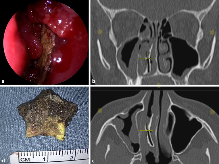
A 16-year-old girl presented to the otorhinolaryngology outpatient clinic with a 10-year history of unilateral nasal airway obstruction and foul-smelling nasal discharge. The anterior rhinoscopy showed a large irregular mass in the nasal cavity consisting of extensive crusting and inflammatory granulation tissue (Fig. 1a). Computed tomography of the paranasal sinuses revealed a vertically situated hyperdense foreign body with surrounding soft tissue density in the right common middle meatus (Fig. 1b,c). Endoscopic sinus surgery with the patient under general anesthesia was further scheduled in order to remove the foreign body. During surgery, a yellowish-black foreign body in the shape of a star was successfully removed in toto (Fig. 1d). The respective foreign body was a foam star that has triggered a local inflammatory reaction and had developed into a rhinolith after some time. The star was probably put in the nose by the patient herself many years ago as a child, which she could not remember. The further postoperative follow-up remained free of complications and the patient no longer reports any sinus symptoms.
Fig. 1.
a Anterior rhinoscopy. b, c Computed tomography imaging of the paranasal sinuses (b coronal plane, c axial plane). The crosswire indicates a vertically situated hyperdense foreign body with surrounding soft tissue density in the right common middle meatus. d Removed foam star from the nasal cavity
Acknowledgments
Funding
No funding was received for conducting this study.
Funding
Open access funding provided by Medical University of Graz.
Declarations
Conflict of interest
A. Andrianakis and P.V. Tomazic declare that they have no competing interests.
Ethical standards
For this article no studies with human participants or animals were performed by any of the authors. All studies performed were in accordance with the ethical standards indicated. All information that could be used to potentially identify the patient was removed. Informed consent for publication was obtained from the patient.
Footnotes
Publisher’s Note
Springer Nature remains neutral with regard to jurisdictional claims in published maps and institutional affiliations.


