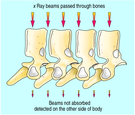
• In bone mineral density (BMD) scanning, the energy of x ray beams that are passed through bones is absorbed, and what is not absorbed is detected on the other side of the body. The more dense the bones (from greater mineral content), the more energy is absorbed, and the less energy detected. 
• The radiation energy per pixel (“picture element”) is detected and converted into an “areal density” measured in g/cm. The number of pixels in the area is summed, then the amount of bone in each pixel is calculated. This allows a bone density to be calculated for the specific bone in question.
• Basic BMD measurement requires the patient to be “on the table” for about 2 minutes, undressed to light clothing, and with no metal piercings. Navel piercings can be a problem, because they cover the vertebra L4, which is a common site to scan.
• In modern scanners, x ray beams of two different energies are used (dual x ray absorptiometry), giving a dose up to that of a chest x ray. If the whole skeleton is being examined a higher dose is received because of the greater area exposed (about 1.5 chest x rays). Two energies allow an estimate to be made for soft tissue absorption separately from that of the bones.
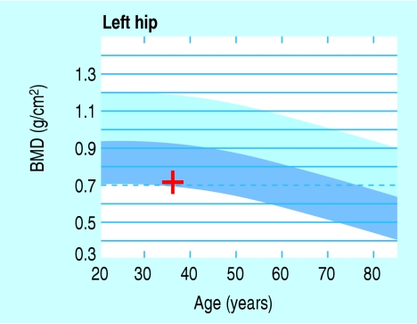
• Values for bone density may be quoted as g/cm2 or converted into values related to the average female (or male) peak bone mass or to the bone mass related to the patient's age. These are T scores and Z scores, and involve the following calculations.
•
•
In women, the peak lumbar spine density is maximal in the fourth decade of life. Osteoporosis is diagnosed if the T score is over -2.5 according to the WHO (1994).1 Z scores may be used to monitor long term follow up of treatment.
• Calibrations for average bone densities are often based on a database of the upper femur called the NHANES database.2
• The x rays are directed anterior-posterior or vice versa, depending on the instrument. There is little deflection of beam to the operator, but with faster fan beam machines deflection is greater. Fan beam and pencil beam machines can scan laterally around the side of a patient, which is useful for measuring the bone density of the lumbar spine.
• In elderly people, where the lowest ribs often cover L2 and the iliac crests cover L4, L3 may be the only vertebra that can be scanned in the lateral position.
• For femoral examinations, the measurement most frequently used at present is known as the “total upper femur.” This is an easy measurement for technicians to reproduce, and includes the femoral neck, trochanteric region, and inter trochanteric region.
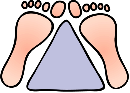
• Femurs are examined with patients lying flat on their back with toes together and a heel separation of 23 cm. 
• The spine is examined with patients lying on their backs and their knees flexed over a block at right angles to flatten out the lumbar lordosis. The maximum weight on a scanning table is 136 kg.
• Future machines will be able to send the data directly from a scan into the clinic for diagnostic or treatment decisions to be made. 
Supplementary Material
Figure.

Bone density scan of upper femur
Figure.
Value of femoral neck bone density scan plotted on chart showing mean and limits of +2 and –2 standard deviations of a health population
Figure.

Ankylosing spondylitis diagnosed from bone density scan with calcification of longitudinal ligament. The diagnosis was later verified by radiographs of the spine sacroiliac joints
References
- 1.Kanis JA, Melton LJ, 3rd, Christiansen C, Johnston CC, Khaltaev N. The diagnosis of osteoporosis. J Bone Miner Res. 1994;9:1137–1141. doi: 10.1002/jbmr.5650090802. [DOI] [PubMed] [Google Scholar]
- 2.Looker AC, Wahner HW, Dunn WL, Calvo MS, Harris TB, Heyse SP, et al. Updated data on proximal femur bone mineral levels of US adults. Osteoporos Int. 1998;8:468–489. doi: 10.1007/s001980050093. [DOI] [PubMed] [Google Scholar]
Associated Data
This section collects any data citations, data availability statements, or supplementary materials included in this article.


