


Abstract
Background
Periodontitis and peri‐implant diseases are chronic inflammatory conditions occurring in the mouth. Left untreated, periodontitis progressively destroys the tooth‐supporting apparatus. Peri‐implant diseases occur in tissues around dental implants and are characterised by inflammation in the peri‐implant mucosa and subsequent progressive loss of supporting bone.
Treatment aims to clean the pockets around teeth or dental implants and prevent damage to surrounding soft tissue and bone, including improvement of oral hygiene, risk factor control (e.g. encouraging cessation of smoking) and surgical interventions. The key aspect of standard non‐surgical treatment is the removal of the subgingival biofilm using subgingival instrumentation (SI) (also called scaling and root planing). Antimicrobial photodynamic therapy (aPDT) can be used an adjunctive treatment to SI. It uses light energy to kill micro‐organisms that have been treated with a light‐absorbing photosensitising agent immediately prior to aPDT.
Objectives
To assess the effects of SI with adjunctive aPDT versus SI alone or with placebo aPDT for periodontitis and peri‐implant diseases in adults.
Search methods
We searched the Cochrane Oral Health Trials Register, CENTRAL, MEDLINE, Embase, two other databases and two trials registers up to 14 February 2024.
Selection criteria
We included randomised controlled trials (RCTs) (both parallel‐group and split‐mouth design) in participants with a clinical diagnosis of periodontitis, peri‐implantitis or peri‐implant disease. We compared the adjunctive use of antimicrobial photodynamic therapy (aPDT), in which aPDT was given after subgingival or submucosal instrumentation (SI), versus SI alone or a combination of SI and a placebo aPDT given during the active or supportive phase of therapy.
Data collection and analysis
We used standard Cochrane methodological procedures, and we used GRADE to assess the certainty of the evidence. We prioritised six outcomes and the measure of change from baseline to six months after treatment: probing pocket depth (PPD), bleeding on probing (BOP), clinical attachment level (CAL), gingival recession (REC), pocket closure and adverse effects related to aPDT. We were also interested in change in bone level (for participants with peri‐implantitis), and participant satisfaction and quality of life.
Main results
We included 50 RCTs with 1407 participants. Most studies used a split‐mouth study design; only 18 studies used a parallel‐group design. Studies were small, ranging from 10 participants to 88. Adjunctive aPDT was given in a single session in 39 studies, in multiple sessions (between two and four sessions) in 11 studies, and one study included both single and multiple sessions. SI was given using hand or power‐driven instrumentation (or both), and was carried out prior to adjunctive aPDT. Five studies used placebo aPDT in the control group and we combined these in meta‐analyses with studies in which SI alone was used.
All studies included high or unclear risks of bias, such as selection bias or performance bias of personnel (when SI was carried out by an operator aware of group allocation). We downgraded the certainty of all the evidence owing to these risks of bias, as well as for unexplained statistical inconsistency in the pooled effect estimates or for imprecision when evidence was derived from very few participants and confidence intervals (CI) indicated possible benefit to both intervention and control groups.
Adjunctive aPDT versus SI alone during active treatment of periodontitis (44 studies)
We are very uncertain whether adjunctive aPDT during active treatment of periodontitis leads to improvement in any clinical outcomes at six months when compared to SI alone: PPD (mean difference (MD) 0.52 mm, 95% CI 0.31 to 0.74; 15 studies, 452 participants), BOP (MD 5.72%, 95% CI 1.62 to 9.81; 5 studies, 171 studies), CAL (MD 0.44 mm, 95% CI 0.24 to 0.64; 13 studies, 414 participants) and REC (MD 0.00, 95% CI ‐0.16 to 0.16; 4 studies, 95 participants); very low‐certainty evidence. Any apparent differences between adjunctive aPDT and SI alone were not judged to be clinically important. Twenty‐four studies (639 participants) observed no adverse effects related to aPDT (moderate‐certainty evidence). No studies reported pocket closure at six months, participant satisfaction or quality of life.
Adjunctive aPDT versus SI alone during supportive treatment of periodontitis (six studies)
We were very uncertain whether adjunctive aPDT during supportive treatment of periodontitis leads to improvement in any clinical outcomes at six months when compared to SI alone: PPD (MD ‐0.04 mm, 95% CI ‐0.19 to 0.10; 3 studies, 125 participants), BOP (MD 4.98%, 95% CI ‐2.51 to 12.46; 3 studies, 127 participants), CAL (MD 0.07 mm, 95% CI ‐0.26 to 0.40; 2 studies, 85 participants) and REC (MD ‐0.20 mm, 95% CI ‐0.48 to 0.08; 1 study, 24 participants); very low‐certainty evidence. These findings were all imprecise and included no clinically important benefits for aPDT. Three studies (134 participants) reported adverse effects: a single participant developed an abscess, though it is not evident whether this was related to aPDT, and two studies observed no adverse effects related to aPDT (moderate‐certainty evidence). No studies reported pocket closure at six months, participant satisfaction or quality of life.
Authors' conclusions
Because the certainty of the evidence is very low, we cannot be sure if adjunctive aPDT leads to improved clinical outcomes during the active or supportive treatment of periodontitis; moreover, results suggest that any improvements may be too small to be clinically important. The certainty of this evidence can only be increased by the inclusion of large, well‐conducted RCTs that are appropriately analysed to account for change in outcome over time or within‐participant split‐mouth study designs (or both). We found no studies including people with peri‐implantitis, and only one study including people with peri‐implant mucositis, but this very small study reported no data at six months, warranting more evidence for adjunctive aPDT in this population group.
Keywords: Adult, Humans, Combined Modality Therapy, Combined Modality Therapy/methods, Dental Implants, Dental Implants/adverse effects, Dental Implants/microbiology, Dental Scaling, Peri-Implantitis, Peri-Implantitis/drug therapy, Peri-Implantitis/therapy, Periodontal Diseases, Periodontal Diseases/drug therapy, Periodontitis, Periodontitis/drug therapy, Periodontitis/microbiology, Periodontitis/therapy, Photochemotherapy, Photochemotherapy/methods, Photosensitizing Agents, Photosensitizing Agents/therapeutic use, Randomized Controlled Trials as Topic, Root Planing
Plain language summary
Antimicrobial photodynamic therapy as an addition to standard treatment for gum disease around natural teeth or dental implants
Key messages
We are very unsure whether adding antimicrobial photodynamic therapy (aPDT) to standard treatment has any important benefits for adults with gum disease when compared to usual treatment alone.
What is gum disease around natural teeth and around dental implants?
Symptoms of gum (periodontal) disease include bleeding gums, swollen gums and bad breath. The infection can damage the soft tissues around the teeth, and in some cases people may lose their teeth. Many people with a dental implant (a false tooth that is fixed in the gum) will be affected by this kind of disease around the implant.
How is gum disease treated?
As well as advice to brush teeth twice a day and regularly floss between teeth, people may need treatment for gum disease from their dentist. Standard periodontal treatment includes scraping any bacteria from the infected areas of the mouth using hand instruments or power‐driven instruments. People may also need to take antibiotics, but because of increasing resistance of bacteria to antibiotics, alternative additional treatments may help.
Antimicrobial photodynamic therapy (aPDT) combines a light‐absorbing dye (applied to the affected areas of the mouth after bacteria have been removed) and a light source (typically a low‐energy diode laser device).
What did we want to find out?
We wanted to find out if aPDT added to standard treatment is more effective than standard treatment alone for people with gum disease. We were interested in the long‐term effects of using additional aPDT so we looked at results six months after treatment. We looked at the difference in the depth of pockets (spaces around the teeth caused by gum disease), bleeding (after gentle probing of the affected sites), attachment of the tooth to the bone, the amount of gum that has pulled away from the tooth (gum recession) and how many pockets had closed after treatment. We also wanted to find out if there were any harms associated with using aPDT.
What did we do?
We searched for studies in adults with gum disease or disease around dental implants. We included studies that compared aPDT given after standard treatment versus standard treatment alone. We compared and summarised the results according to whether the treatment was given to people who had never been treated for gum disease (active treatment) or people who were receiving long‐lasting care (supportive treatment). We rated our confidence in the evidence based on factors such as study methods and sizes.
What did we find?
We included 50 studies involving 1407 adults. In most studies, aPDT was carried out in a single session. In 11 studies, people had multiple sessions of aPDT (two, three or four sessions). Most studies involved non‐smokers. None of the people in the studies had taken antibiotics in the six months before they enrolled in the study. One very small study involved people who had swelling around dental implants. All other studies involved people with gum disease around natural teeth.
What were the main results?
During active treatment of gum disease, we are very unsure whether additional aPDT has any important benefits at six months compared with standard treatment. This includes change in the depth of pockets, bleeding, attachment of the tooth to the bone and gum recession.
We are also very unsure about the findings for the same measurements at six months during the supportive phase of gum disease treatment.
In one study, a participant developed an abscess (a swelling at one tooth), but it is not clear if this was related to aPDT. Other studies observed no harms related to aPDT. No studies reported information about how many pockets had closed six months after treatment.
What are the limitations of the evidence?
We are not confident in the evidence because some studies may not have been well conducted and they included only very small numbers of people. We also found that there were differences between the results of many of the studies and we could not explain what caused this variation.
How up‐to‐date is this evidence?
This evidence is current to 14 February 2024.
Summary of findings
Summary of findings 1. Adjunctive antimicrobial photodynamic therapy (aPDT) versus control during active treatment of periodontitis.
| Adjunctive antimicrobial photodynamic therapy (aPDT) versus control during active treatment of periodontitis | ||||||
| Outcomes | Anticipated absolute effects* (95% CI) | Relative effect (95% CI) | Number of participants (studiesa) | Certainty of the evidence (GRADE) | Comments | |
| Risk with control | Risk with aPDT | |||||
|
Population: adults with untreated periodontitis
Setting: university dental departments or private dental practice Intervention: aPDT carried out after subgingival instrumentation (either in a single session or in multiple sessions) Comparison: subgingival instrumentation alone using hand or power‐driven instrumentation, or a combination of both | ||||||
| Change in PPD from baseline to 6 months after initial treatment | Mean reduction in PPD from baseline to follow‐up ranged from 0.25 mm to 2.56 mm | MD 0.52 mm more reduction in PPD (0.31 mm to 0.74 mm more reduction) | ‐ | 452 (15 RCTs) | ⊕⊝⊝⊝ Very lowb | The reduction in PPD after treatment with adjunctive aPDT was not clinically important |
| Change in BOP from baseline to 6 months after initial treatment | Mean reduction in BOP from baseline to follow‐up ranged from 7.75% to 39% | MD 5.72% more reduction in BOP (1.62% to 9.81% more reduction) | ‐ | 171 (5 RCTs) | ⊕⊝⊝⊝ Very lowb | The reduction in BOP after treatment with adjunctive aPDT was not clinically important |
| Change in CAL from baseline to 6 months after initial treatment | Mean gain in CAL from baseline to follow‐up ranged from ‐0.12 mm to 2.63 mm | MD 0.44 mm higher gain in CAL (0.24 mm to 0.64 mm higher gain) | ‐ | 414 (13 RCTs) | ⊕⊝⊝⊝ Very lowb | The gain in CAL after treatment with adjunctive aPDT was not clinically important |
| Change in REC from baseline to 6 months after initial treatment | Mean change in recession from baseline to follow‐up ranged from ‐0.77 mm to 0.2 mm | MD 0.00 mm change in REC (0.16 mm less recession to 0.16 mm more recession) | ‐ | 95 (4 RCTs) | ⊕⊝⊝⊝ Very lowc | The result was imprecise and included no clinical benefit after treatment with adjunctive aPDT |
| Change in pocket depth from baseline to 6 months after initial treatment | ‐ | ‐ | ‐ | ‐ | ‐ | No studies measured this outcome at 6 months |
| Any adverse effect related to aPDT | 23 studies reported that no adverse effects of aPDT were observed | ‐ | 639 (24 studies) |
⊕⊕⊕⊝d Moderate |
‐ | |
| *The risk in the intervention group (and its 95% CI) is based on the assumed risk in the comparison group and the relative effect of the intervention (and its 95% CI). aPDT: antimicrobial photodynamic therapy; BOP: bleeding on probing; CAL: clinical attachment level; CI: confidence interval; MD: mean difference; mm: millimetres; PPD: probing pocket depth; REC: gingival recession; RCT: randomised controlled trial | ||||||
| GRADE Working Group grades of evidence High certainty: we are very confident that the true effect lies close to that of the estimate of the effect. Moderate certainty: we are moderately confident in the effect estimate: the true effect is likely to be close to the estimate of the effect, but there is a possibility that it is substantially different. Low certainty: our confidence in the effect estimate is limited: the true effect may be substantially different from the estimate of the effect. Very low certainty: we have very little confidence in the effect estimate: the true effect is likely to be substantially different from the estimate of effect. | ||||||
aEvidence in the review was derived from RCTs with a split‐mouth and a parallel‐group design. bDowngraded by 1 level for risk of bias in the included studies and 2 levels for very substantial (and unexplained) statistical heterogeneity. cDowngraded by 1 level for risk of bias in the included studies, and 2 levels for imprecision because the effect estimate was derived from very few participants and included the possibility of no benefit for either treatment option. dDowngraded by 1 level for risk of bias in the included studies.
Summary of findings 2. Adjunctive antimicrobial photodynamic therapy (aPDT) versus control during supportive treatment of periodontitis.
| Outcomes | Anticipated absolute effects* (95% CI) | Relative effect (95% CI) | Number of participants (studies) | Certainty of the evidence (GRADE) | Comments | |
| Risk with control | Risk with aPDT | |||||
| Adjunctive antimicrobial photodynamic therapy (aPDT) versus control during supportive treatment of periodontitis | ||||||
|
Population: adults with recurrent periodontitis
Setting: university dental departments or private dental practice Intervention: aPDT carried out after subgingival instrumentation (either in a single session or multiple sessions) Comparison: subgingival instrumentation alone using hand or power‐driven instrumentation, or a combination of both | ||||||
| Change in PPD from baseline to 6 months after treatment | Mean reduction in PPD from baseline to follow‐up ranged from 0.26 mm to 0.9 mm | MD 0.04 mm less reduction in PPD (0.19 mm less reduction to 0.10 mm more reduction) | ‐ | 125 (3 RCTs) | ⊕⊝⊝⊝ Very lowa | The result was imprecise and included no clinical benefit after treatment with adjunctive aPDT |
| Change in BOP from baseline to 6 months after treatment | Mean reduction in BOP from baseline to follow‐up ranged from 1% to 10% | MD more reduction in BOP 4.98% (2.51% less reduction to 12.46% more reduction) | ‐ | 127 (3 RCTs) | ⊕⊝⊝⊝ Very lowb | The result was imprecise and included no clinical benefit after treatment with adjunctive aPDT |
| Change in CAL from baseline to 6 months after treatment | Mean gain in CAL from baseline to follow‐up ranged from 0.15 mm to 0.5 mm | MD higher gain in CAL 0.07 mm (0.26 mm lower gain to 0.40 mm higher gain) | ‐ | 85 (2 RCTs) | ⊕⊝⊝⊝ Very lowa | The result was imprecise and included no clinical benefit after treatment with adjunctive aPDT |
| Change in REC from baseline to 6 months after treatment | Mean change in REC was 0.4 mm | MD less recession 0.2 mm (0.48 mm less recession to 0.08 mm more recession) | ‐ | 24 (1 RCT) | ⊕⊝⊝⊝ Very lowa | The result was imprecise and included no clinical benefit after treatment with adjunctive aPDT |
| Change in pocket depth from baseline to 6 months after treatment | ‐ | ‐ | ‐ | ‐ | ‐ | No studies measured this outcome at 6 months |
| Any adverse effect related to aPDT | In 1 study, 1 participant had an abscess (not specified if this was related to aPDT) In 2 other studies, no adverse effects of aPDT were observed |
‐ | 134 (3 RCTs) |
⊕⊕⊕⊝ Moderatec |
‐ | |
| *The risk in the intervention group (and its 95% CI) is based on the assumed risk in the comparison group and the relative effect of the intervention (and its 95% CI). aPDT: antimicrobial photodynamic therapy; BOP: bleeding on probing; CAL: clinical attachment level; CI: confidence interval; MD: mean difference; mm: millimetres; PPD: probing pocket depth; REC: recessions; RCT: randomised controlled trial | ||||||
| GRADE Working Group grades of evidence High certainty: we are very confident that the true effect lies close to that of the estimate of the effect. Moderate certainty: we are moderately confident in the effect estimate: the true effect is likely to be close to the estimate of the effect, but there is a possibility that it is substantially different. Low certainty: our confidence in the effect estimate is limited: the true effect may be substantially different from the estimate of the effect. Very low certainty: we have very little confidence in the effect estimate: the true effect is likely to be substantially different from the estimate of effect. | ||||||
aDowngraded by 1 level for risk of bias in the included studies and 2 levels for imprecision because the effect estimate was derived from very few participants and included the possibility of no benefit for either treatment option. bDowngraded by 1 level for risk of bias in the included studies and two levels for very substantial statistical heterogeneity, and we noted that the imprecise effect estimate was derived from very few participants and included the possibility of no benefit for either treatment option. cDowngraded by 1 level for risk of bias in the included studies.
Background
Description of the condition
Periodontal diseases
Periodontal health is defined by the absence of clinically detectable inflammation of the tissues around the teeth (Chapple 2018). Periodontal diseases are classified into gingivitis and periodontitis. Gingival diseases include non‐dental plaque biofilm‐induced gingival diseases and dental plaque‐induced gingivitis (Chapple 2018). Periodontitis is a chronic multifactorial inflammatory disease associated with dysbiotic dental plaque biofilm, affecting the tissues surrounding the teeth. It is characterised by clinical attachment loss (CAL) and radiographically assessed alveolar bone loss, presence of periodontal pocketing and gingival bleeding (Papapanou 2018; Sanz 2020). The chronic character of the disease offers a wide time frame for potential interventions, yet periodontitis remains one of the most common causes of tooth loss (Slots 2013). Although it is generally accepted that bacteria are the most important aetiological agents for the initiation and progression of periodontal diseases, the immune response shows remarkable inter‐individual differences (Dahlén 1998; Seymour 2004). A correlation between the microbiological composition of the oral cavity and sustained periodontal health has been observed (Dahlén 1998).
In 2017, the World Workshop for Classification of Periodontal Diseases and Conditions (co‐sponsored by the American Academy of Periodontology (AAP) and the European Federation of Periodontology (EFP)) presented a new Classification for Periodontal and Peri‐implant Diseases and Conditions (Caton 2018). Key changes from the previous version were descriptions of periodontal health, peri‐implant health and peri‐implant diseases, as well as the introduction of 'staging' and 'grading' of periodontitis (Caton 2018). A case definition of periodontitis is based on its 'stage' and 'grade', taking into account severity, complexity and extent, as well as direct and indirect evidence of progression rate (Tonetti 2018).
In the previous International Workshop for a Classification of Periodontal Diseases and Conditions in 1999, the terms 'chronic periodontitis' and 'aggressive periodontitis' were introduced (Armitage 1999). These terms are not used any more, but this review still used the terminology to select studies for inclusion.
Peri‐implant diseases
Peri‐implant diseases affect oral implants and can be separated into peri‐implant mucositis, which corresponds to gingivitis, and peri‐implantitis, which corresponds to periodontitis (Berglundh 2011; Berglundh 2018; Lang 2011a). While the lesion of peri‐implant mucositis is characterised by the presence of inflammation in the mucosa but no loss of supporting bone, peri‐implantitis is characterised by the loss of supporting bone in addition to the mucosal inflammation (Berglundh 2018; Lindhe 2008; Zitzmann 2008).
Like periodontal diseases, peri‐implant diseases are thought to be caused by bacteria, hence the clinical features and therapeutic strategies are similar (Berglundh 2011; Berglundh 2018; Mombelli 2011b). It is believed that both peri‐implant mucositis and peri‐implantitis have an infectious aetiology associated with a biofilm of pathogenic bacteria (Renvert 2018a).
In both periodontitis and peri‐implantitis, there is bone loss mediated through inflammatory reactions. However, in periodontitis, there is a protective connective‐tissue capsule separating the lesion from the alveolar bone. This is not found in peri‐implantitis, which results in an extension of the lesion to the bony crest (Lang 2011b). Additionally, findings from human biopsy materials from people with peri‐implantitis indicate a more pronounced extension of inflammatory cell infiltrate around implants, often apical of the pocket epithelium (Berglundh 2011).
Description of the intervention
Standard initial treatment of periodontal diseases is known as 'cause‐related non‐surgical therapy' (Badersten 1984a; Badersten 1984b). According to current clinical practice guidelines, there is good evidence for cause‐related non‐surgical therapy as the standard of treatment for periodontitis (Suvan 2020), and also as the first step in the treatment of peri‐implantitis (Herrera 2023). Various antimicrobial strategies have been proposed as adjuncts to subgingival/submucosal instrumentation, among them antimicrobial photodynamic therapy (aPDT). Here, we investigate the effects of adjunctive aPDT in the non‐surgical treatment of periodontitis and peri‐implant diseases.
Cause‐related non‐surgical therapy
Mechanical treatment of periodontitis
Periodontal treatment aims to establish long‐term infection control. An important factor for a successful treatment outcome is the individual's own efforts at supragingival plaque control (Axelsson 2004). In addition to the self‐care of the person with disease, professional supra‐ and subgingival instrumentation is regarded as one of the most effective procedures for the treatment of infectious periodontal diseases (Cobb 2002). This instrumentation leads to disruption of the biofilm and removes hard deposits on the tooth surface supra‐ and subgingivally; in this review, we describe this mechanical treatment as subgingival instrumentation (SI). It can be performed by hand instruments (e.g. scalers, curettes) or ultrasonic devices (Suvan 2020). In clinical practice, hand instruments and ultrasonic devices are mostly combined (Suvan 2020). This anti‐infective periodontal therapy results in resolution of inflammation, leading to a reduction of pocket probing depths and bleeding on probing, as well as gain in clinical attachment. A literature review of microbiological goals of periodontal therapy reported that SI substantially alters the bacterial community and may lead to a change in the biofilm composition, with a shift from pathogenic to beneficial species (Teles 2006). The review also described the immediate effect of SI as a dramatic reduction in the total mass of the subgingival biofilm, up to 90% (Teles 2006). Anatomical structures such as deep pockets, furcations, grooves or concavities on the root surface may prevent complete removal of the subgingival biofilm, which again might result in rapid bacterial regrowth and a recurrence of the disease (Heitz‐Mayfield 2002; Umeda 2004).
Mechanical treatment of peri‐implant diseases
Non‐surgical therapy appears effective in the management of peri‐implant mucositis (Herrera 2023); the adjunctive use of antimicrobial mouth rinses may be considered (Gennai 2023). Mechanical sub‐marginal instrumentation of implants is recommended (Herrera 2023), however, if the goals of the non‐surgical therapy have not been achieved, surgical therapy is recommended (Herrera 2023).
Adjunctive therapy
Antimicrobial agents
To reduce the number of micro‐organisms in areas of the mouth that are difficult to access with hand or ultrasonic instruments (e.g. deep pockets, grooves or root furcations), adjunctive antimicrobial therapy may be considered (Jepsen 2016). The systemic or local use of such agents may reduce periodontal surgical needs or improve clinical periodontal conditions (Loesche 1992; Mombelli 2011a; Slots 2002; Walker 2002). For systemic use, the combination of amoxicillin and metronidazole has been recommended, but other systemic antibiotics were included in a recent systematic review (Teughels 2020). Despite reports of significant benefits in terms of clinical outcomes, frequent adverse events with systemic antimicrobials are known to occur (Teughels 2020). Another systematic review looked at antimicrobial compounds for local use (Herrera 2020). Significant improvements in the reduction of pocket depths were noted without relevant side effects. The use of antimicrobials has limitations: on the one hand, bacteria grow as complex biofilm on tooth and implant surfaces, which leads to a higher tolerance against antiseptics or antimicrobials (Marsh 2005; Socransky 2002). Another limitation is the increasing development of resistance by the target organisms (Carlet 2015; Jepsen 2016).
Antimicrobial photodynamic therapy
This review evaluates antimicrobial photodynamic therapy as an adjunctive treatment to SI. Adjunctive aPDT combines three components: a light source of specific wavelength (light emitting diode (LED) or laser), a photosensitiser and oxygen (Takasaki 2009). It is based on the ability of bacteria to incorporate a light‐absorbing photosensitiser, which can be activated by light at a particular wavelength. This activation generates singlet oxygen and free radicals, which kill a micro‐organism through damage to its cytoplasmic membrane, cytoplasmic proteins, organelles and DNA (Cobb 2017; Pazzanezi 2015). Some of the benefits of using aPDT in the treatment of periodontitis may include avoidance of microbial resistance, inactivation/neutralisation of virulence factors, like lipopolysaccharides and proteases, associated with gram‐negative bacteria, and the ability to reach deeper parts of the pocket (Cobb 2017; Soukos 2011).
Gram‐negative and gram‐positive bacteria can directly be targeted by positively charged photosensitisers like porphyrins, phthalocyanines and phenothiazines (e.g. toluidine blue O or methylene blue) or tricarbocyanines (e.g. indocyanine green) (Nagahara 2013; Parker 2013; Sarkar 1993; Soukos 2011). Reactive oxygen species produced by the photonic absorption in the photosensitiser are toxic to the target bacterial cells (Mizutani 2016). The ability to induce cytokine synthesis by lipopolysaccharides is reduced after exposure to toluidine blue O and He‐Ne laser (Kömerik 2000). To obtain any effect, the photosensitiser and the energy source (i.e. wavelength of the laser/LED) must be compatible (Cobb 2017). Phenothiazine dyes have an intense absorption at a wavelength of 620 nanometres (nm) to 660 nm, toluidine blue O shows good effects when exposed to 630 nm, and methylene blue at 660 nm (Pazzanezi 2015). Tricarbocyanines, such as indocyanine green, have their main peak absorption at 800 nm to 805 nm (Nagahara 2013).
Studies have found aPDT to be a safe adjunctive treatment to conventional instrumentation of periodontitis because of the low energy dose of the laser light, the low non‐toxic concentration of the photosensitiser and the ability of human cells to undergo repair after irradiation (Kömerik 2002; Luan 2009; Soukos 1996; Soukos 2011). Its use for peri‐implant disease has shown beneficial clinical and microbiological outcomes (Mizutani 2016; Soukos 2011).
Why it is important to do this review
Periodontitis affects a large proportion of the population. Attachment loss of 4 mm or more is found in 18% to 60% of young adults worldwide and 56% to 85% of European adults 60 years or older (Dye 2012). The prevalence of peri‐implantitis is estimated to be at least 56% in people with dental implants (Derks 2015; Zitzmann 2008). Periodontitis causes tooth loss and impairs the possibility of restoring a functional dentition; both diseases affect quality of life and are costly to treat. Whilst non‐surgical periodontal therapy, in general, is highly effective, its effectiveness could be improved in some conditions, such as for the deep pockets found in advanced disease. The use of aPDT as an adjunctive treatment to mechanical therapy aims to enhance the elimination of pathogenic bacteria, without the problem of inducing bacterial resistance to antimicrobial drugs, which is not expected when using aPDT. Until now, there has been no Cochrane systematic review to summarise and assess the available evidence about whether standard periodontal treatment can be improved by the adjunctive use of aPDT.
Objectives
To assess the effects of subgingival instrumentation with adjunctive antimicrobial photodynamic therapy (aPDT) versus subgingival instrumentation alone or with placebo aPDT for periodontitis and peri‐implant diseases in adults.
Methods
Criteria for considering studies for this review
Types of studies
We included randomised controlled trials (RCTs) with at least one month of follow‐up. We included split‐mouth study designs (where the mouth is divided into two or more experimental segments that are randomly assigned to different treatments) and parallel‐group study designs. The inclusion of the split‐mouth study design was legitimate as neither the photosensitiser nor light alone can induce an efficient cytotoxic effect on the cells. The necessary irradiation with laser or light will have a local effect in the test sites only, and no effects in the control‐treated sites (Takasaki 2009). We excluded cross‐over studies because we assumed that the order of the interventions would influence the outcomes.
Types of participants
We included studies treating adults (> 18 years of age) with a clinical diagnosis of periodontitis, peri‐implant mucositis or peri‐implantitis. For diagnosis, we included studies using either the older definitions of aggressive and chronic periodontitis according to the 1999 International Workshop for a Classification of Periodontal Diseases and Conditions (Armitage 1999), or the newer typology of Stage II and Stage III periodontitis developed by the 2017 World Workshop (Papapanou 2018; Tonetti 2018). For peri‐implant diseases, we accepted studies employing the peri‐implant disease definitions developed at the 2017 World Workshop (Berglundh 2018; Renvert 2018a). We included participants regardless of smoking status.
We excluded studies of participants with systemic diseases or who were taking any medication that may affect periodontal disease or treatment (for example, diabetes mellitus, rheumatoid arthritis, human immunodeficiency virus (HIV), obesity). We also excluded participants who had taken, or were allowed to take, antibiotics in the six months prior to the study's start; thus, for example, we could include a study in which participants had taken antibiotics seven months prior to their enrolment. We excluded studies that combined the intervention with local or systemic antibiotics. We excluded participants who had necrotising periodontitis.
Types of interventions
We included studies comparing adjunctive antimicrobial photodynamic therapy (aPDT) (in which subgingival aPDT was performed after subgingival or submucosal instrumentation (SI) in all participants) with SI alone. We included interventions given during the active stage of treatment (also defined as non‐surgical anti‐infective therapy) in which participants had not previously been treated for periodontitis, or interventions given for supportive non‐surgical treatment (also defined as the maintenance period). We included aPDT performed in a single session or in multiple sessions with any interval of time between sessions. The intervention may be performed immediately after SI, or within an appropriate time interval of less than one week.
For SI delivered to the intervention and control groups, we included hand or power‐driven instrumentation, or a combination of both.
For the control group, we included studies in which SI alone was used (also described in this review as a 'negative control') or in which a sham aPDT procedure was carried out after SI (or 'placebo control'). In the case of a sham procedure, we included a sham laser device (e.g. with the laser switched off) and a sham photosensitising agent. However, we excluded studies in which participants were given a genuine photosensitising agent (e.g. methylene blue) with a sham laser device because we could not be certain of the antimicrobial effect of the photosensitising agent (Dörtbudak 2001; Pazzanezi 2015; Usacheva 2001).
We included aPDT in which any photosensitising agent was used, and the laser or light‐emitting diode (LED) was used at any wavelength; in the event that photosensitising agents and lasers were likely to be incompatible, we noted this in the Results section of the review. We excluded studies in which a laser was used without a photosensitising agent or in which a laser was used in addition to aPDT. In this review, we included aPDT used subgingivally; we excluded studies investigating transgingival irradiation.
We only included additional treatments (e.g. chlorhexidine mouthrinse) if they were given in both arms of the study.
We included the following comparisons in the review:
Adjunctive aPDT versus control during active treatment of periodontitis.
Adjunctive aPDT versus control during supportive treatment of periodontitis.
Adjunctive aPDT versus control during the active treatment of peri‐implant mucositis.
We also planned to include comparisons for supportive treatment of peri‐implant mucositis, and for the active and supportive treatment of peri‐implantitis.
Types of outcome measures
Critical outcomes
Change in probing pocket depths (PPD) in teeth, or change in probing depths (PD) in implants. Measured from baseline to three months or six months, in mm.
Change in bleeding on probing (BOP). Measured from baseline to three months or six months, in percentages.
Change in clinical attachment level (CAL/RAL). Measured from baseline to three months or six months, in mm.
Change in gingival recession (REC). Measured from baseline to three months or six months, in mm.
Pocket closure. Measured as the proportion of pockets changing from ≥ 5 mm to ≤ 4 mm.
Any adverse effect related to aPDT.
Other important outcomes
Change in bone level for implants from baseline to the end of the study. Measured only in studies of participants with peri‐implantitis.
Patient satisfaction.
Quality of life.
We expected that measurements would be taken using appropriate periodontal probes or stents (as described in Appendix 1).
Search methods for identification of studies
Electronic searches
We searched the following databases:
Cochrane Oral Health Trials Register (26 July 2022; because of changes to Cochrane Oral Health, this registry was not up‐to‐date when we ran additional searches in February 2024).
Cochrane Central Register of Controlled Trials (CENTRAL) (14 February 2024).
MEDLINE Ovid and Epub Ahead of Print, In‐Process, In‐Data‐Review & Other Non‐Indexed Citations, Daily and Versions (1946 to 14 February 2024).
Embase Ovid (1980 to 14 February 2024).
CINAHL EBSCO (Cumulative Index to Nursing and Allied Health Literature; 1937 to 14 February 2024).
AMED Ovid (Allied and Complementary Medicine; from 1985 to 14 February 2024).
We modelled subject strategies on the search strategy designed for MEDLINE Ovid. Where appropriate, we combined subject‐specific terms with the highly sensitive search strategies designed by Cochrane for identifying RCTs and controlled clinical trials (as described in the Cochrane Handbook for Systematic Reviews of Interventions, Version 6.3 (Lefebvre 2022)). We did not apply any language restrictions. The search strategies for all databases are reported in Appendix 2.
To identify ongoing trials, we searched the following trial registries.
US National Institutes of Health ongoing trials register, ClinicalTrials.gov (clinicaltrials.gov) (searched 29 April 2024) (Appendix 2).
World Health Organization (WHO) International Clinical Trials Registry Platform (apps.who.int/trialsearch/default.aspx) (searched 29 April 2024) (Appendix 2).
Searching other resources
We checked the reference lists of included studies and relevant systematic reviews for further studies. In the early preparation of this review, we also searched for unpublished evidence in conference proceedings of the International Association for Dental Research (IADR), American Academy of Periodontology (AAP) and European Federation of Periodontology (EFP) up to June 2021. At the same time, we sought relevant 'in press' manuscripts from the Journal of Clinical Periodontology, Journal of Periodontology, Journal of Dental Research and Journal of Periodontal Research via contact with journal editors. We also attempted to contact editors of the following journals to identify relevant manuscripts accepted through a peer review process but not yet published: Journal of Clinical Periodontology; Journal of Periodontology; Journal of Periodontal Research; Journal of Dental Research; Lasers in Medical Science; Lasers in Surgery and Medicine; Clinical Oral Investigations; Clinical Oral Implants Research; Photomedicine and Laser Surgery; Photodiagnosis and Photodynamic Therapy; Journal of Photochemistry and Photobiology B. Due to limited resources, we did not repeat attempts to source unpublished materials in February 2024.
Data collection and analysis
Selection of studies
Two review authors (PS, and either RC, JB or SL) independently and in duplicate screened the titles and abstracts of all records identified from the searches. We obtained the full text for all studies that appeared to meet the inclusion criteria, and two review authors (PS, RC, JB or SL) independently assessed the full texts to establish whether studies met the inclusion criteria. We contacted study authors when necessary for clarification of eligibility. If necessary, we resolved any disagreements through discussion with another review author (SJ).
Data extraction and management
Two review authors (PS, and either JB or SL), independently and in duplicate, extracted information from the included study reports using a data extraction form designed for this review. We resolved any disagreement by discussion with other review authors (RC, SJ, HW or IN). Review authors did not extract data (or resolve disagreements) from studies in which they were involved; these studies were evaluated by other review authors. We collected the following information from each study.
Methods: study design, year of study, study duration, population, country and city of origin, funding and declarations of interest, study inclusion and exclusion criteria.
Participants: total number, diagnostic criteria, sex, age, study setting (university/private practice).
-
Interventions:
For SI: type of instrumentation (hand/powered), duration of instrumentation, experience level of clinicians.
For adjunctive aPDT: type of photosensitiser and exposure time, type of irradiation device, power settings, irradiation time); phase of treatment (active or supportive phase); number of applications and sessions (single or multiple).
For control: details of any placebo treatment.
Additional information: oral hygiene interventions before study start, level of oral hygiene by study start, oral hygiene interventions during study (e.g. recall); concomitant treatments given to both groups during study period.
Outcomes (type of outcome, measurement tool, timing of data collection).
We contacted study authors for clarification of data or missing data.
Assessment of risk of bias in included studies
We assessed the risk of bias in the included studies using the Cochrane risk of bias tool (Higgins 2011). We assessed the following domains.
Sequence generation (selection bias)
Allocation concealment (selection bias)
Blinding of participants and personnel (performance bias)
Blinding of outcome assessment (detection bias)
Incomplete outcome data (attrition bias)
Selective reporting (reporting bias)
Other sources of bias
For each domain, two review authors (PS, JB or SL) judged whether study authors made sufficient attempts to minimise bias in their design. For each domain, we made judgements using three measures ‐ high, low or unclear risk of bias ‐ and we recorded these judgements in risk of bias tables. We made these judgements independently and in duplicate. We reached consensus through discussion and, if necessary, included a third review author for resolution.
We made an overall assessment of the risk of bias in each study as follows:
If all domains were at low risk, we judged the study to be at low risk of bias overall.
If any domains had an unclear risk, we judged the study as having an unclear risk of bias overall.
If any had a high risk of bias, we judged the study to be at high risk of bias overall.
Measures of treatment effect
For continuous outcomes, we used mean differences (MD) and 95% confidence intervals (CI) to summarise the data for each group and between groups. Had studies measured the same outcomes using different scales, we would have expressed the effect estimates as standardised mean differences (with 95% CIs). For dichotomous outcomes, we planned to express the effect estimates as risk ratios (RR) and 95% CIs.
According to meta‐analyses by Suvan 2020 and Hung 2002, it is reasonable to judge treatment success after SI according to the value of reductions in PPD, BOP and CAL, as well as an increase in pockets ≤ 4 mm (pocket closure). For a successful outcome in this review, we expected the mean PPD reduction to be between 1 mm (for initially moderate pockets of 4 to 6 mm) and 2.6 mm (for initially deep pockets of 7 mm or more) (Hung 2002; Suvan 2020), and the mean reduction in CAL to be between 0.5 mm (for initially moderate pockets) and 1.2 mm (for initially deep pockets) (Hung 2002). For BOP and pocket closure, expected values to judge success are available for all sites together, regardless of initial pocket depth. Therefore, we expected BOP to decrease by approximately 63% and pocket closure to increase by approximately 74% for us to judge a successful outcome (Suvan 2020).
A clinically relevant difference between control and test treatments can only be seen as a higher or lower percentage of obtained values in the test group relative to the results in the control group.
Such values are not specified for implants, since there are no anatomically defined probing depths around an implant. In the case of peri‐implant mucositis, the endpoint is defined as no BOP at more than one point at the implant, and no suppuration. For peri‐implantitis, these criteria are also at implant level, and residual probing depths of ≤ 5 mm should be achieved (Herrera 2023).
Unit of analysis issues
For parallel‐group study designs, we used the participant as the unit of analysis. For split‐mouth studies, in which areas of the mouth are allocated to treatment or control, the site of investigation is the unit of analysis; in these studies, the participant acts as their own control. For the analysis of split‐mouth studies, we attempted to address unit of analysis issues using estimations (as described in Data synthesis).
Dealing with missing data
We attempted to contact study authors to obtain any missing data. We calculated missing standard deviations (SDs) using the methods described in the Cochrane Handbook for Systematic Reviews of Interventions (Higgins 2020).
Assessment of heterogeneity
We assessed statistical heterogeneity in any pooled analyses using the Chi2 test and the I2 statistic. We used the following interpretation of I2 values: 0% to 40% might not be important; 30% to 60% may represent moderate heterogeneity; 50% to 90% may represent substantial heterogeneity; and 75% to 100%, considerable heterogeneity.
Assessment of reporting biases
We had planned to evaluate the possibility of publication bias through assessment of funnel plot asymmetry through Egger's test. However, most of the studies in large meta‐analyses were from split‐mouth study designs and included estimations for the standard errors (SEs). Therefore, we expected that tests for publication bias would provide results that were not meaningful.
Data synthesis
We followed the guidelines in the Cochrane Handbookfor Systematic Reviews of Interventions for statistical analysis of results (Higgins 2020). For studies in the same comparison reporting the same outcome measures, we performed a meta‐analysis.
We used means and standard deviations reported in the included studies for change measurements (from baseline to follow‐up) in each of the study groups. If these data were not available, we calculated change measurements (from baseline to three months or six months) in each group. For these change measurements, we used an intraclass correlation coefficient (ICC) of 0.5 to calculate the SDs associated with the MDs; this accounts for the dependency within groups for the change measurements. For split‐mouth studies, we then calculated the MD and associated standard error (SE) between the intervention and control groups; again, we used an ICC of 0.5 to account for the dependent relationship of the data in these split‐mouth study designs. For parallel‐group study designs, we used the calculator in RevMan Web to calculate the MD in change measurements between the intervention and control group (Review Manager 2020). Calculations using imputed data are available upon request (cochraneoralhealth@manchester.ac.uk). When combining data from split‐mouth study designs with data from parallel study designs, we used the generic inverse variance approach, and combined the MDs (and associated 95% CIs) from each parallel‐group study with the MD and SE from each split‐mouth study. We used a random‐effects model for all the meta‐analysis.
Although we reported the pooled results of studies in each comparison regardless of type of study design, or population and intervention components, we stratified the presentation of all pooled results according to whether adjunctive aPDT was performed in a single session or multiple sessions.
We found that some studies reported outcome data separately according to initial pocket depth. In these cases, we selected the data for the pocket depth that was most commonly reported across all studies.
Subgroup analysis and investigation of heterogeneity
We had intended to conduct a subgroup analysis according to the stage of treatment, but we decided that studies conducted during active treatment should be considered separately from studies conducted during the supportive care stage.
For analyses in which there were at least 10 studies, we conducted subgroup analyses to identify differences in results according to the type of control group (placebo control versus negative control or SI alone).
We also conducted post hoc sensitivity analyses to explore whether there were any differences in results according to whether aPDT was delivered in a single session or in multiple sessions. We considered this an important distinction between studies, and we therefore stratified the presentation of results according to this criterion, regardless of the results of formal tests of subgroup interactions.
We only conducted subgroup analyses for comparisons and outcomes included in the summary of findings tables.
Sensitivity analysis
We planned to conduct sensitivity analyses by excluding any studies that did not have an overall low risk of bias. However, we found that this was not feasible following our assessment of the risk of bias in the included studies. Therefore, we adjusted this sensitivity analysis to explore the impact of studies at high or unclear risk of selection bias or at high risk of performance bias. We also explored the impact of decisions made during the review process to manage split‐mouth studies, reported funding sources in included studies, and the impact of including studies in which all participants were smokers. Therefore, we conducted the following sensitivity analyses.
-
Risk of bias:
We excluded studies judged to have unclear or high risk of selection bias (sequence generation or allocation concealment).
We excluded studies judged to have a high risk of performance bias (for personnel).
-
Approximations of SDs during analysis:
We excluded all split‐mouth studies.
We excluded split‐mouth studies with 2 approximations of SDs.
We excluded all split‐mouth studies, and all parallel‐group designs with 1 approximation of SD.
-
Sources of funding:
We excluded studies with industry funding.
We excluded studies with industry funding or with no description of the funding source.
We excluded studies in which all participants were smokers.
We compared the results of the sensitivity analyses with the results of our primary analyses and reported the effect estimates in Appendices.
Summary of findings and assessment of the certainty of the evidence
Two review authors (PS and SL) used the GRADE system to assess the certainty of the body of evidence associated with the six critical outcomes at a six‐month follow‐up (Schünemann 2020).
The GRADE approach assesses the certainty of a body of evidence based on the extent to which we can be confident that an estimate of effect or association reflects the item being assessed. Evaluation of the certainty of a body of evidence considers within‐study risks of bias, directness of the evidence (indirectness), heterogeneity of the data (inconsistency), precision of the effect estimates (imprecision) and risk of publication bias. The certainty of the evidence could be high, moderate, low or very low, downgraded by one or two levels depending on the presence and extent of concerns in each of the five GRADE domains. We used footnotes to describe the reasons for downgrading the certainty of the evidence for each outcome and used these judgements when drawing conclusions in the review.
We used GRADEpro GDT software to prepare the following summary of findings tables (GRADEpro GDT).
Adjunctive aPDT versus control during active treatment of periodontitis.
Adjunctive aPDT versus control during supportive treatment of periodontitis.
Results
Description of studies
Results of the search
We retrieved a total of 4961 records from searches of databases and trials registers; we found no records from other sources. After removal of duplicates, we screened 2900 records. We excluded 2776 records based on title and abstract and obtained the full text of the remaining 124 records. We excluded 62 full‐text records (Characteristics of excluded studies). We categorised 10 studies (with 10 records) as awaiting classification (Characteristics of studies awaiting classification) and found two ongoing studies (Characteristics of ongoing studies). We included 50 studies (59 records). For a detailed description of our screening process, see Figure 1.
1.

Study selection process
Included studies
See Characteristics of included studies. We contacted one study author who provided additional data to allow us to include the review in the analyses (Braun 2008).
Design
Of our 50 included studies, 18 used a parallel‐group design (Al Rifaiy 2022; Betsy 2014; Borekci 2019; Chondros 2009; Christodoulides 2008; Cosgarea 2021; de Araújo Silva 2020; Derikvand 2020; El Mobadder 2023; Elsadek 2022; Grzech‐Leśniak 2019; Monzavi 2016; Patyna 2021; Petelin 2015; Polansky 2009; Raut 2018; Rühling 2010; Sethi 2019). The remaining studies used a split‐mouth design in which the unit of randomisation was the area of the mouth (sides, quadrants or teeth) and each participant acted as their own control.
Location and setting
Nine of the 50 included studies were conducted in Brazil (Bechara Andere 2018; Campos 2013; Coelho 2023; da Siva 2020; de Araújo Silva 2020; Moreira 2015; Queiroz 2015; Rodrigues 2023; Theodoro 2012); 10 in India (Annaji 2016; Arya 2023; Betsy 2014; Joshi 2020; Karmakar 2021; Malgikar 2016; Mallineni 2020; Raut 2018; Sethi 2019; Srikanth 2015); seven in Germany (Berakdar 2012; Braun 2008; Cosgarea 2021; Hill 2019; Patyna 2021; Romanos 2010; Rühling 2010); seven in Iran (Amini 2014; Bassir 2013; Chitsazi 2014; Derikvand 2020; Monzavi 2016; Pourabbas 2014; Talebi 2016); five in Saudi Arabia (Al‐Kheraif 2022a; Al‐Kheraif 2022b; Al Rifaiy 2022; Al‐Zahrani 2011; Elsadek 2022); two in the Netherlands (Chondros 2009; Christodoulides 2008); two in Poland (El Mobadder 2023; Grzech‐Leśniak 2019); two in Romania (Cosgarea 2021; Munteanu 2022); and one each in Austria (Polansky 2009); France (Courval 2020); Jordan (Alwaeli 2015); Malaysia (Pulikkotil 2016); Slovenia (Petelin 2015); Turkey (Borekci 2019); and the USA (Gandhi 2019).
Most studies were conducted in a university hospital setting. Only three studies were conducted in a private practice setting (Al Rifaiy 2022; Cosgarea 2021; Romanos 2010). Most studies were conducted from a single centre; one did not report how many centres were involved (Grzech‐Leśniak 2019), although most likely it was one centre.
Duration
Most of the studies (56%) had a duration of three months. Nineteen studies lasted six months (Al‐Kheraif 2022a; Al‐Kheraif 2022b; Bechara Andere 2018; Berakdar 2012; Betsy 2014; Chondros 2009; Christodoulides 2008; Cosgarea 2021; Courval 2020; de Araújo Silva 2020; Derikvand 2020; Elsadek 2022; Grzech‐Leśniak 2019; Hill 2019; Malgikar 2016; Patyna 2021; Raut 2018; Srikanth 2015; Theodoro 2012). Three studies lasted longer than six months: nine months (Gandhi 2019); 12 months (Alwaeli 2015; Petelin 2015). Only one study lasted less than three months (Borekci 2019).
Sources of funding
Nine studies received industry funding (Braun 2008; Chondros 2009; Christodoulides 2008; Cosgarea 2021; Courval 2020; Hill 2019; Patyna 2021; Petelin 2015; Rühling 2010), and 10 studies did not report details of financial support (Alwaeli 2015; Annaji 2016; Arya 2023; Berakdar 2012; Gandhi 2019; Joshi 2020; Karmakar 2021; Romanos 2010; Srikanth 2015; Talebi 2016). The remaining studies were funded through the study authors' own institution (university), had received non‐commercial (e.g. government) grants, or declared that they had received no funding.
Participants
In total, 1407 adult participants were included in the studies, aged between 18 and 74 years of age. The size of studies ranged from 10 to 88 participants.
In one study, participants had peri‐implant mucositis (Al Rifaiy 2022). In the remaining studies, all participants had periodontitis. The inclusion criteria for 38 studies used the former classification from 1999 (Armitage 1999): five trials included only participants with aggressive periodontitis (Annaji 2016; Bechara Andere 2018; Borekci 2019; Chitsazi 2014; Moreira 2015); one study included participants with either chronic or aggressive periodontitis (Pulikkotil 2016) and in the remaining studies, participants were diagnosed with chronic periodontitis. Nine studies used the new classification from 2018 (Caton 2018): two studies included participants with stage II periodontitis (Al‐Kheraif 2022a; Arya 2023); four studies included participants with stage III periodontitis (Al‐Kheraif 2022b; Coelho 2023; Elsadek 2022; Rodrigues 2023); one study included participants with stage II and III (Patyna 2021); one study included participants with stage III and IV (El Mobadder 2023); and one study included participants with stage I to IV (Cosgarea 2021). Three studies did not give detailed information about participants' diagnoses (da Siva 2020; de Araújo Silva 2020; Romanos 2010).
Two‐thirds of studies excluded smokers from participation. Eight studies involved a mix of smokers and non‐smokers (Al‐Kheraif 2022a; Al‐Kheraif 2022b; Bassir 2013; Chondros 2009; Christodoulides 2008; Cosgarea 2021; Courval 2020; Polansky 2009), and three studies treated smokers only (Al‐Zahrani 2011; Al Rifaiy 2022; Queiroz 2015), but in Al Rifaiy 2022, this was limited only to smokers of e‐cigarettes. Five studies did not clearly report smoking status (Alwaeli 2015; Amini 2014; El Mobadder 2023; Grzech‐Leśniak 2019; Munteanu 2022).
Forty‐four studies evaluated adjunctive aPDT in the active phase of treatment (step 2) of periodontitis. Three of these studies described participants as untreated (Alwaeli 2015; Braun 2008; Courval 2020), one study performed aPDT after six weeks (Romanos 2010), seven studies set the limit for previous periodontal treatment at one year (Arya 2023; Bassir 2013; Bechara Andere 2018; Pourabbas 2014; Raut 2018; Sethi 2019; Theodoro 2012), three studies had a limit of two years after previous periodontal treatment (Amini 2014; Christodoulides 2008; Polansky 2009), and nine studies did not give detailed information about previous periodontal treatment (Al‐Zahrani 2011; Berakdar 2012; Betsy 2014; da Siva 2020; Derikvand 2020; El Mobadder 2023; Karmakar 2021; Mallineni 2020; Petelin 2015). The remaining studies in active phase of treatment set the limit for previous periodontal treatment at six months (Al‐Kheraif 2022a; Al‐Kheraif 2022b; Annaji 2016; Borekci 2019; Chitsazi 2014; Chondros 2009; Coelho 2023; Cosgarea 2021; de Araújo Silva 2020; Elsadek 2022; Gandhi 2019; Hill 2019; Joshi 2020; Malgikar 2016; Moreira 2015; Munteanu 2022; Patyna 2021; Pulikkotil 2016; Queiroz 2015; Rodrigues 2023; Srikanth 2015; Talebi 2016). In addition, one study evaluated adjunctive aPDT during the active treatment phase of peri‐implant mucositis (Al Rifaiy 2022).
Six studies examined the effect of adjunctive aPDT in the supportive phase (step 4 of treatment) (Campos 2013; Chondros 2009; Cosgarea 2021; Grzech‐Leśniak 2019; Monzavi 2016; Rühling 2010). Only two of these studies described the interval from participants' last treatment (six months, Cosgarea 2021; 12 months, Rühling 2010).
Twenty‐four of 49 studies predefined the sites of interest based on an initial probing pocket depth (PPD) of at least 5 mm. One study included an initial PPD of 3 mm (Amini 2014), eight studies set the limit for inclusion at a PPD of ≥ 4 mm (Campos 2013; Chitsazi 2014; Chondros 2009; Cosgarea 2021; Courval 2020; Munteanu 2022; Petelin 2015; Pourabbas 2014), two studies limited the range to 5 mm (Al‐Kheraif 2022a; Arya 2023), one study included only sites within an initial range of 4 mm to 6 mm (Betsy 2014), one study within a range of 5 mm to 7 mm (Joshi 2020), one within a range of 5 mm to 8 mm (Polansky 2009), and one with a range of 5 mm to 9 mm (Theodoro 2012).
Interventions
Subgingival instrumentation (SI) was carried out on all participants prior to aPDT; studies used hand instruments, ultrasonic instruments, or both. Additionally, studies provided oral hygiene instruction to participants. One study performed re‐treatment with SI after three and six months follow‐up (Courval 2020). For the purpose of analysis, we only used the three‐month data before retreatment with SI; this ensured comparability with other studies in meta‐analyses.
In most studies, adjunctive aPDT was carried out in a single application. However, 11 studies included multiple applications of aPDT, with a range of two to four sessions (Al‐Kheraif 2022a; Bassir 2013; Borekci 2019; Coelho 2023; Cosgarea 2021; Courval 2020; Grzech‐Leśniak 2019; Monzavi 2016; Moreira 2015; Petelin 2015; Rodrigues 2023). One multi‐arm study included a group with multiple applications (three sessions) as well as another group with a single application (Annaji 2016). In three studies in which multiple applications of aPDT were used, SI was carried out in each session of aPDT (Bassir 2013; Monzavi 2016; Petelin 2015). However, in all other studies, SI was carried out only once, irrespective of the number of aPDT sessions.
Twenty‐two studies used methylene blue as a photosensitiser. Of these, 21 used a diode laser (Al Rifaiy 2022; Alwaeli 2015; Al‐Zahrani 2011; Bechara Andere 2018; Berakdar 2012; Betsy 2014; Braun 2008; Campos 2013; Chondros 2009; Christodoulides 2008; Coelho 2023 ; Cosgarea 2021; Derikvand 2020; Elsadek 2022; Malgikar 2016; Moreira 2015; Petelin 2015; Queiroz 2015; Rodrigues 2023; Romanos 2010), with an energy source of 660 nm to 670 nm; one study used a light‐emitting diode (LED) source with 628 nm (Pulikkotil 2016); one used a diode laser with 680 nm as energy source (Polansky 2009) and one used a diode laser with 980 nm (Malgikar 2016). Fourteen studies combined toluidine blue with either a 628 nm to 640 nm LED or diode laser (Amini 2014; Bassir 2013; Borekci 2019; Courval 2020; El Mobadder 2023; Grzech‐Leśniak 2019; Mallineni 2020; Munteanu 2022; Patyna 2021; Pourabbas 2014; Rühling 2010), two with a 660 nm to 680 nm diode laser (Chitsazi 2014; Theodoro 2012) and one with a 810 nm diode laser (Annaji 2016). All these photosensitisers were accepted as phenothiazine dyes (e.g. toluidine blue O and methylene blue). They are described as having the best absorption in the region of 620 nm to 670 nm (Cobb 2017; Pazzanezi 2015). Four studies used chloro‐aluminium phthalocyanine dye with a 685 nm (Al‐Kheraif 2022a; Al‐Kheraif 2022b) or 660 nm diode laser (da Siva 2020; de Araújo Silva 2020). Eight studies described the effect of indocyanine green activated with an 810 nm diode laser (Arya 2023; Gandhi 2019; Hill 2019; Joshi 2020; Karmakar 2021; Monzavi 2016; Raut 2018; Srikanth 2015), one used indocyanine green as the photosensitiser, but did not give information about the wavelength used (Sethi 2019).
We noted that two studies used a combination of a photosensitiser and a diode laser with a possible non‐compatible wavelength. One study combined toluidine blue with a wavelength of 810 nm (Annaji 2016), the other combined methylene blue with a wavelength of 980 nm (Malgikar 2016).
The majority of the studies used the energy source in a continuous mode, with only two studies using the laser in a pulsed mode (Hill 2019; Malgikar 2016); two other studies reported no information about the working mode (de Araújo Silva 2020; Talebi 2016).
One study used chlorhexidine 0.12% irrigation for 10 seconds in both groups immediately after SI (El Mobadder 2023). Otherwise, all interventions were carried out using a sufficiently similar approach, and we expected that any variations were unlikely to be clinically important.
Outcomes
We included three studies that met the review eligibility criteria but reported outcome data in a way that we were unable to use in the review (da Siva 2020; de Araújo Silva 2020; Talebi 2016), and another study that only reported microbiological parameters that we did not report in this review (Romanos 2010). We reported only adverse effects data from Betsy 2014 and Munteanu 2022. Clinical data were available in all the remaining studies. However, no studies reported change in bone level for implants, or participant‐reported measures of satisfaction or quality of life.
We prioritised data reported at six months in this review, and 18 studies provided data for our primary outcomes at this time point (Al‐Kheraif 2022a; Al‐Kheraif 2022b; Alwaeli 2015; Bechara Andere 2018; Berakdar 2012; Chondros 2009; Christodoulides 2008; Cosgarea 2021; Derikvand 2020; Elsadek 2022; Gandhi 2019; Grzech‐Leśniak 2019; Hill 2019; Malgikar 2016; Patyna 2021; Raut 2018; Srikanth 2015; Theodoro 2012). For the remaining studies, in which clinical data were available, these data were reported at three months. Although three studies reported data at later time points of nine months (Gandhi 2019), and 12 months (Alwaeli 2015; Petelin 2015), we did not include these data in the review.
Excluded studies
We excluded 62 studies after inspection of the full‐text reports (Characteristics of excluded studies). Most of these studies excluded participants that had used antibiotics in recent months (most typically within three months). This meant that some participants may have used antibiotics within six months of the start of the study (e.g. between three and six months), and therefore were not eligible for this review.
We also excluded studies because the type of study design was ineligible, or because the intervention or comparison was ineligible, as follows.
Five studies were not randomised (Al Deeb 2020; Ahmed 2023; Losev 2023; Mangalekar 2022; Pinheiro 2010).
Four studies included an inappropriately long interval between SI and aPDT (Bundidpun 2018; Carvalho 2015; da Cruz Andrade 2017; Sigusch 2010).
Two studies carried out transgingival rather than subgingival irradiation during aPDT (Katsikanis 2020; Yamashita 2022).
One study assessed the effects of the photosensitising agent rather than aPDT and therefore used SI and adjunctive aPDT without a photosensitising agent in the control group (AlSarhan 2021).
One study did not include a control group that only had SI (Alvarenga 2019).
In three studies, the sham aPDT in the control group included an active photosensitising agent but without use of an active laser (Correa 2016; Luchesi 2013; Kassa 2023).
Studies awaiting classification
We were unable to fully assess eligibility for 10 studies; see Characteristics of studies awaiting classification. We were unable to source the full‐text article for Lopez 2020, and we await translation of another study in Persian (Farhad 2015). For the other studies, we attempted to contact study authors to request additional information to allow us to assess eligibility but, at the time of publication, we received no reply from these studies; for the majority of studies, we were interested in the time since participants may have taken antibiotics.
Ongoing studies
We found two ongoing studies (CTRI/2023/02/050002; CTRI/2023/04/051300). Both studies are recruiting participants to adjunctive aPDT or SI alone for the active treatment of periodontitis. In total, these studies have an estimated sample size of 46 participants. See Characteristics of ongoing studies.
Risk of bias in included studies
We summarise the risk of bias for each domain for each study in Figure 2 and Figure 3. We did not complete risk of bias assessment for four studies because we did not include any data from these studies in the review (da Siva 2020; de Araújo Silva 2020; Romanos 2010; Talebi 2016); hence, the figures include blank spaces because of the missing risk of bias assessment.
2.
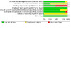
Risk of bias graph: review authors' judgements about each risk of bias item presented as percentages across all included studies. We did not complete risk of bias assessment for 3 studies (6%), indicated by blank (white) space at the right of the graph
3.
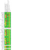
Risk of bias summary: review authors' judgements about each risk of bias item for each included study. Blank spaces indicate that risk of bias assessment was not completed; we did not use any outcome data for these studies in the review.
We judged no studies to have a low risk of bias overall. Twenty studies had an unclear risk of bias overall (Al‐Kheraif 2022a; Al‐Kheraif 2022b; Bassir 2013; Bechara Andere 2018; Braun 2008; Chitsazi 2014; Christodoulides 2008; Coelho 2023; Cosgarea 2021; Derikvand 2020; Hill 2019; Malgikar 2016; Mallineni 2020; Monzavi 2016; Moreira 2015; Patyna 2021; Petelin 2015; Pourabbas 2014; Rodrigues 2023; Srikanth 2015). The remaining studies had a high risk of bias overall.
Allocation
In five studies, the method of random sequence generation was not described and we were uncertain of the risk of selection bias in these studies (Chitsazi 2014; Karmakar 2021; Munteanu 2022; Petelin 2015; Sethi 2019). The remaining studies all used an appropriate method, such as computer‐generated randomisation, coin toss or a lottery method, and we judged the risk of selection bias to be low in these studies.
Generally, methods of allocation concealment were poorly reported, and two‐thirds of studies either reported no information or inadequate information; we judged the risk of selection bias for allocation concealment in these studies to be unclear. In one study, non‐sealed envelopes were used to conceal allocation and we judged the risk of bias to be high (Elsadek 2022). The remaining 13 studies used methods to describe allocation concealment that we judged to be at low risk of bias (Al‐Kheraif 2022a; Al‐Kheraif 2022b; Al Rifaiy 2022; Bassir 2013; Bechara Andere 2018; Betsy 2014; Cosgarea 2021; Courval 2020; Mallineni 2020; Monzavi 2016; Moreira 2015; Patyna 2021; Rodrigues 2023).
Performance bias
It was not possible to blind any personnel to those participants (or areas of the mouth, in the case of split‐mouth studies) in which aPDT was used. We judged that the most important method to reduce risk of performance bias for personnel was if SI was carried out by a separate operator who was unaware of allocation to aPDT, or if SI was carried out prior to randomisation. We believed that this would increase the likelihood that SI was carried out comparably in both test and control groups. We judged that this study approach was adequately conducted in 22 studies and we judged all of these to be at low risk of personnel‐related performance bias (Al‐Kheraif 2022a; Al‐Kheraif 2022b; Alwaeli 2015; Bassir 2013; Braun 2008; Chitsazi 2014; Christodoulides 2008; Coelho 2023; Cosgarea 2021; Courval 2020; Derikvand 2020; Elsadek 2022; Hill 2019; Malgikar 2016; Mallineni 2020; Monzavi 2016; Moreira 2015; Patyna 2021; Petelin 2015; Pourabbas 2014; Rodrigues 2023; Srikanth 2015). In the remaining studies, SI was carried out after randomisation (sometimes using the same operator); we judged these studies to be at high risk of personnel‐related performance bias.
For participants, some studies attempted to mask participants to treatment allocation by using a sham procedure for aPDT (Coelho 2023; Derikvand 2020; Monzavi 2016; Moreira 2015; Raut 2018). However, even when a sham procedure was not used, we expected that all participants recruited to an included study would follow oral hygiene advice during the study period regardless of treatment allocation; therefore, we judged all studies to be at low risk of participant‐related performance bias.
Detection bias
We judged two studies to be at high risk of detection bias (Borekci 2019; Sethi 2019). In Borekci 2019, an unblinded examiner was used. In Sethi 2019, this was not stated explicitly, but we assumed this to be the case because the study included insufficient study authors to allow for this. In 13 studies, methods used to blind examiners were not described and we were uncertain whether these studies were at risk of detection bias (Al‐Kheraif 2022a; Al‐Kheraif 2022b; Amini 2014; Coelho 2023; El Mobadder 2023; Elsadek 2022; Grzech‐Leśniak 2019; Hill 2019; Karmakar 2021; Malgikar 2016; Munteanu 2022; Petelin 2015; Pulikkotil 2016). We judged the remaining studies to be at low risk of detection bias because they described adequate methods to blind examiners to group allocation.
Incomplete outcome data
We judged four studies to be at high risk of attrition bias (Alwaeli 2015; Al‐Zahrani 2011; Courval 2020; Pulikkotil 2016). In these studies, at least 15% of participants dropped out during the study period. We judged the remaining studies to be at low risk of attrition bias because participant loss was less than 15%, and we noted no concerns about the reasons for losses or any imbalance of losses between study groups.
Selective reporting
Only Courval 2020 and Arya 2023 were prospectively registered with a clinical trials register. We judged Courval 2020 to be at low risk of reporting bias because the reported outcomes were consistent with those in the trials register documents. However, outcomes were unclearly specified in the trials register documents for Arya 2023 and, therefore, we were uncertain whether this study included any risk of reporting bias.
Fifteen studies were also registered with a clinical trials register, but registration in these studies was completed retrospectively (Amini 2014; Bassir 2013; Bechara Andere 2018; Borekci 2019; Chitsazi 2014; Cosgarea 2021; Joshi 2020; Mallineni 2020; Moreira 2015; Patyna 2021; Pourabbas 2014; Srikanth 2015), or we were unable to access the clinical trials registration documents (Al‐Kheraif 2022a; Betsy 2014; Pulikkotil 2016); therefore, we judged the risk of reporting bias to be unclear in these studies. We also judged the risk of reporting bias to be unclear in the remaining studies in which a protocol or clinical trials registration was not described.
Other potential sources of bias
We noted no other sources of bias in the included studies.
Effects of interventions
Adjunctive antimicrobial photodynamic therapy (aPDT) versus control during active treatment of periodontitis
One study was a multi‐arm study including a single application and multiple applications of aPDT; in all the relevant analyses, we included data for both study arms (Annaji 2016). Five studies reported data according to the size of the initial pocket depth (Borekci 2019; Braun 2008; Hill 2019; Moreira 2015; Rodrigues 2023). In the primary analyses, we included the following data for these studies:
Borekci 2019: we used data for pocket depths of ≥ 5 mm rather than all pocket depths.
Braun 2008: we used data supplied by study authors for all pocket sizes rather than incomplete data in the study report for pocket depths of < 3 mm, > 3 mm to ≤ 5 mm, and > 5 mm.
Hill 2019: we used data for pocket depths of > 6 mm rather than pocket depths of 1 mm to 3 mm, or 4 mm to 6 mm.
Moreira 2015: we used data for pocket depths of 5 mm and 6 mm rather than pocket depths of ≥ 7 mm.
Rodrigues 2023: we used data for pocket depths > 4 mm (rather than all pocket depths).
Additionally, data in Courval 2020 were presented for furcation sites and non‐furcation sites (called 'other sites'). In the primary analyses, we used data from non‐furcation sites.
For the number of participants in each effect estimate, we report the number of participants in the study; this accounts for split‐mouth study designs in which the same participants are allocated to both treatment and control groups.
For each result that appeared to favour aPDT, we considered the minimal clinically important difference (MCID) for the outcome and used these values to judge whether the difference between treatments was likely to be clinically meaningful (Measures of treatment effect). For critical outcomes reported at six months, we also report the certainty of the evidence alongside the effect estimate; see Table 1.
Change in probing pocket depths (PPD)
We found a very small difference in the reduction of PPD at three months when adjunctive aPDT was used compared with SI alone and the difference between treatments was not clinically important (MD 0.44 mm, 95% CI 0.29 to 0.59; 35 studies, 873 participants; I2 = 97%; Analysis 1.1). This analysis included 26 studies with a split‐mouth design. We noted that this analysis included very substantial levels of statistical heterogeneity, which we were unable to explain.
1.1. Analysis.
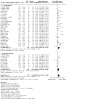
Comparison 1: Adjunctive aPDT versus control during active periodontal treatment (3‐month and 6‐month follow‐up), Outcome 1: PPD (change from baseline (mm)); 3 months
We were very uncertain of the effects of adjunctive aPDT on PPD at six months. Although we found a very small difference in the reduction in PPD at six months when adjunctive aPDT was used, the difference was also not clinically important (MD 0.52 mm, 95% CI 0.31 to 0.74; 15 studies, 452 participants; I2 = 97%; very low‐certainty evidence; Analysis 1.2). This analysis included 10 studies with a split‐mouth design. We downgraded the certainty of the evidence by one level for risk of bias in the included studies, and two levels for very substantial statistical heterogeneity.
1.2. Analysis.
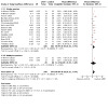
Comparison 1: Adjunctive aPDT versus control during active periodontal treatment (3‐month and 6‐month follow‐up), Outcome 2: PPD (change from baseline (mm)); 6 months
Change in bleeding on probing (BOP)
We found a very small difference in the reduction of BOP when adjunctive aPDT was used compared with SI alone and the difference between treatments was not clinically important (MD 2.19%, 95% CI 1.00 to 3.38; 11 studies, 282 participants; I2 = 88%; Analysis 1.3). This analysis included six studies with a split‐mouth design. We noted that this analysis included very substantial levels of statistical heterogeneity, which we were unable to explain.
1.3. Analysis.
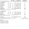
Comparison 1: Adjunctive aPDT versus control during active periodontal treatment (3‐month and 6‐month follow‐up), Outcome 3: BOP (change from baseline (%)); 3 months
We were very uncertain of the effects of adjunctive aPDT on BOP at six months. Although we found a very small difference in the reduction in BOP at six months when adjunctive aPDT was used, the difference was not clinically important (MD 5.72%, 95% CI 1.62 to 9.81; 5 studies, 171 studies; I2 = 98%; very low‐certainty evidence; Analysis 1.4). This analysis included two studies with a split‐mouth design. We downgraded the certainty of the evidence by one level for risk of bias in the included studies, and two levels for very substantial statistical heterogeneity.
1.4. Analysis.
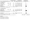
Comparison 1: Adjunctive aPDT versus control during active periodontal treatment (3‐month and 6‐month follow‐up), Outcome 4: BOP (change from baseline (%)); 6 months
Change in clinical attachment level (CAL)
We found a very small difference in the gain of CAL when adjunctive aPDT was used compared with SI alone and the difference between treatments was not clinically important (MD 0.36 mm, 95% CI 0.24 to 0.48; 34 studies, 875 participants; I2 = 89%; Analysis 1.5). This analysis included 26 studies with a split‐mouth design. We noted that this analysis included very substantial levels of statistical heterogeneity, which we were unable to explain.
1.5. Analysis.
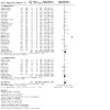
Comparison 1: Adjunctive aPDT versus control during active periodontal treatment (3‐month and 6‐month follow‐up), Outcome 5: CAL (change from baseline (mm)); 3 months
We were very uncertain of the effects of adjunctive aPDT on CAL at six months. Although we found a very small difference in gain in CAL at six months when adjunctive aPDT was used, the difference was not clinically important (MD 0.44 mm, 95% CI 0.24 to 0.64; 13 studies, 414 participants; I2 = 92%; very low‐certainty evidence; Analysis 1.6). This analysis included nine studies with a split‐mouth design. We downgraded the certainty of the evidence by one level for risk of bias in the included studies, and two levels for very substantial statistical heterogeneity.
1.6. Analysis.
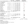
Comparison 1: Adjunctive aPDT versus control during active periodontal treatment (3‐month and 6‐month follow‐up), Outcome 6: CAL (change from baseline (mm)); 6 months
Change in gingival recession (REC)
The difference in change of REC at three months between participants treated with the adjunctive aPDT and those treated with SI alone was imprecise and included no benefits for either treatment (MD ‐0.01 mm, 95% CI ‐0.06 to 0.05; 13 studies, 284 participants; I2 = 8%; Analysis 1.7). This analysis included 10 studies with a split‐mouth design.
1.7. Analysis.
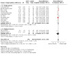
Comparison 1: Adjunctive aPDT versus control during active periodontal treatment (3‐month and 6‐month follow‐up), Outcome 7: REC (change from baseline (mm)); 3 months
We were very uncertain of the effects of adjunctive aPDT on REC at six months. The difference in change of REC between groups at this time point was imprecise and included no clinically important differences in the change in REC for either treatment (MD 0.00, 95% CI ‐0.16 to 0.16; 4 studies, 95 participants; I2 = 0%; very low‐certainty evidence; Analysis 1.8). This analysis included three studies with a split‐mouth design. We downgraded the certainty of the evidence by one level for risk of bias in the included studies, and two levels for imprecision because the effect estimate was derived from very few participants and included the possibility of no benefit for either treatment option.
1.8. Analysis.
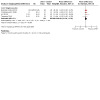
Comparison 1: Adjunctive aPDT versus control during active periodontal treatment (3‐month and 6‐month follow‐up), Outcome 8: REC (change from baseline (mm)); 6 months
Pocket closure
Three studies evaluated the proportion of closed pockets three months after treatment (Al‐Zahrani 2011; Courval 2020; Rodrigues 2023); studies used different criteria to measure this outcome (e.g. which pockets to evaluate depending on initial depth). All studies reported an increase in the proportion of closed pockets within treatment groups three months after treatment. We summarise the data in Appendix 3.
No studies reported this outcome at six months.
Any adverse effect related to aPDT
Twenty‐four studies reported adverse effects, with all studies stating that no adverse effects were observed. We include statements on adverse effects for each study in Appendix 4. We judged the certainty of the evidence for adverse effects to be moderate; we downgraded by one level for risk of bias in the included studies.
Patient satisfaction
No studies reported data for this outcome.
Quality of life
No studies reported data for this outcome.
Subgroup analyses
We conducted formal tests for subgroup interactions on outcomes reported in the summary of findings tables, and in which data were available from at least 10 studies; for this comparison, we report data for PPD and CAL at six months. We found no important differences in findings for these outcomes according to whether a placebo control was used (rather than a negative control of SI alone), or aPDT was given in a single session or multiple sessions. See Appendix 5.
Sensitivity analyses
We conducted sensitivity analyses on all the pooled results in this comparison group. We found variation in effect estimates when comparing the results of the primary analyses described above with the results of sensitivity analyses across all outcomes. For some sensitivity analyses, we found that effect estimates were no longer precise (with CIs indicating possible benefit to both treatments) or point estimates that favoured the control group. See Appendix 6.
Adjunctive antimicrobial photodynamic therapy (aPDT) versus control during supportive periodontal care
For the number of participants in each effect estimate, we report the number of participants in the study; this accounts for split‐mouth study designs in which the same participants are allocated to both treatment and control groups.
For each result that appeared to favour aPDT, we considered the minimal clinically important difference (MCID) for the outcome and used these values to judge whether the difference between treatments was likely to be clinically meaningful (Measures of treatment effect). For critical outcomes reported at six months, we also report the certainty of the evidence alongside the effect estimate; see Table 2.
Change in PPD
The difference in change of PPD at three months between participants treated with the adjunctive aPDT and those treated with SI alone was imprecise and included no clinical benefit for either treatment (MD 0.44 mm, 95% CI ‐0.20 to 1.08; 6 studies, 244 participants; I2 = 95%; Analysis 2.1). This analysis included one study with a split‐mouth design. We noted that this analysis included very substantial levels of statistical heterogeneity, which we were unable to explain.
2.1. Analysis.
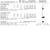
Comparison 2: Adjunctive aPDT versus control during supportive periodontal care (3‐month and 6‐month follow‐up), Outcome 1: PPD (change from baseline (mm)); 3 months
We were very uncertain of the effects of adjunctive aPDT on PPD at six months. The difference in change of PPD at six months between participants treated with the adjunctive aPDT and those treated with SI alone included no clinical benefit for either treatment (MD ‐0.04 mm, 95% CI ‐0.19 to 0.10; 3 studies, 125 participants; I2 = 0%; very low‐certainty evidence; Analysis 2.2). We downgraded the certainty of the evidence by one level for risk of bias in the included studies, and two levels for imprecision because the effect estimate was derived from very few participants and included the possibility of no benefit for either treatment option.
2.2. Analysis.
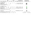
Comparison 2: Adjunctive aPDT versus control during supportive periodontal care (3‐month and 6‐month follow‐up), Outcome 2: PPD (change from baseline (mm)); 6 months
Change in BOP
The difference in change of BOP at three months between participants treated with the adjunctive aPDT and those treated with SI alone was imprecise and included no clinical benefit for either treatment (MD 2.38%, 95% CI ‐1.34 to 6.10; 5 studies, 231 participants; I2 = 81%; Analysis 2.3). We noted that this analysis included very substantial levels of statistical heterogeneity, which we were unable to explain.
2.3. Analysis.
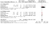
Comparison 2: Adjunctive aPDT versus control during supportive periodontal care (3‐month and 6‐month follow‐up), Outcome 3: BOP (change from baseline (%)); 3 months
We were very uncertain of the effects of adjunctive aPDT on BOP at six months. The difference in change of BOP at six months between participants treated with the adjunctive aPDT and those treated with SI alone included no clinical benefit for either treatment (MD 4.98%, 95% CI ‐2.51 to 12.46; 3 studies, 127 participants; I2 = 87%; very low‐certainty evidence; Analysis 2.4). We downgraded the certainty of the evidence by one level for risk of bias in the included studies, and two levels for very substantial statistical heterogeneity, and we noted that the imprecise effect estimate was derived from very few participants and included the possibility of no benefit for either treatment option.
2.4. Analysis.
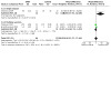
Comparison 2: Adjunctive aPDT versus control during supportive periodontal care (3‐month and 6‐month follow‐up), Outcome 4: BOP (change from baseline (%)); 6 months
Change in CAL
The difference in change of CAL at three months between participants treated with the adjunctive aPDT and those treated with SI alone was imprecise and included no clinical benefit for either treatment (MD 0.07 mm, 95% CI ‐0.21 to 0.36; 5 studies, 204 participants; I2 = 40%; Analysis 2.5). This analysis included one study with a split‐mouth design.
2.5. Analysis.
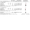
Comparison 2: Adjunctive aPDT versus control during supportive periodontal care (3‐month and 6‐month follow‐up), Outcome 5: CAL (change from baseline (mm)); 3 months
We were very uncertain of the effects of adjunctive aPDT on CAL at six months. The difference in change of CAL at six months between participants treated with the adjunctive aPDT and those treated with SI alone included no clinical benefit for either treatment (MD 0.07 mm, 95% CI ‐0.26 to 0.40; 2 studies, 85 participants; I2 = 0%; very low‐certainty evidence; Analysis 2.6). We downgraded the certainty of the evidence by one level for risk of bias in the included studies, and two levels for imprecision because the effect estimate was derived from very few participants and included the possibility of no benefit for either treatment option.
2.6. Analysis.
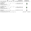
Comparison 2: Adjunctive aPDT versus control during supportive periodontal care (3‐month and 6‐month follow‐up), Outcome 6: CAL (change from baseline (mm)); 6 months
Change in REC
The difference in change of REC at three months between participants treated with the adjunctive aPDT and those treated with SI alone was imprecise and included no clinical benefit for either treatment (MD ‐0.01 mm, 95% CI ‐0.27 to 0.25; 2 studies, 37 participants; I2 = 0%; Analysis 2.7).
2.7. Analysis.
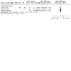
Comparison 2: Adjunctive aPDT versus control during supportive periodontal care (3‐month and 6‐month follow‐up), Outcome 7: REC (change from baseline (mm)); 3 months
Only one very small study reported data for REC at six months (Chondros 2009), and we were very uncertain of the effects of adjunctive aPDT on REC at this time point. The result included no clinical benefit for either treatment (MD ‐0.20 mm, 95% CI ‐0.48 to 0.08; 1 study, 24 participants; very low‐certainty evidence; Analysis 2.8). We downgraded the certainty of the evidence by one level for risk of bias in the included study, and two levels for imprecision because the effect estimate was derived from very few participants and included the possibility of no benefit for either treatment option.
2.8. Analysis.
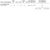
Comparison 2: Adjunctive aPDT versus control during supportive periodontal care (3‐month and 6‐month follow‐up), Outcome 8: REC (change from baseline (mm)); 6 months
Pocket closure
Three studies evaluated the proportion of closed pockets three months after treatment (Campos 2013; Grzech‐Leśniak 2019; Rühling 2010); studies used different criteria to measure this outcome (e.g. which pockets to evaluate depending on initial depth). All studies reported an increase in the proportion of closed pockets within treatment groups three months after treatment. We summarise the data in Appendix 3.
No studies reported these data at six months after treatment.
Any adverse effect related to aPDT
Only three of the seven included studies reported any information about adverse effects (Chondros 2009; Monzavi 2016; Rühling 2010). In Rühling 2010, one participant in the aPDT group developed an abscess, with no other adverse effects in the control group; however, there were no additional details about this event and we could not be certain whether it was directly related to the intervention. The remaining two studies reported that healing was uneventful. See Appendix 7. We judged the certainty of the evidence for adverse effects to be moderate; we downgraded by one level for risk of bias in the included studies.
Patient satisfaction
No studies reported data for this outcome.
Quality of life
No studies reported data for this outcome.
Subgroup analyses
Because all pooled analyses included fewer than 10 studies, all of which had very few participants, we conducted no formal tests for subgroup interactions in this comparison.
Sensitivity analyses
We conducted sensitivity analyses on all the pooled results in this comparison group. For some sensitivity analyses, we found variation in effect estimates when comparing the results of the primary analyses described above with the results of sensitivity analyses; these included effect estimates in which point estimates favoured the alternative treatment option. See Appendix 8.
Adjunctive antimicrobial photodynamic therapy (aPDT) versus control during active treatment of peri‐implant disease
Only one study reported evidence for adjunctive aPDT in people with peri‐implant disease (Al Rifaiy 2022). Data were only available for two outcomes, both reported only at three months. No studies reported data for CAL, REC, pocket closure, any adverse effects, participant satisfaction or quality of life. Because we found no evidence at six months for this comparison, we did not prepare a summary of findings table for this evidence.
Change in PPD
The difference in change of PPD at three months between participants treated with adjunctive aPDT and those treated with SI alone was imprecise and included no clinical benefit for either treatment (MD ‐0.10 mm, 95% CI ‐0.57 to 0.37; 1 study, 38 participants; Analysis 3.1).
3.1. Analysis.
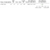
Comparison 3: Adjunctive aPDT versus SI alone during treatment of peri‐implant mucositis, Outcome 1: PPD (change from baseline(mm)); 3 months
Change in BOP
We found a very small difference in the reduction of BOP when adjunctive aPDT was used compared with SI alone and the difference between treatments was not clinically important (MD 1.60%, 95% CI 0.27 to 2.93; 1 study, 38 participants; Analysis 3.2).
3.2. Analysis.
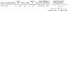
Comparison 3: Adjunctive aPDT versus SI alone during treatment of peri‐implant mucositis, Outcome 2: BOP (change from baseline (%)); 3 months
Discussion
Summary of main results
We included 50 RCTS with 1407 adults. Most of these used a split‐mouth study design in which an area of the mouth was randomly assigned to the treatment or the control. Only 18 studies were parallel design studies.
Only one study included people with peri‐implant mucositis and the other studies all evaluated the effects of the intervention in people with periodontitis. All participants received subgingival instrumentation (SI) as part of standard non‐surgical treatment. The intervention consisted of adjunctive antimicrobial photodynamic therapy (aPDT) in which a light source, typically a diode laser, was used with a photosensitising agent on the affected sites of the mouth. A fifth of studies applied aPDT over more than one session (two to four separate sessions) and the other studies used aPDT in a single session; only one multi‐arm study included groups for a single session and for multiple sessions of application. Most studies evaluated the effects of aPDT during the active treatment phase, with only six studies using aPDT during the supportive phase of treatment.
Adjunctive aPDT versus SI alone during active treatment of periodontitis
Because we found very low‐certainty evidence for all the clinical outcomes in this comparison, we could not be certain whether adjunctive aPDT has any long‐term benefits (at six months) in reducing the clinical symptoms of periodontitis during active treatment of periodontitis. These measures include probing pocket depth (PPD), bleeding on probing (BOP), clinical attachment level (CAL), gingival recession (REC) and pocket closure. We note that any effect estimates that could infer an apparent benefit in favour of aPDT were very small and were not clinically important. More than half of the studies observed no adverse effects of aPDT use (moderate‐certainty evidence).
Adjunctive aPDT versus SI alone during supportive treatment of periodontitis
Because we found very low‐certainty evidence for all the clinical outcomes in this comparison, we could not be certain whether adjunctive aPDT has any long‐term benefits (at six months) in reducing the clinical symptoms of periodontitis during active treatment of periodontitis. These measures include the same measures as above (PPD, BOP, CAL, REC and pocket closure). The effect estimates were all imprecise and included no clinically important benefits in favour of aPDT. Three of seven studies reported adverse effects (moderate‐certainty evidence). Although one of these studies reported an abscess in a single participant, it was not clear if this was an adverse effect of aPDT. The other two studies observed no adverse effects.
Overall completeness and applicability of evidence
The evidence in this review was largely limited to adults with periodontitis, and therefore includes a clear lack of evidence for the effectiveness of the intervention for people with peri‐implant disease. Most studies excluded smokers and, as we had specified in our own criteria, no participants had systemic diseases that may impact the prognosis of periodontitis. Because we excluded studies in which people had systemic diseases (and found that most studies also excluded people with systemic diseases), we note that the evidence in this review is only representative of an otherwise healthy population with periodontitis. This included people with periodontitis with pockets that are moderate or semi‐moderate in depth and are suitable for non‐surgical treatment. In this review, we did not fully explore the impact of aPDT in smokers, where there is likely to be variation within and between studies regarding smoking of tobacco products and e‐cigarettes.
Whilst we found a large number of eligible studies, these were often very small and overall sample sizes in our pooled estimates are often also small. Thus, the evidence is less complete than could be expected given the large number of studies. In addition, we found that more evidence was available for outcomes at three months. We prioritised data at six months in this review and this decision was based on the European Federation of Periodontology S3 Guidelines for the treatment of stage I‐III periodontitis (Sanz 2020), and the general consensus that six months is a meaningful endpoint for step 2 of periodontal therapy (Loos 2020; Suvan 2020). However, we found that fewer studies reported data at the later time point of six months. Few studies reported pocket closure and these studies only reported data at three months. No studies reported the participant satisfaction with the treatment or their quality of life and data for these outcomes are therefore incomplete.
We only conducted one subgroup analysis as planned in the review protocol, and an additional post hoc subgroup analysis related to the number of sessions in which aPDT was applied. We did not explore other differences in treatment approaches, such as the type of photosensitiser or the wavelengths used in diode lasers.
We note that aPDT includes equipment that may not be readily available in all dental care settings globally. In addition, adjunctive treatment with aPDT may be more costly than standard treatment alone, and this may limit the applicability of any evidence for this treatment worldwide. We did not set out to assess the cost‐benefit of adjunctive aPDT in this review, and noted that this was not formally addressed in any of our included studies.
Sustainability of the intervention
The delivery of good oral health care results in fewer subsequent interventions and a reduced environmental impact. This review evaluates interventions for the management of periodontal and peri‐implant diseases. Following Cochrane Oral Health policy (https://oralhealth.cochrane.org/about-us/sustainability), we conducted a brief search for health care sustainability science research for aPDT using the search strategy in Appendix 9 (MEDLINE Ovid; 1946 to 22 May 2024); one person (SL) screened the results of this search. We found no sustainability science research for this intervention. As noted above, there are cost implications for the procurement of the technology. We cannot comment on whether there are sustainability benefits for the individual needing treatment, the oral healthcare setting, or the wider environment that outweigh the likely costs; this is beyond the scope of this Cochrane review. Similarly, we are unaware of the sustainability implications related to the chemical components of photosensitising agents used with the intervention. We encourage users to explore other resources on this topic to understand, learn and promote sustainable actions in their practice.
Certainty of the evidence
We assessed the evidence for all clinical outcomes at six months to be of very low certainty. We found that many studies reported insufficient methods and we judged some domains to be at unclear or high risk of bias. For example, we could not always determine whether adequate methods were used to conceal group allocation after randomisation. We also believed that studies included a high risk of performance bias if they did not make attempts to blind the operator who carried out SI to group allocation. We conducted sensitivity analysis to explore these risk of bias decisions and found that the evidence for some outcomes was not sufficiently robust (i.e. excluding studies at high or unclear risk of bias reduced the size of the effect estimate). Therefore, we downgraded all the evidence by one level for risk of bias.
We also downgraded the certainty of the evidence for inconsistency when we noted substantial statistical heterogeneity in the pooled results. This affected the evidence for aPDT during active treatment of periodontitis for change in PPD, change in BOP and change in CAL with levels of statistical heterogeneity in pooled estimates ranging from 92% to 98%. As this statistical heterogeneity was so substantial, we downgraded this evidence by two levels. Subgroup analysis when we had sufficient studies (change in PPD and change in CAL) according to the type of control or whether the intervention was delivered in a single session or multiple sessions did not explain this heterogeneity. Similarly, variations in some results when we conducted sensitivity analysis did not indicate a single explanation for this.
Some of the effect estimates were imprecise, with confidence intervals that indicated a possible (but not clinically important) benefit for either treatment option. These results (change in REC in the active treatment comparison, and all the evidence for aPDT in the supportive phase of treatment) were all derived from very small sample sizes and, in these cases, we downgraded by two levels for imprecision. Change in BOP at six months in the supportive phase of treatment included both an imprecise effect estimate, a sample size of only 127 participants, and very substantial levels of statistical heterogeneity (I2 = 87%).
We had more confidence in the reports of adverse effects in the studies. Although we downgraded this evidence owing to the risk of bias in all studies, we judged that these results ‐ of no observed adverse effects related to aPDT ‐ were consistent across studies and were from moderate‐certainty evidence.
We did not downgrade for indirectness as the populations were appropriate for this review. We did not assess publication bias, and therefore we could not rule out the possibility of this risk in these review findings.
Potential biases in the review process
We conducted a thorough search, and two review authors independently assessed study eligibility, extracted data and assessed risk of bias in the included studies before reaching consensus. One review author (RC) was the study author of one of the included studies; we ensured that assessments of this study were undertaken independently by other members of the review team.
During the review process, we made some changes to the methods in the protocol. In particular, we re‐organised the outcomes into critical and other important outcomes, and these changes did not impact the reporting of data in the review. However, we also chose not to report data for microbiological parameters in the review. This decision was based on changes to international guidelines that no longer advise the use of microbiological sampling for diagnosis or treatment (Sanz 2020). We included studies in this review regardless of outcomes reported, and in order to direct interested readers to these data, we cite here all identified studies otherwise eligible for this review that report microbiological data (Al‐Kheraif 2022a; Al‐Kheraif 2022b; Borekci 2019; Chitsazi 2014; Chondros 2009; Cosgarea 2021; Gandhi 2019; Grzech‐Leśniak 2019; Karmakar 2021; Mallineni 2020; Moreira 2015; Munteanu 2022; Patyna 2021; Petelin 2015; Polansky 2009; Pulikkotil 2016; Queiroz 2015; Raut 2018; Romanos 2010; Rühling 2010; Sethi 2019; Srikanth 2015; Talebi 2016; Theodoro 2012). We also made a post hoc decision to report the data separately for active and supportive treatment of periodontitis, rather than exploring differences between these groups in subgroup analysis. Additionally, we presented data in forest plots according to the number of applications of aPDT. We conducted post hoc subgroup analyses according to the number of applications, and although we found no meaningful differences between single and multiple sessions, we believed that it was informative to provide a visual presentation of all the data according to these two different application approaches. In reporting the overall review findings, we did not describe effect estimates for each approach. It was not our intention to infer any differences between application approaches.
We included studies that used diode lasers with any wavelength and photosensitiser combinations. Because of this decision, we included two studies that did not use combinations that were known to be compatible (Annaji 2016; Malgikar 2016). We evaluated this decision in sensitivity analysis but found that the results of these studies did not impact the overall interpretation of the meta‐analyses.
The evidence in this review is limited only to adults who had not taken antibiotics in the previous six months. We adopted these criteria in order to reduce the risk of a sustained antibiotic effect. Similarly, we excluded studies in which a photosensitising agent was used in the control group as part of placebo aPDT (i.e. photosensitiser but with diode laser) because it is possible that some concentrations of these agents include antibacterial affects that may have impacted our results (Dörtbudak 2001; Pazzanezi 2015). However, we acknowledge the large number of studies that we subsequently excluded from the review, particularly studies in which participants were excluded if they had taken antibiotics within a three‐month period before the start of the review; in these latter studies, it is possible that some participants will not have had antibiotics within six months and would otherwise have been eligible for the review.
This review included a large number of studies that had not reported data sufficiently ‐ or in the manner that we required for use in our own analyses ‐ such that we needed to carry out additional calculations on the data reported in study reports. Therefore, when studies did not report SDs for the change from baseline to follow‐up in each group, we estimated these in accordance with the Cochrane Handbook for Systematic Reviews of Interventions version 6.1 (Higgins 2020). Similarly, we used the same approach to estimate SDs between test and control groups in split‐mouth studies. For both of these calculations, we used an intraclass correlation coefficient (ICC) of 0.5. We used the data from one split‐mouth study that had published paired and unpaired data for PPD, BOP and CAL at three months to calculate the ICC to be in the range of 0.3 to 0.6 for these outcomes (Joshi 2020); therefore, we believed that an ICC value of 0.5 was appropriate. However, we acknowledge that these two sets of approximations may have impacted the overall effect estimates. We attempted to address this by using sensitivity analyses, excluding all split‐mouth studies from analysis, as well as exploring the impact of excluding any study with two approximations and excluding any parallel study design with one approximation. We could not rule out the possibility that using a different ICC would reduce the effect sizes even further. However, as we had judged the clinical evidence for this review to be very low certainty, and all effect estimates were not clinically important, we felt that a more cautious approach to analysis would not impact our overall interpretation of the evidence for aPDT.
Agreements and disagreements with other studies or reviews
The recently published guideline by Sanz and colleagues (Treatment of stage I–III periodontitis—The EFP S3 level clinical practice guideline; Sanz 2020) includes a systematic review evaluating non‐surgical approaches to manage untreated periodontitis (Salvi 2020). The authors found limited advantage of aPDT due to heterogeneity between eight included studies, and could not draw any firm conclusions. Their findings are consistent with this Cochrane review, despite our own review including many more trials. We similarly found unexplained statistical heterogeneity and studies with high and unclear risks of bias. Some differences in the number of studies between these reviews are due to different inclusion criteria. Salvi 2020 included only studies with at least 20 participants per treatment arm, and with a minimum of six months follow‐up, whereas we have included less conservative criteria.
Chambrone and colleagues have previously published an American Academy of Periodontology best evidence review on aPDT including 26 studies for analysis (Chambrone 2018). The inclusion criteria were similar to ours, including adults ≥ 18 years old with the (former) diagnosis of aggressive or chronic periodontitis. But in contrast to our review, they also included aPDT in combination with root debridement with or without surgical flap access. We included only trials with non‐surgical periodontal treatment. Chambrone 2018 concluded that aPDT provided similar clinical results to conventional periodontal therapy. Similarly, although we were very uncertain about the interpretation of the evidence in this review, we noted that none of the effect estimates indicated a clinical benefit of aPDT above standard non‐surgical treatment of periodontitis (with SI alone).
A narrative review focusing on laser treatment including aPDT concluded that aPDT in periodontal therapy provided clinically insignificant reductions in PPD and CAL compared with conventional periodontal treatment (Cobb 2017). This was based on several systematic reviews. Cobb 2017 commented on considerable heterogeneity between studies, both in design and outcome measures, which limited the number of studies that could be entered into a meta‐analysis. Mizutani 2016 reported a reduction in BOP in favour of aPDT as an adjunct to SI.
A recently published review included 31 studies; 18 studies were used for meta‐analysis (Dalvi 2021). Inclusion and exclusion criteria were relatively similar to ours, but light‐emitting diodes (LEDs) for the light source was one of their exclusion criteria, whereas use of antibiotics was not an exclusion criterion. Additionally, they included studies regardless of combinations of photosensitiser/wavelength, comparisons between aPDT alone (monotherapy) and SI or aPDT combined with SI compared with SI and antibiotics. Dalvi and colleagues found inconsistencies and methodological bias in the included studies, assessing 18 to be at high risk of bias. Of the 31 studies included, only nine were included in this Cochrane review. Whilst Dalvi and colleagues found that most studies demonstrated the effectiveness of adjunctive aPDT, they concluded that the overall efficacy of aPDT in the non‐surgical management of periodontitis remained debatable (Dalvi 2021).
Another recently published review included 22 articles, and excluded treatment with antibiotics in the six months before the start of the study and excluded possible incompatible combinations of photosensitiser/wavelength (Moro 2021). Results were reported as meta‐analyses pooled with regard to photosensitiser or risk of bias. Nine of their 22 studies are included in this Cochrane review. Moro and colleagues found adjunctive aPDT to have a positive impact on CAL gain and PPD reduction. In addition, they noted favourable clinical effects for indocyanine green‐mediated aPDT as well as for high concentrations of phenothiazine chloride (Moro 2021).
The treatment with aPDT is well investigated for its safety and has widespread use in medicine and dentistry (Soukos 2011). We found moderate‐certainty evidence from the studies included in this review that aPDT probably causes no adverse effects when used as an adjunctive treatment for periodontitis.
Finally, a narrative review suggested that the use of aPDT showed clinically advantageous results in people with mild to moderate periodontitis (Sculean 2021). Use of aPDT in people with stage III and IV grade C periodontitis would provide some improvements, but not as a replacement for systemic antibiotics. In supportive therapy, aPDT could be a valuable tool in moderate residual pockets (Sculean 2021).
Very recently, a systematic review included in the guidelines for the treatment of peri‐implant diseases (Herrera 2023) focused on the adjunctive use of aPDT in the treatment of peri‐implant mucositis, including five studies (Dommisch 2023); here, it was suggested not to use photodynamic therapy adjunctively to submucosal instrumentation.
The discussion of appropriate photosensitiser and wavelength combinations was recently addressed in a multicentre study using an aPDT‐system based on a chromophore (photosensitiser) and a non‐corresponding wavelength (Preshaw 2021). The study was not included in the present review, as our inclusion criteria of no antibiotics within the last six months was not met. The photosensitiser was activated at a wavelength of 430 nm; the energy source was the standard light curing dental device mounted with a special plastic probe‐like tip for subgingival application of energy. According to the study authors, this multi‐LED light source emits a broadband spectrum of 385 nm to 515 nm, with a main peak at 465 nm and a secondary peak at 405 nm (Preshaw 2021). One of the concerns of Preshaw and colleagues was that activation of the chromophore was considered to require light at wavelength 430 nm; the wavelengths with energy transfer maxima of the dental curing light (i.e. 405 nm and 465 nm) did not correspond to those needed for chromophore activation. Furthermore, the study authors found it questionable if the known power density of the LED light (referred to a distance of 3 mm to 5 mm between the light source and the target) could be transferred by the working tip to the periodontal pocket. If this did not happen, it would lead to significant lack of energy, which would prevent sufficient power density in the periodontal pocket at the tip to activate the chromophores (Preshaw 2021). In the study, we noted that no significant differences were found between adjunctive aPDT and SI alone.
Authors' conclusions
Implications for practice.
We are very uncertain whether adjunctive antimicrobial photodynamic therapy (aPDT), applied after subgingival instrumentation (SI) as part of non‐surgical treatment of periodontitis, has any more important clinical benefits over that of SI alone, when measured at six months. This finding was similar whether aPDT was used during active treatment or supportive treatment. Other than one participant reporting an abscess (which may or may not have been caused by aPDT), studies that reported adverse effects observed no adverse effects associated with aPDT use.
Implications for research.
There is currently insufficient evidence for this treatment option in people with peri‐implant diseases and future research should address this population.
In this review, we found that most studies had very small sample sizes, and we found some indications that studies with high or unclear risks of bias may have impacted the overall findings. The reliability of the evidence for this review question would be improved by future trials of aPDT that are prospectively registered with clinical trials registers and sufficiently large and well‐conducted to reduce risks of bias. As we are interested in the differences between adjunctive aPDT and SI alone in terms of the change in clinical outcomes, we encourage future studies to report the results of paired analyses for change from baseline to end of study follow‐up. For split‐mouth studies, we would similarly encourage reporting of paired analyses between groups for these change scores. This would mean that our own analyses would include fewer approximations.
Studies should include consideration of participant‐relevant outcomes. In particular, pocket closure should be included within the investigation plan, as well as quality of life and participant satisfaction with treatment. A cost‐benefit analysis would also allow for exploration of whether any clinical benefits can be weighed against the cost of using this technology.
There is currently no standard treatment protocol for studies using aPDT. The questions about the optimal combination of photosensitiser and energy source, parameters of the energy source, irradiation time and application devices are still unanswered. Other open questions include how to apply the energy, for example, whether it should be subgingival, transgingival or in combination, as well as the optimal time point for the use of aPDT in the context of periodontal therapy.
What's new
| Date | Event | Description |
|---|---|---|
| 14 August 2024 | Amended | Correction of a minor typographical error in the Abstract that does not impact the data, findings or interpretation of the review. |
History
Protocol first published: Issue 6, 2015 Review first published: Issue 7, 2024
Acknowledgements
We sincerely thank Anne Littlewood (Cochrane Oral Health) for her useful help and for preparing the search strategy and Joanne Weldon (Cochrane Oral Health) for her valuable support. We also cordially thank Laura MacDonald, Tanya Walsh, Sunil Kumar Nettemu and Mariano Sanz for their valuable comments and help to optimise the protocol.
For the review, we sincerely want to thank Luisa M Fernandez Mauleffinch (Managing Editor, Cochrane Oral Health) and Anne Littlewood (Information Specialist, Cochrane Oral Health) for their useful help, excellent support during the preparation and writing of this review, and for performing the initial searches.
We want to acknowledge the valuable comments from the peer‐reviewers Philip Riley (The University of Manchester, UK; methods peer‐review) and Sunil Kumar Nettemu (University of Manipal, Malaysia; clinical peer‐review). We sincerely thank Anne‐Marie Glenny (Co‐ordinating Editor, Cochrane Oral Health) for her help with editing the review in response to feedback and preparing the review for publication.
Cochrane Oral Health supported the authors in the development of this review. The following people conducted the editorial process for this article.
Sign‐off Editor (final editorial decision): Anne‐Marie Glenny (Co‐ordinating Editor, Cochrane Oral Health, The University of Manchester, UK).
Managing Editor (selected peer‐reviewers, collated peer‐reviewer comments, provided editorial guidance to authors, conducted editorial policy checks): Luisa M Fernandez Mauleffinch (Managing Editor, Cochrane Oral Health, The University of Manchester, UK).
Information Specialist (checked accuracy of search sections of the review): Anne Littlewood (Information Specialist, Cochrane Oral Health, The University of Manchester, UK).
Copy Editor (copy edited final draft according to Cochrane style manual): Faith Armitage and Jenny Bellorini, Cochrane Central Production Service.
Appendices
Appendix 1. Dental probes for clinical measurements
For clinical measurements such as probing pocket depth (PPD), clinical attachment level (CAL), bleeding on probing (BOP) and gingival recession (REC), periodontal probes are used. A periodontal probe is a tapered, rod‐shaped, blunt instrument calibrated in millimetres. It is used to measure, locate and mark the depths of pockets around a tooth/implant to establish the health of the tissue around the tooth/implant. Calibration can be done in 1 mm steps from 1 mm to 15 mm (PCP‐15 or UNC‐15 (University of North Carolina)), with colour codings in 3 mm steps from 3 mm to 12 mm (PCP‐12) or in varying steps at 1 ‐ 2 ‐ 3 ‐ 5 ‐ 7 ‐ 8 ‐ 9 ‐ 10 mm markings (Williams probe). A further development is the pressure calibrated probe. The markings are identical to the instruments listed above, but the pressure for the measurement is controlled by a computer. This results in a standardised force for the measurements in the pocket.
In addition, a stent can be used for the measurements. It is a small, custom‐made acrylic splint placed over the teeth during clinical measurements. It is trimmed in such a way that a marginal edge near the gingival margin ensures a fixed point for the clinical measurements. As a result, the reactions of the periodontal tissue have no influence on the measurements. Moreover, small grooves can be cut into the stent to ensure a reproducible position of the probe around the tooth. If a stent is not used, the cementoenamel junction is used as a fixpoint for the measurements.
Appendix 2. Search strategies
Cochrane Oral Health’s Trials Register
1 (periodont*:ti,ab) AND (INREGISTER) 2 (("peri implantitis" or periimplantitis or peri‐implantitis):ti,ab) AND (INREGISTER) 3 ((implant* and (inflamm* or infect* or diseas* or mucositis)):ti,ab) AND (INREGISTER) 4 (#1 or #2 or #3) AND (INREGISTER) 5 ((photodynamic* or photochemo* or photoradiat* or photolysis* or photosensiti* or photo‐dynamic* or photo‐chemo* or photo‐radiat* or photo‐lysis or photo‐sensitiv*):ti,ab) AND (INREGISTER) 6 ((photochemotherap* or chemoprophylaxis or photo‐chemotherap* or chemo‐prophylaxis):ti,ab) AND (INREGISTER) 7 ((APDT or PDT):ti,ab) AND (INREGISTER) 8 (light*:ti,ab) AND (INREGISTER) 9 (#5 or #6 or #7 or #8) AND (INREGISTER) 10 (#4 and #9) AND (INREGISTER)
Cochrane Central Register of Controlled Trials (CENTRAL)
#1 [mh "periodontal diseases"] #2 periodont* #3 [mh ^peri‐implantitis] #4 ("peri implantitis" or periimplantitis or peri‐implantitis) #5 (implant* near/5 (inflamm* or infect* or diseas* or mucositis)) #6 {or #1‐#5} #7 [mh ^photochemotherapy] #8 [mh ^phototherapy] #9 (photodynamic* or photochemo* or photoradiat* or photolysis* or photosensiti* or photo‐dynamic* or photo‐chemo* or photo‐radiat* or photo‐lysis or photo‐sensitiv*) #10 (photochemotherap* or chemoprophylaxis or photo‐chemotherap* or chemo‐prophylaxis) #11 (APDT or PDT):ti,ab #12 light* #13 {or #7‐#12} #14 #6 and #13
MEDLINE (via Ovid)
1. exp Periodontal Diseases/ 2. periodont$.mp. 3. Peri‐implantitis/ 4. ("peri implantitis" or periimplantitis or peri‐implantitis).mp. 5. (implant$ adj5 (inflamm$ or infect$ or diseas$ or mucositis)).mp. 6. or/1‐5 7. Photochemotherapy/ 8. Phototherapy/ 9. (photodynamic$ or photochemo$ or photoradiat$ or photolysis$ or photosensiti$ or photo‐dynamic$ or photo‐chemo$ or photo‐radiat$ or photo‐lysis or photo‐sensiti$).mp. 10. (photochemotherap$ or chemoprophylaxis or photo‐chemotherap$ or chemo prophylaxis).mp. 11. (APDT or PDT).ti,ab. 12. light$.mp. 13. or/7‐12 14. 6 and 13
The above subject search was linked with the highly sensitive search strategy designed by Cochrane for identifying randomised controlled trials and controlled clinical trials in MEDLINE (as described in Lefebvre 2022, box 3c).
1. randomized controlled trial.pt. 2. controlled clinical trial.pt. 3. randomized.ab. 4. placebo.ab. 5. drug therapy.fs. 6. randomly.ab. 7. trial.ab. 8. groups.ab. 9. or/1‐8 10. exp animals/ not humans.sh. 11. 9 not 10
Embase (via Ovid)
1. exp Periodontal Disease/ 2. periodont$.mp. 3. Periimplantitis/ 4. ("peri implantitis" or periimplantitis or peri‐implantitis).mp. 5. (implant$ adj5 (inflamm$ or infect$ or diseas$ or mucositis)).mp. 6. or/1‐5 7. Photochemotherapy/ 8. Phototherapy/ 9. (photodynamic$ or photochemo$ or photoradiat$ or photolysis$ or photosensiti$ or photo‐dynamic$ or photo‐chemo$ or photo‐radiat$ or photo‐lysis or photo‐sensiti$).mp. 10. (photochemotherap$ or chemoprophylaxis or photo‐chemotherap$ or chemo‐ prophylaxis).mp. 11. (APDT or PDT).ti,ab. 12. light$.mp. 13. or/7‐12 14. 6 and 13
The above subject search was linked with the highly sensitive search strategy designed by Cochrane for identifying randomised controlled trials and controlled clinical trials in Embase (as described in Lefebvre 2022, box 3e).
Randomized controlled trial/
Controlled clinical study/
random$.ti,ab.
randomization/
intermethod comparison/
placebo.ti,ab.
(compare or compared or comparison).ti.
((evaluated or evaluate or evaluating or assessed or assess) and (compare or compared or comparing or comparison)).ab.
(open adj label).ti,ab.
((double or single or doubly or singly) adj (blind or blinded or blindly)).ti,ab.
double blind procedure/
parallel group$1.ti,ab.
(crossover or cross over).ti,ab.
((assign$ or match or matched or allocation) adj5 (alternate or group$1 or intervention$1 or patient$1 or subject$1 or participant$1)).ti,ab.
(assigned or allocated).ti,ab.
(controlled adj7 (study or design or trial)).ti,ab.
(volunteer or volunteers).ti,ab.
human experiment/
trial.ti.
or/1‐19
(random$ adj sampl$ adj7 ("cross section$" or questionnaire$1 or survey$ or database$1)).ti,ab. not (comparative study/ or controlled study/ or randomi?ed controlled.ti,ab. or randomly assigned.ti,ab.)
Cross‐sectional study/ not (randomized controlled trial/ or controlled clinical study/ or controlled study/ or randomi?ed controlled.ti,ab. or control group$1.ti,ab.)
(((case adj control$) and random$) not randomi?ed controlled).ti,ab.
(Systematic review not (trial or study)).ti.
(nonrandom$ not random$).ti,ab.
"Random field$".ti,ab.
(random cluster adj3 sampl$).ti,ab.
(review.ab. and review.pt.) not trial.ti.
"we searched".ab. and (review.ti. or review.pt.)
"update review".ab.
(databases adj4 searched).ab.
(rat or rats or mouse or mice or swine or porcine or murine or sheep or lambs or pigs or piglets or rabbit or rabbits or cat or cats or dog or dogs or cattle or bovine or monkey or monkeys or trout or marmoset$1).ti. and animal experiment/
Animal experiment/ not (human experiment/ or human/)
or/21‐33
20 not 34
CINAHL (Cumulative Index to Nursing and Allied Health Literature) (via EBSCO)
S13 S5 and S12 S12 S6 or S7 or S8 or S9 or S10 or S11 S11 light* S10 TI ( (APDT or PDT) ) OR AB ( (APDT or PDT) ) S9 (photochemotherap* or chemoprophylaxis or photo‐chemotherap* or chemo‐prophylaxis) S8 (photodynamic* or photochemo* or photoradiat* or photolysis* or photosensiti* or photo‐dynamic* or photo‐chemo* or photo‐radiat* or photo‐lysis or photo‐sensitiv*) S7 (MH Phototherapy) S6 (MH Photochemotherapy) S5 S1 or S2 or S3 or S4 S4 (implant* N5 (inflamm* or infect* or diseas* or mucositis)) S3 ("peri implantitis" or periimplantitis or peri‐implantitis) S2 periodont* S1 (MH Periodontal Diseases+)
AMED Ovid (Allied and Complementary Medicine)
1. exp Periodontal Disease/ 2. periodont$.mp. 3. ("peri implantitis" or periimplantitis or peri‐implantitis).mp. 4. (implant$ adj5 (inflamm$ or infect$ or diseas$ or mucositis)).mp. 5. or/1‐4 6. Phototherapy/ 7. (photodynamic$ or photochemo$ or photoradiat$ or photolysis$ or photosensiti$ or photo‐dynamic$ or photo‐chemo$ or photo‐radiat$ or photo‐lysis or photo‐sensiti$).mp. 8. (photochemotherap$ or chemoprophylaxis or photo‐chemotherap$ or chemo‐prophylaxis).mp. 9. (APDT or PDT).ti,ab. 10. light$.mp. 11. or/6‐10 12. 5 and 11
US National Institutes of Health Ongoing Trials Register (ClinicalTrials.gov)
periodontitis and photodynamic
periodontitis and phototherapy
World Health Organization International Clinical Trials Registry Platform
periodontitis and photodynamic
periodontitis and phototherapy
Appendix 3. Pocket closure (number/proportion of sites with PPD ≤ 4 mm after treatment); 3 months
| Comparison | Study | Design | Number of applications | aPDT baseline | aPDT 3 months | aPDT reduction | Control baseline | Control 3 months | Control reduction | Sites with PPD | Notes |
| Adjunctive aPDT vs control during active treatment of periodontitis | Al‐Zahrani 2011 | Split‐mouth | Single | 100 | 26.6 | 73.4 | 100 | 33.4 | 66.6 | ≥ 5 mm | PPD reduction ≥ 2mm: aPDT: 50% Control: 47% |
| Courval 2020 | Split‐mouth | Multiple | 20.47 | 7.31 | 13.16 | 21.07 | 10.41 | 10.66 | > 5 mm | Data for non‐furcation sites | |
| Rodrigues 2023 | Split‐mouth | Multiple | 100 | 8.9 | 91.1 | 100 | 13.7 | 86.3 | > 5 mm | Data only for sites with no BOP | |
| Adjunctive aPDT vs control during supportive treatment of periodontitis | Campos 2013 | Split‐mouth | Single | 100 | 20.22 | 79.78 | 100 | 60 | 40 | > 5 mm | Only sites with no BOP |
| Grzech‐Leśniak 2019 | Parallel‐group | Multiple | 38 | 26.3 | 11.7 | 36.5 | 23.4 | 13.1 | > 4 mm | — | |
| Rühling 2010 | Parallel‐group | Single | 29.7 | 23.3 | 6.4 | 32.7 | 28.7 | 4 | > 4 mm | — |
aPDT: antimicrobial photodynamic therapy; BOP: bleeding on probing; PPD: probing pocket depth
Appendix 4. Summary of adverse effects: aPDT vs control during active treatment of periodontis
| Study | Outcome |
| Alwaeli 2015 | Adverse events or side effects: "No adverse effects of aPDT were observed or reported by the patients." |
| Arya 2023 | "There were no reports of any adverse events throughout the study period" |
| Bassir 2013 | "The post‐operative healing was uneventful in all cases and no adverse events or complications were observed throughout the study." |
| Bechara Andere 2018 | "No adverse reactions were observed due to the debridement or the aPDT." |
| Berakdar 2012 | "No undesirable effects were observed, and both therapies were tolerated well by the patients." |
| Betsy 2014 | "Healing was uneventful in all cases and no adverse effects, such as discomfort, burning sensation, or pain related to the laser irradiation, were reported by any of the subjects" |
| Chitsazi 2014 | "No complications, such as pain or infection, were observed in this study." |
| Christodoulides 2008 | "Healing was uneventful in all cases. No adverse effects, such as discomfort, burning sensation, or pain related to the laser irradiation, were reported by any of the subjects." |
| Coelho 2023 | "No adverse effects of aPDT or the sham procedure were observed" |
| Courval 2020 | "No adverse effects after therapies was reported." |
| Derikvand 2020 | "The healing was uneventful and no adverse reactions were observed." |
| Hill 2019 | "In neither case were there any particular indications for side effects upon using indocyanine green in this clinical study" |
| Joshi 2020 | "Healing was uneventful in all the cases and no discomfort or adverse effects were reported." |
| Karmakar 2021 | "Uneventful postoperative healing was observed in all the individuals, any kind of adverse effect to the photosensitiser or the laser irradiation was not reported by any of the individuals." |
| Malgikar 2016 | "No patients reporting any postoperative pain, discomfort or complications at any of the follow‐up appointments" |
| Moreira 2015 | "The postoperative healing was uneventful in all cases. No adverse effects were observed with the use of aPDT." |
| Munteanu 2022 | "All patients completed the study and healing was uneventful for all of them. There were no levels of pain or any other discomfort reported through the patient questionnaire" |
| Patyna 2021 | "No adverse events or complications were recorded during the study." |
| Pourabbas 2014 | "There were no complications related to treatment during the follow‐up period." |
| Pulikkotil 2016 | "No patients reported any discomfort or complications related to the trial." |
| Raut 2018 | "Healing took place without any complications. No side effects like staining of adjacent mucosa or teeth were reported from the subjects enrolled in the test group." |
| Rodrigues 2023 | "No adverse effects of aPDT or of the sham procedure were observed" |
| Theodoro 2012 | "No postoperative complications, abscesses or infections were observed during the whole study period." |
Appendix 5. Subgroup analyses: adjunctive aPDT vs control during active treatment of periodontitis
| Analyses table | Subgroups | Results of test for subgroup interactions |
| PPD at 6 months | Single session vs multiple sessions | No evidence of a difference between subgroups (P = 0.87) |
| PPD at 6 months | Placebo control vs negative control | No evidence of a difference between subgroups (P = 0.12) |
| CAL at 6 months | Single session vs multiple sessions | No evidence of a difference between subgroups (P = 0.76) |
| CAL at 6 months | Placebo control or negative control | The test of subgroup differences indicated the possibility of a difference in effect (P = 0.02). However, this subgroup analysis included only one small study using a placebo control and we considered this sample size to be insufficient to infer any meaningful difference between results. |
Appendix 6. Sensitivity analyses: adjunctive aPDT vs control during active treatment of periodontitis
Results in bold text indicate that the effect estimates in sensitivity analyses demonstrated a different finding to the primary analyses, either in the direction of effect or the precision of the effect estimate, or both.
| Analyses | Primary analysis results | Results of sensitivity analysis |
| Analysis 1.1 | MD 0.44 mm, 95% CI 0.29 to 0.59; 35 studies; I2 = 97% | Excluding studies in which all participants were smokersa: MD 0.42, 95% CI 0.28 to 0.56; 33 studies; I2 = 96% Excluding all split‐mouth studies: MD 0.25 mm, 95% CI ‐0.04 to 0.54; 9 studies; I2 = 75% Excluding split‐mouth studies with 2 approximations of SD: MD 0.23 mm, 95% CI 0.07 to 0.38; 14 studies; I2 = 74% Excluding all split‐mouth studies, and parallel‐group designs with 1 approximation of SD: MD 0.00 mm, 95% CI ‐0.33 to 0.33; 1 study Excluding studies at high risk of performance bias (personnel): MD 0.38 mm, 95% CI 0.21 to 0.56; 20 studies; I2 = 98% Excluding studies with unclear or high risk of selection bias (sequence generation or allocation concealment): MD 0.40 mm, 95% CI 0.09 to 0.72; 11 studies; I2 = 99% Excluding studies with industry funding: MD 0.52 mm, 95% CI 0.35 to 0.68; 29 studies; I2 = 98% Excluding studies with industry funding or with no description of funding source: MD 0.38 mm, 95% CI 0.20 to 0.56; 22 studies; I2 = 98% Excluding studies with possible incompatible diode laser wavelengths and photosensitiser: MD 0.42 mm, 95% CI 0.26 to 0.57; 33 studies; I2 = 97% |
| Analysis 1.2 | MD 0.52 mm, 95% CI 0.31 to 0.74; 15 studies; I2 = 97% | Excluding studies in which all participants were smokersa: MD 0.46 mm, 95% CI 0.27 to 0.65; 15 studies, I2 = 91% Excluding all split‐mouth studies: MD 0.59 mm, 95% CI ‐0.01 to 1.20; 5 studies; I2 = 88% Excluding split‐mouth studies with 2 approximations of SD: MD 0.58 mm, 95% CI.13 to 1.04; 7 studies; I2 = 88% Excluding parallel‐group designs with 1 approximation of SD, and all split‐mouth studies: MD 0.20 mm, 95% CI ‐0.23 to 0.63; 1 study Excluding studies at high risk of performance bias (personnel): MD 0.48 mm, 95%CI 0.24 to 0.73; 9 studies; I2 = 98% Excluding studies with unclear or high risk of selection bias (sequence generation or allocation concealment): MD 0.40 mm, 95% CI 0.06 to 0.75; 6 studies; I2 = 99% Excluding studies with industry funding: MD 0.57 mm, 95% CI 0.33 to 0.81; 12 studies; I2 = 98% Excluding studies with commercial funding or with no description of funding source: MD 0.47 mm, 95% CI 0.18 to 0.76; 8 studies; I2 = 98% Excluding studies with possible incompatible diode laser wavelengths and photosensitiser: MD 0.56 mm, 95% CI 0.33 to 0.78; 14 studies; I2 = 97% |
| Analysis 1.3 | MD 2.19%, 95% CI 1.00 to 3.38; 11 studies; I2 = 88% | Excluding studies in which all participants were smokersa: MD 2.32%, 95% CI 0.54 to 4.11; 14 studies Excluding all split‐mouth studies: MD 2.94%, 95% CI ‐2.15 to 8.03; 5 studies; I2 = 70% Excluding split‐mouth studies with 2 approximations of SD: MD 3.43%, 95% CI 0.34 to 6.51; 6 studies; I2 = 66% Excluding parallel‐group designs with 1 approximation of SD, and all split‐mouth studies: MD 4.00%, 95% CI ‐0.93 to 17.31; 1 study Excluding studies at high risk of performance bias (personnel): MD 2.09%, 95% CI 0.91 to 3.27; 8 studies; I2 = 90% Excluding studies with unclear or high risk of selection bias (sequence generation or allocation concealment): MD 1.09, 95% CI 0.02 to 2.16; 4 studies; I2 = 88% Excluding studies with industry funding: MD 1.92, 95% CI 0.72 to 3.12; 8 studies; I2 = 89% Excluding studies with industry funding or with no description of funding source: MD 1.92, 95% CI 0.72 to 3.12; 8 studies; I2 = 89% Excluding studies with possible incompatible diode laser wavelengths and photosensitiser: n/a (no studies with these criteria) |
| Analysis 1.4 | MD 5.72%, 95% CI 1.62 to 9.81; 5 studies; I2 = 98% | Excluding studies in which all participants were smokersa: MD 1.29%, 95% CI 0.13 to 2.44; 5 studies, I2 = 0% Excluding all split‐mouth studies: MD 3.86, 95% CI ‐0.37 to 8.08; 3 studies; I2 = 0% Excluding split‐mouth studies with 2 approximations of SD: MD 3.86, 95% CI ‐0.37 to 8.08; 3 studies; I2 = 0% Excluding parallel‐group designs with 1 approximation of SD, and all split‐mouth studies: MD 5.00 mm, 95% CI ‐8.56 to 18.56; 1 study Excluding studies at high risk of performance bias (personnel): n/a ‐ all studies at low risk of performance bias Excluding studies with unclear or high risk of selection bias (sequence generation or allocation concealment): MD 5.98, 95% 1.39 to 10.56; 3 studies; I2 = 99% Excluding studies with industry funding: MD 6.23, 95% CI 1.74 to 10.72; 3 studies; I2 = 99% Excluding studies with industry funding or with no description of funding source: MD 6.23, 95% CI 1.74 to 10.72; 3 studies; I2 = 99% Excluding studies with possible incompatible diode laser wavelengths and photosensitiser: n/a (no studies with these criteria) |
| Analysis 1.5 | MD 0.36 mm, 95% CI 0.24 to 0.48; 34 studies; I2 = 89% | Excluding studies in which all participants were smokersa: MD 0.36, 95% CI 0.23 to 0.50; 32 studies; I2 = 89% Excluding all split‐mouth studies: MD 0.07 mm, 95% CI ‐0.37 to 0.17; 7 studies, I2 = 22% Excluding split‐mouth studies with 2 approximations of SD: MD 0.11, 95% CI ‐0.00 to 0.22; 13 studies, I2 = 45% Excluding parallel‐group designs with 1 approximation of SD, and all split‐mouth studies: MD 0.10 mm, 95% CI ‐0.17 to 0.37; 1 study Excluding studies at high risk of performance bias (personnel): MD 0.31 mm, 95% CI 0.18 to 0.45; 19 studies; I2 = 89% Excluding studies with unclear or high risk of selection bias (sequence generation or allocation concealment): MD 0.36 mm, 95% CI 0.17 to 0.56; 9 studies; I2 = 92% Excluding studies with industry funding: MD 0.41 mm, 95% CI 0.28 to 0.55; 28 studies; I2 = 90% Excluding studies with industry funding or with no description of funding source: MD 0.26, 95% CI 0.13 to 0.39; 20 studies, I2 = 86% Excluding studies with possible incompatible diode laser wavelengths and photosensitiser: MD 0.35 mm, 95% CI 0.23 to 0.47; 32 studies; I2 = 88% |
| Analysis 1.6 | MD 0.44 mm, 95% CI 0.24 to 0.64; 13 studies; I2 = 92% | Excluding studies in which all participants were smokersa: MD 0.40 mm, 95% CI 0.18 to 0.61; 13 studies, I2 = 88% Excluding all split‐mouth studies: MD 0.28 mm, 95% CI ‐0.20 to 0.75; 4 studies, I2 = 76% Excluding split‐mouth studies with 2 approximations of SD: MD 0.35 mm, 95% CI ‐0.07 to 0.77; 6 studies; I2 = 85% Excluding parallel‐group designs with 1 approximation of SD, and all split‐mouth studies: MD 0.20, 95% CI ‐0.13 to 0.53; 1 study Excluding studies at high risk of performance bias (personnel): MD 0.42 mm, 95% CI 0.20 to 0.63; 10 studies; I2 = 93% Excluding studies with unclear or high risk of selection bias (sequence generation or allocation concealment): MD 0.35 mm, 95% CI 0.10 to 0.60; 4 studies; I2 = 94% Excluding studies with industry funding: MD 0.47 mm, 95% CI 0.25 to 0.69; 10 studies; I2 = 93% Excluding studies with industry funding or with no description of funding source: MD 0.30 mm, 95% CI 0.08 to 0.53; 7 studies, I2 = 93% Excluding studies with possible incompatible diode laser wavelengths and photosensitiser: MD 0.49 mm, 95% CI 0.29 to 0.69; 12 studies; I2 = 91% |
| Analysis 1.7 | MD ‐0.01, 95% CI ‐ 0.06 to 0.05; 13 studies; I2 = 8% | Excluding studies in which all participants were smokersa: MD ‐0.00 mm, 95% CI ‐0.08 to 0.07; 11 studies; I2 = 20% Excluding all split mouth studies: MD ‐0.10 mm, 95% CI ‐0.25 to 0.06; 3 studies; I2 = 0% Excluding split‐mouth studies with 2 approximations of SD: MD ‐0.10 mm, 95% CI ‐0.25 to 0.06; 3 studies; I2 = 0% Excluding parallel‐group designs with 1 approximation of SD, and all split‐mouth studies: MD 0.10 mm, 95% CI ‐0.30 to 0.10; 1 study Excluding studies at high risk of performance bias (personnel): MD 0.01, 95% CI ‐0.08 to 0.11; 8 studies; I2 = 42% Excluding studies with unclear or high risk of selection bias (sequence generation or allocation concealment): MD 0.06, 95% CI ‐0.19 to 0.30; 3 studies; I2 = 59% Excluding studies with industry funding: MD ‐0.01, 95% CI ‐0.06 to 0.04; 10 studies; I2 = 6% Excluding studies with industry funding or with no description of funding source: MD ‐0.01, 95% CI ‐0.06 to 0.04; 10 studies; I2 = 6% Excluding studies with possible incompatible diode laser wavelengths and photosensitiser: n/a (no studies with these criteria) |
| Analysis 1.8 | MD 0.00 mm, 95% CI ‐0.16 to 0.16; 4 studies; I2 0% | Excluding studies in which all participants were smokersa: n/a (primary analysis included no studies in which all participants were smokers) Excluding all split mouth studies: MD 0.00 mm, 95% CI ‐0.27 to 0.27; 1 study Excluding split‐mouth studies with 2 approximations of SD: MD 0.00 mm, 95% CI ‐0.27 to 0.27; 1 study Excluding parallel‐group designs with 1 approximation of SD, and all split‐mouth studies: MD 0.00 mm, 95% CI ‐0.27 to 0.27; 1 study Excluding studies at high risk of performance bias (personnel): MD 0.02 mm, 95% CI ‐0.15 to 0.19; 3 studies; I2 = 0% Excluding studies with unclear risk of selection bias (sequence generation or allocation concealment): MD 0.00, 95% CI ‐0.25 to 0.25; 1 study Excluding studies with industry funding: MD ‐0.03, 95% CI ‐0.25 to 0.18; 2 studies; I2 = 0% Excluding studies with commercial funding or with no description of funding source: MD ‐0.03, 95% CI ‐0.25 to 0.18; 2 studies; I2 = 0% Excluding studies with possible incompatible diode laser wavelengths and photosensitiser: n/a (no studies with these criteria) |
aIn this sensitivity analysis, we excluded a subset of data for participants that were smokers in Al‐Kheraif 2022a and Al‐Kheraif 2022b, as well as data from studies in which all participants were smokers. CI: confidence interval; MD: mean difference; n/a: not applicable; SD: standard deviation
Appendix 7. Summary of adverse effects: aPDT vs control during supportive periodontal care
| Study ID | Adverse effects data as described in study reports |
| Chondros 2009 | "Healing was uneventful in all patients. Neither pain nor any other discomfort was reported by any of the patients following both treatments." |
| Monzavi 2016 | "Healing took place without complications or adverse events in all patients (“uneventful healing”)." |
| Rühling 2010 | "In the UST group, no adverse events or side effects (e.g., redness, swelling, or abscesses) were observed; in the PDT group, one abscess developed." |
Appendix 8. Sensitivity analyses: adjunctive aPDT vs control during supportive periodontal care
Results in bold text indicate that the effect estimates in sensitivity analyses demonstrated a different finding to the primary analyses, either in the direction of effect or the precision of the effect estimate, or both.
| Analyses | Primary analysis results | Results of sensitivity analysis |
| Analysis 2.1 | MD 0.44 mm, 95% CI ‐0.20 to 1.08; 6 studies; I2 = 95% | Excluding studies in which all participants were smokers: n/a (no studies with this criterion) Excluding all split‐mouth studies: MD 0.33 mm, 95% CI ‐0.36 to 1.03; 5 studies; I2 = 96% Excluding split‐mouth studies with 2 approximations of SD: n/a (no studies with this criterion) Excluding all split‐mouth studies, and all parallel‐group designs with 1 approximation of SD: n/a (no studies with this criterion) Excluding studies at high risk of performance bias (personnel): MD 0.91 mm, 95% CI ‐1.03 to 2.85; 2 studies; I2 = 99% Excluding studies with unclear or high risk of selection bias (sequence generation or allocation concealment): MD 0.91 mm, 95% CI ‐1.03 to 2.85; 2 studies; I2 = 99% Excluding studies with industry funding: MD 1.00 mm, 95% CI ‐0.27 to 2.26; 3 studies; I2 = 95% Excluding studies with industry funding or with no description of funding source: MD 1.00 mm, 95% CI ‐0.27 to 2.26; 3 studies; I2 = 95% |
| Analysis 2.2 | MD ‐0.04 mm, 95% CI ‐0.19 to 0.10; 3 studies; I2 = 0% | Excluding studies in which all participants were smokers: n/a (no studies including only smokers) Excluding all split‐mouth studies: n/a (no studies with this criterion) Excluding split‐mouth studies with 2 approximations of SD: n/a (no studies with this criterion) Excluding all split‐mouth studies, and all parallel‐group designs with 1 approximation of SD: n/a (no studies with these criteria) Excluding studies at high risk of performance bias (personnel): MD ‐0.05 mm, 95% CI ‐0.21 to 0.11; 1 study Excluding studies with unclear or high risk of selection bias (sequence generation or allocation concealment): MD ‐0.05 mm, 95% CI ‐0.21 to 0.11; 1 study Excluding studies with industry funding: MD 0.03 mm, 95% CI ‐0.39 to 0.45; 1 study Excluding studies with industry funding or with no description of funding source: MD 0.03 mm, 95% CI ‐0.39 to 0.45; 1 study |
| Analysis 2.3 | MD 2.38%, 95% CI ‐1.34 to 6.10; 5 studies; I2 = 81% | Excluding studies in which all participants were smokers: n/a (no studies including only smokers) Excluding all split‐mouth studies: n/a (no studies with this criterion) Excluding split‐mouth studies with 2 approximations of SD: n/a (no studies with this criterion) Excluding all split‐mouth studies, and all parallel‐group designs with 1 approximation of SD: n/a (no studies with these criteria) Excluding studies at high risk of performance bias (personnel): MD ‐0.29%, 95% CI ‐0.68 to 0.11; 2 studies; I2 = 0% Excluding studies with unclear or high risk of selection bias (sequence generation or allocation concealment): MD ‐0.29%, 95% CI ‐0.68 to 0.11; 2 studies; I2 = 0% Excluding studies with industry funding: MD 1.13%, 95% CI ‐2.10 to 4.36; 2 studies, I2 = 0% Excluding studies with industry funding or with no description of funding source: MD 1.13%, 95% CI ‐2.10 to 4.36; 2 studies, I2 = 0% |
| Analysis 2.4 | MD 4.98%, 95% CI ‐2.51 to 12.46; 3 studies; I2 = 87% | Excluding studies in which all participants were smokers: n/a (no studies with this criterion) Excluding all split‐mouth studies: n/a (no studies with this criterion) Excluding split‐mouth studies with 2 approximations of SD: n/a (no studies with this criterion) Excluding all split‐mouth studies, and all parallel‐group designs with 1 approximation of SD: n/a (no studies with these criteria) Excluding studies at high risk of performance bias (personnel): MD ‐0.18%, 95% CI ‐0.56 to 0.20; 1 study Excluding studies with unclear or high risk of selection bias (sequence generation or allocation concealment): MD ‐0.18%, 95% CI ‐0.56 to 0.20; 1 study Excluding studies with industry funding: MD 4.30%, 95% CI 0.06 to 8.54; 1 study Excluding studies with industry funding or with no description of funding source: MD 4.30%, 95% CI 0.06 to 8.54; 1 study |
| Analysis 2.5 | MD ‐0.07 mm, 95% CI ‐0.21 to 0.36; 5 studies; I2 = 40% | Excluding studies in which all participants were smokers: n/a (no studies with this criterion) Excluding all split‐mouth studies: MD ‐0.03 mm, 95% CI ‐0.25 to 0.19; 4 studies; I2 = 0% Excluding split‐mouth studies with 2 approximations of SD: MD ‐0.03 mm, 95% CI ‐0.25 to 0.19; 4 studies; I2 = 0% Excluding all split‐mouth studies, and all parallel‐group designs with 1 approximation of SD: n/a (no studies with these criteria) Excluding studies at high risk of performance bias (personnel): MD ‐0.14 mm, 95% CI ‐0.44 to 0.16; 2 studies; I2 = 0% Excluding studies with unclear or high risk of selection bias (sequence generation or allocation concealment): MD ‐0.14 mm, 95% CI ‐0.44 to 0.16; 2 studies; I2 = 0% Excluding studies with industry funding: MD 0.32 mm, 95% CI ‐0.77 to 1.40; 2 studies; I2 = 84% Excluding studies with industry funding or with no description of funding source: MD 0.32 mm, 95% CI ‐0.77 to 1.40; 2 studies; I2 = 84% |
| Analysis 2.6 | MD 0.07 mm, 95% CI ‐0.26 to 0.40; 2 studies; I2 = 0% | Excluding studies in which all participants were smokers: n/a (no studies with this criterion) Excluding all split‐mouth studies: n/a (no studies with this criterion) Excluding split‐mouth studies with 2 approximations of SD: n/a (no studies with this criterion) Excluding all split‐mouth studies, and all parallel‐group designs with 1 approximation of SD: n/a (no studies with these criteria) Excluding studies at high risk of performance bias (personnel): MD ‐0.01 mm, 95% CI ‐0.44 to 0.42; 1 study Excluding studies with unclear or high risk of selection bias (sequence generation or allocation concealment): MD ‐0.01 mm, 95% CI ‐0.44 to 0.42; 1 study Excluding studies with industry funding: n/a (this criterion applied to both studies) Excluding studies with industry funding or with no description of funding source: n/a (this criterion applied to both studies) |
| Analysis 2.7 | MD ‐0.01 mm, 95% CI ‐0.27 to 0.25; 2 studies; 50% | Excluding studies in which all participants were smokers: n/a (no studies including only smokers) Excluding all split‐mouth studies: MD 0.00 mm, 95% CI ‐0.27 to 0.25; 1 study Excluding split‐mouth studies with 2 approximations of SD: MD 0.00 mm, 95% CI ‐0.27 to 0.25; 1 study Excluding all split‐mouth studies, and all parallel‐group designs with 1 approximation of SD: n/a (no studies with these criteria) Excluding studies at high risk of performance bias (personnel): n/a (this criterion applied to both studies) Excluding studies with unclear or high risk of selection bias (sequence generation or allocation concealment): n/a (this criterion applied to both studies) Excluding studies with industry funding: MD ‐0.07, 95% CI ‐0.85 to 0.71; 1 study Excluding studies with industry funding or with no description of funding source: MD ‐0.07, 95% CI ‐0.85 to 0.71; 1 study |
| Analysis 2.8 | MD ‐ 0.20 mm, 95% CI ‐0.48 to 0.08; 1 study | n/a (only 1 study in the primary analysis) |
CI: confidence interval; MD: mean difference; n/a: not applicable; SD: standard deviation
Appendix 9. Sustainability of the intervention: search strategy
MEDLINE (via Ovid; 1946 to 22 May 2024)
antimicrobial photodynamic therapy.mp.
(aPDT or PDT).mp.
(photodynamic$ or phototherap$).mp.
or/1‐3
exp Sustainable Development/
Environmental Monitoring/
Carbon Footprint/
"Conservation of Natural Resources"/
Waste Management/
Air Pollution/
Climate Change/
(life cycle adj3 (assess* or analys*)).mp.
"cradle to grave".mp.
sustainab*.mp.
(environment* adj3 impact).mp.
carbon footprint.mp.
sustainable development.mp.
waste management.mp.
climate change.mp.
circular economy.mp.
or/5‐20
4 and 21
Data and analyses
Comparison 1. Adjunctive aPDT versus control during active periodontal treatment (3‐month and 6‐month follow‐up).
| Outcome or subgroup title | No. of studies | No. of participants | Statistical method | Effect size |
|---|---|---|---|---|
| 1.1 PPD (change from baseline (mm)); 3 months | 35 | 1450 | Mean Difference (IV, Random, 95% CI) | 0.44 [0.29, 0.59] |
| 1.1.1 Single session | 27 | 1148 | Mean Difference (IV, Random, 95% CI) | 0.50 [0.30, 0.70] |
| 1.1.2 Multiple sessions | 9 | 302 | Mean Difference (IV, Random, 95% CI) | 0.29 [0.10, 0.49] |
| 1.2 PPD (change from baseline (mm)); 6 months | 15 | 693 | Mean Difference (IV, Random, 95% CI) | 0.52 [0.31, 0.74] |
| 1.2.1 Single session | 14 | 641 | Mean Difference (IV, Random, 95% CI) | 0.53 [0.32, 0.74] |
| 1.2.2 Multiple sessions | 1 | 52 | Mean Difference (IV, Random, 95% CI) | 0.46 [‐0.33, 1.26] |
| 1.3 BOP (change from baseline (%)); 3 months | 11 | 418 | Mean Difference (IV, Random, 95% CI) | 2.19 [1.00, 3.38] |
| 1.3.1 Single session | 8 | 316 | Mean Difference (IV, Random, 95% CI) | 2.62 [0.77, 4.48] |
| 1.3.2 Multiple sessions | 3 | 102 | Mean Difference (IV, Random, 95% CI) | 1.86 [0.19, 3.53] |
| 1.4 BOP (change from baseline (%)); 6 months | 5 | 226 | Mean Difference (IV, Random, 95% CI) | 5.72 [1.62, 9.81] |
| 1.4.1 Single session | 4 | 174 | Mean Difference (IV, Random, 95% CI) | 5.01 [‐1.87, 11.90] |
| 1.4.2 Multiple sessions | 1 | 52 | Mean Difference (IV, Random, 95% CI) | 6.73 [‐3.98, 17.44] |
| 1.5 CAL (change from baseline (mm)); 3 months | 34 | 1468 | Mean Difference (IV, Random, 95% CI) | 0.36 [0.24, 0.48] |
| 1.5.1 Single session | 26 | 1166 | Mean Difference (IV, Random, 95% CI) | 0.41 [0.26, 0.56] |
| 1.5.2 Multiple sessions | 9 | 302 | Mean Difference (IV, Random, 95% CI) | 0.26 [0.04, 0.48] |
| 1.6 CAL (change from baseline (mm)); 6 months | 13 | 667 | Mean Difference (IV, Random, 95% CI) | 0.44 [0.24, 0.64] |
| 1.6.1 Single session | 12 | 615 | Mean Difference (IV, Random, 95% CI) | 0.43 [0.20, 0.65] |
| 1.6.2 Multiple sessions | 1 | 52 | Mean Difference (IV, Random, 95% CI) | 0.52 [‐0.07, 1.12] |
| 1.7 REC (change from baseline (mm)); 3 months | 13 | 502 | Mean Difference (IV, Random, 95% CI) | ‐0.01 [‐0.06, 0.05] |
| 1.7.1 Single session | 11 | 434 | Mean Difference (IV, Random, 95% CI) | ‐0.02 [‐0.04, 0.00] |
| 1.7.2 Multiple sessions | 2 | 68 | Mean Difference (IV, Random, 95% CI) | ‐0.06 [‐0.47, 0.35] |
| 1.8 REC (change from baseline (mm)); 6 months | 4 | Mean Difference (IV, Random, 95% CI) | Subtotals only | |
| 1.8.1 Single session | 4 | 166 | Mean Difference (IV, Random, 95% CI) | 0.00 [‐0.16, 0.16] |
| 1.9 PPD at 6 months. Subgroup analysis according to type of control (placebo or negative control) | 15 | 693 | Mean Difference (IV, Random, 95% CI) | 0.52 [0.31, 0.74] |
| 1.9.1 Negative control | 13 | 598 | Mean Difference (IV, Random, 95% CI) | 0.45 [0.23, 0.68] |
| 1.9.2 Placebo control | 2 | 95 | Mean Difference (IV, Random, 95% CI) | 1.12 [0.30, 1.93] |
| 1.10 CAL at 6 months. Subgroup analysis according to type of control (placebo or negative control) | 13 | 667 | Mean Difference (IV, Random, 95% CI) | 0.44 [0.24, 0.64] |
| 1.10.1 Negative control | 12 | 622 | Mean Difference (IV, Random, 95% CI) | 0.41 [0.20, 0.61] |
| 1.10.2 Placebo control | 1 | 45 | Mean Difference (IV, Random, 95% CI) | 0.96 [0.53, 1.39] |
1.9. Analysis.
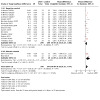
Comparison 1: Adjunctive aPDT versus control during active periodontal treatment (3‐month and 6‐month follow‐up), Outcome 9: PPD at 6 months. Subgroup analysis according to type of control (placebo or negative control)
1.10. Analysis.
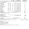
Comparison 1: Adjunctive aPDT versus control during active periodontal treatment (3‐month and 6‐month follow‐up), Outcome 10: CAL at 6 months. Subgroup analysis according to type of control (placebo or negative control)
Comparison 2. Adjunctive aPDT versus control during supportive periodontal care (3‐month and 6‐month follow‐up).
| Outcome or subgroup title | No. of studies | No. of participants | Statistical method | Effect size |
|---|---|---|---|---|
| 2.1 PPD (change from baseline (mm)); 3 months | 6 | 257 | Mean Difference (IV, Random, 95% CI) | 0.44 [‐0.20, 1.08] |
| 2.1.1 Single session | 3 | 104 | Mean Difference (IV, Random, 95% CI) | 0.20 [‐0.40, 0.79] |
| 2.1.2 Multiple sessions | 3 | 153 | Mean Difference (IV, Random, 95% CI) | 0.62 [‐0.60, 1.84] |
| 2.2 PPD (change from baseline (mm)); 6 months | 3 | 125 | Mean Difference (IV, Random, 95% CI) | ‐0.04 [‐0.19, 0.10] |
| 2.2.1 Single session | 1 | 24 | Mean Difference (IV, Random, 95% CI) | ‐0.10 [‐0.63, 0.43] |
| 2.2.2 Multiple sessions | 2 | 101 | Mean Difference (IV, Random, 95% CI) | ‐0.04 [‐0.19, 0.11] |
| 2.3 BOP (change from baseline (%)); 3 months | 5 | 231 | Mean Difference (IV, Random, 95% CI) | 2.38 [‐1.34, 6.10] |
| 2.3.1 Single session | 2 | 78 | Mean Difference (IV, Random, 95% CI) | 22.06 [‐17.91, 62.04] |
| 2.3.2 Multiple sessions | 3 | 153 | Mean Difference (IV, Random, 95% CI) | ‐0.28 [‐0.67, 0.11] |
| 2.4 BOP (change from baseline (%)); 6 months | 3 | 127 | Mean Difference (IV, Random, 95% CI) | 4.98 [‐2.51, 12.46] |
| 2.4.1 Single session | 1 | 24 | Mean Difference (IV, Random, 95% CI) | 40.00 [15.92, 64.08] |
| 2.4.2 Multiple sessions | 2 | 103 | Mean Difference (IV, Random, 95% CI) | 1.54 [‐2.73, 5.81] |
| 2.5 CAL (change from baseline (mm)); 3 months | 5 | 217 | Mean Difference (IV, Random, 95% CI) | 0.07 [‐0.21, 0.36] |
| 2.5.1 Single session | 3 | 104 | Mean Difference (IV, Random, 95% CI) | 0.31 [‐0.18, 0.79] |
| 2.5.2 Multiple sessions | 2 | 113 | Mean Difference (IV, Random, 95% CI) | ‐0.14 [‐0.44, 0.16] |
| 2.6 CAL (change from baseline (mm)); 6 months | 2 | 85 | Mean Difference (IV, Random, 95% CI) | 0.07 [‐0.26, 0.40] |
| 2.6.1 Single session | 1 | 24 | Mean Difference (IV, Random, 95% CI) | 0.20 [‐0.32, 0.72] |
| 2.6.2 Multiple sessions | 1 | 61 | Mean Difference (IV, Random, 95% CI) | ‐0.01 [‐0.44, 0.42] |
| 2.7 REC (change from baseline (mm)); 3 months | 2 | Mean Difference (IV, Random, 95% CI) | Subtotals only | |
| 2.7.1 Single session | 2 | 50 | Mean Difference (IV, Random, 95% CI) | ‐0.01 [‐0.27, 0.25] |
| 2.8 REC (change from baseline (mm)); 6 months | 1 | Mean Difference (IV, Fixed, 95% CI) | Totals not selected | |
| 2.8.1 Single session | 1 | Mean Difference (IV, Fixed, 95% CI) | Totals not selected |
Comparison 3. Adjunctive aPDT versus SI alone during treatment of peri‐implant mucositis.
| Outcome or subgroup title | No. of studies | No. of participants | Statistical method | Effect size |
|---|---|---|---|---|
| 3.1 PPD (change from baseline(mm)); 3 months | 1 | Mean Difference (IV, Fixed, 95% CI) | Totals not selected | |
| 3.2 BOP (change from baseline (%)); 3 months | 1 | Mean Difference (IV, Fixed, 95% CI) | Totals not selected |
Characteristics of studies
Characteristics of included studies [ordered by study ID]
Al Rifaiy 2022.
| Study characteristics | ||
| Methods | Study design: parallel Study period: May 2016 to October 2017 Setting: private dental clinic; Saudi Arabia Number of centres: 1 Funding source: Deanship Of Scientific Research, King Saud University Declarations of interest: "The authors declare that they have no conflict of interest" |
|
| Participants | Diagnosis (periodontal or peri‐implant): peri‐implant mucositis Inclusion criteria: people who reported vaping e‐cigs for at least the past year, with ≥ 1 dental implant that has been in place for ≥ 36 months, with peri‐implant mucositis Exclusion criteria: dual smokers (cigarettes and vaping e‐cigs); waterpipe smokers; smokeless tobacco users; suffering from debilitating systemic disease such as diabetes mellitus, AIDS/HIV, cardiovascular disorders and renal diseases; edentulous; had used antibiotics, steroidal or non‐steroidal anti‐inflammatory drugs within past 6 months Pocket depth at baseline, mean (SD): test group 4.3 (± 0.8) mm; control group 4.5 (± 0.9) mm (using UNC‐15) Age, mean (SD): test group 33.6 (± 2.8) years; control group 35.4 (± 2.1) years Sex, M/F: test group 20/0; control group 18/0 Smokers, n: 38 participants (e‐cigs) Number of randomised participants: 38 (with 65 implants); test group 20 participants; control group 18 participants Number of evaluated participants: 38 |
|
| Interventions | Test group: SI and aPDT (single session) Control group: SI OHI before study start: yes Instruments used for SI: mechanical debridement; no additional details Photodynamic irradiation source: diode laser (wavelength 670 nm; output power 150 mW; manufacturer not specified) Photosensitiser: methylene blue (0.005%) applied into periodontal pocket and left in place for 10 s Phase of treatment: active Duration of study: 12 weeks |
|
| Outcomes | Outcomes included in the review: PPD, BOP Time point(s) used for review outcomes: 3 months Other outcomes reported in the study but not used in the review: plaque index |
|
| Notes | — | |
| Risk of bias | ||
| Bias | Authors' judgement | Support for judgement |
| Random sequence generation (selection bias) | Low risk | Quote: "Randomisation was done by tossing a coin" |
| Allocation concealment (selection bias) | Low risk | Quote: "Sealed non‐transparent envelopes were used for allocation concealment and opened just before the interventions" |
| Blinding of personnel (performance bias) | High risk | It is not feasible to blind personnel to treatment allocation. Study authors do not report whether SI was carried out on all participants before randomisation. |
| Blinding of participants (performance bias) | Low risk | No details of whether participants were aware of allocation. However, we expected that lack of blinding would not influence oral hygiene performance during the study period. |
| Blinding of outcome assessment (detection bias) All outcomes | Low risk | Quote: "A trained and calibrated examiner, who was blinded to the study groups performed the clinical examinations at baseline and 12‐week follow‐up" |
| Incomplete outcome data (attrition bias) All outcomes | Low risk | No apparent participant losses |
| Selective reporting (reporting bias) | Unclear risk | Study authors do not cite a protocol or clinical trials registration. We are unable to assess the risk of selective outcome reporting without these documents. |
| Other bias | Low risk | We identified no other sources of bias. |
Al‐Kheraif 2022a.
| Study characteristics | ||
| Methods | Study design: split‐mouth (randomly divided by upper and lower quadrants, with 2 quadrants in each group) Study period: unclear Setting: University Hospital, Riyadh, Saudi Arabia Number of centres: 1 Funding source: King Saud University Declaration of interests: "The authors declare no conflict of interest in the present study" |
|
| Participants | Diagnosis (periodontal or peri‐implant): untreated chronic periodontitis (Stage II) Inclusion criteria: "stage‐II generalized chronic periodontitis" with PD ≤ 5 mm, clinical attachment loss of 3 mm to 4 mm and horizontal alveolar bone loss in contralateral teeth; smoking habit with smoking ≥ 10 cigarettes per day for at least 5 years; and systemically healthy Exclusion criteria: systemic diseases, medication including antiphlogistics, bleeding‐stimulating pharmaceuticals, or intake of systemic antibiotics within the last 6 months, pregnancy Pocket depth at baseline: ≤ 5 mm; UNC‐15 no stent Age, mean (SD): non‐smokers 43.5 (± 3.6) years; smokers 45.2 (± 4.8) years Sex, M/F: 23/3 Smokers: 13 Number of randomised participants: 26 Number of evaluated participants: 26 (non‐smokers (n = 13): test group 178 sites, control group 186 sites; smokers (n = 13): test group 169 sites, control group 180 sites) |
|
| Interventions | Test group: SI (deep scaling) and aPDT (3 sessions at 4th and 7th day) Control group: SI OHI before study start: yes Instruments used for SI: hand and ultrasonic instruments Photodynamic irradiation source: diode laser (wavelength 685 nm, energy fluence 2.5 J/cm2, output power 35 mW, Thera Lase DMC) Photosensitiser: chloro‐aluminium phthalocyanine gel (30 mg/mL) Phase of treatment: active Duration of study: 6 months |
|
| Outcomes | Outcomes included in the review: PPD, BOP, CAL, adverse effects Time point(s) used for review outcomes: 3 and 6 months Other outcomes reported in the study but not used in the review: PI, GCF, ABL as analysed on intraoral radiographs, microbiological parameters (Pg, Tf) |
|
| Notes | — | |
| Risk of bias | ||
| Bias | Authors' judgement | Support for judgement |
| Random sequence generation (selection bias) | Low risk | Computer‐generated random number table using permuted blocks through computer‐generated numbers |
| Allocation concealment (selection bias) | Low risk | The sequence was masked in closed and opaque envelopes for both assessors and participants. |
| Blinding of personnel (performance bias) | Low risk | Split‐mouth design, with SI applied to all participants prior to randomisation |
| Blinding of participants (performance bias) | Low risk | No details of whether participants were aware of allocation. However, we expected that lack of blinding would not influence oral hygiene performance during the study period. |
| Blinding of outcome assessment (detection bias) All outcomes | Unclear risk | No information |
| Incomplete outcome data (attrition bias) All outcomes | Low risk | All participants completed the study |
| Selective reporting (reporting bias) | Unclear risk | Study is registered with a clinical trial registry of Saudi Arabia. However, the trial registration number is not reported and therefore we were unable to effectively assess the risk of selective reporting bias. |
| Other bias | Low risk | We identified no other sources of bias. |
Al‐Kheraif 2022b.
| Study characteristics | ||
| Methods | Study design: split‐mouth (randomly divided by upper and lower quadrants, with 2 quadrants in each group) Study period: unclear Setting: University Hospital, Riyadh, Saudi Arabia Number of centres: 1 Funding source: King Saud University; Grant number: RSP‐2021/31 Declaration of interests: "The authors declare no conflicts of interest in the present study" |
|
| Participants | Diagnosis (periodontal or peri‐implant): untreated generalised periodontitis (Stage III) Inclusion criteria: "stage‐III generalized chronic periodontitis" with PD ≥ 6 mm, CAL ≥ 5 mm and marginal bone loss in contralateral teeth, tooth loss ≤ 4 teeth; smoking habit with smoking ≥ 10 cigarettes per day for at least 5 years; and systemically healthy Exclusion criteria: systemic diseases, medication including antiphlogistics, bleeding‐stimulating pharmaceuticals or intake of systemic antibiotics within the last 6 months, pregnancy Pocket depth at baseline: ≥ 6 mm; UNC‐15 no stent Age, mean (SD): non‐smokers 40.5 (± 2.6) years; smokers 42.2 (± 3.8) years Sex, M/F: 23/6 Smokers: 15 Number of randomised participants: 30 Number of evaluated participants: 29 (non‐smokers (n = 14): test group 178 sites, control group 186 sites; smokers (n = 15): test group 169 sites, control group 180 sites). We noted an inconsistency in the study report, with the main text stating that 30 participants were involved in the study, but figures and tables show that 29 finished the study, and we have used this number here. |
|
| Interventions | Test group: SI (root surface debridement) and aPDT (single session) Control group: SI OHI before study start: yes Instruments used for SI: hand and US instruments Photodynamic irradiation source: diode laser (wavelength 685 nm, energy fluence 3 J/cm2, output power 29 mW, Thera Lase DMC) Photosensitiser: chloro‐aluminium phthalocyanine gel (25 mg/mL) Phase of treatment: active Duration of study: 6 months |
|
| Outcomes | Outcomes included in the review: PPD, BOP, CAL, adverse effects Time point(s) used for review outcomes: 3 months and 6 months Other outcomes reported in the study but not used in the review: PI, GCF, MBL as analysed on intraoral radiographs, microbiological data for Pg and Tf |
|
| Notes | — | |
| Risk of bias | ||
| Bias | Authors' judgement | Support for judgement |
| Random sequence generation (selection bias) | Low risk | Computer‐generated random number table using permuted blocks through computer‐generated numbers |
| Allocation concealment (selection bias) | Low risk | The sequence was masked in closed and opaque envelopes. |
| Blinding of personnel (performance bias) | Low risk | Split‐mouth design, with SI applied to all participants prior to randomisation |
| Blinding of participants (performance bias) | Low risk | No details of whether participants were aware of allocation. However, we expected that lack of blinding would not influence oral hygiene performance during the study period. |
| Blinding of outcome assessment (detection bias) All outcomes | Unclear risk | No information |
| Incomplete outcome data (attrition bias) All outcomes | Low risk | Unclear if all participants completed the study. The text in the study report states that 30 participants were involved in the study, but information in the figures and tables of the study report indicates that 29 participants finished the trial. However, we judged overall participant loss to be low in this study. |
| Selective reporting (reporting bias) | Unclear risk | Study authors do not cite a protocol or clinical trials registration. We are unable to assess the risk of selective outcome reporting without these documents. |
| Other bias | Low risk | We identified no other sources of bias. |
Al‐Zahrani 2011.
| Study characteristics | ||
| Methods | Study design: split‐mouth (randomly divided by tooth; a tooth, from a pair of teeth with PD ≥ 5 mm, was allocated to each group) Study period: May 2010 to March 2011 Setting: University Hospital, Jeddah, Saudi Arabia Number of centres: 1 Funding source: grant from King Abdulaziz City for Science and Technology, Riyadh, Kingdom of Saudi Arabia (Grant No. LGP‐14‐32) Declaration of interests: unclear |
|
| Participants | Diagnosis (periodontal or peri‐implant): generalised moderate to severe chronic periodontitis Inclusion criteria: smokers (at least 10 cigarettes/day for at least 5 years), male, > 30 years, at least a pair of teeth with PD ≥ 5 mm Exclusion criteria: systemic diseases, systemic antibiotic treatment in the preceding 6 months, and those who require antibiotics prophylaxis before periodontal examination Pocket depth at baseline: ≥ 5 mm; UNC‐15 no stent Age, mean (SD), range: 41.6 (± 9.6) years, 30 to 56 years Sex: all male Smokers: 20 Number of randomised participants: 20 (54 teeth) Number of evaluated participants: 17 (48 teeth, 24 in each group) |
|
| Interventions | Test group: SI and aPDT (single session) Control group: SI OHI before study start: yes Instruments used for SI: hand instruments Photodynamic irradiation source: diode laser (wavelength 670 nm; output power 150 mW*; Periowave) Photosensitiser: 0.01% methylene blue Phase of treatment: active Duration of study: 3 months |
|
| Outcomes | Outcomes included in the review: PPD, BOP, CAL, adverse effects Time point(s) used for review outcomes: 3 months Other outcomes reported in the study but not used in the review: PI |
|
| Notes | 3 participants (6 teeth) lost to follow‐up and not included in data analysis * From manufacturer |
|
| Risk of bias | ||
| Bias | Authors' judgement | Support for judgement |
| Random sequence generation (selection bias) | Low risk | Computer‐generated random number table |
| Allocation concealment (selection bias) | Unclear risk | Method of concealment not described |
| Blinding of personnel (performance bias) | High risk | Split mouth design. SI was carried out after randomisation. |
| Blinding of participants (performance bias) | Low risk | No details of whether participants were aware of allocation. However, we expected that lack of blinding would not influence oral hygiene performance during the study period. |
| Blinding of outcome assessment (detection bias) All outcomes | Low risk | The same examiner, who was blinded to the therapies, carried out all clinical evaluation measurements. |
| Incomplete outcome data (attrition bias) All outcomes | High risk | Dropouts 3/20 participants (15%), unclear time point of dropouts or the reasons for dropout |
| Selective reporting (reporting bias) | Unclear risk | Study authors do not cite a protocol or clinical trials registration. We are unable to assess the risk of selective outcome reporting without these documents. |
| Other bias | Low risk | We identified no other sources of bias. |
Alwaeli 2015.
| Study characteristics | ||
| Methods | Study design: split‐mouth (randomly divided by quadrant, with 2 quadrants in each group) Study period: February 2011 to November 2012 Setting: University Hospital, Irbid, Jordan Number of centres: 1 Funding source: unclear Declaration of interests: unclear |
|
| Participants | Diagnosis (periodontal or peri‐implant): untreated chronic periodontitis Inclusion criteria: previously untreated chronic periodontitis, at least 1 premolar and 1 molar in every quadrant with a minimum of 4 teeth each, and at least 1 tooth with attachment loss of ≥ 4 mm in every quadrant Exclusion criteria: systemic diseases, including antiphlogistics, bleeding‐stimulating pharmaceuticals or intake of systemic antibiotics within the last 6 months Pocket depth at baseline: > 6 mm; pressure‐calibrated probe (manufacturer unknown) with stent Age, mean (SD), for analysed participants only: 40.9 (± 13.34) years Sex, M/F, for analysed participants only: 5/11 Smokers: unclear Number of randomised participants: 21 Number of evaluated participants: 16 (test group 73 teeth, control group 63 teeth) |
|
| Interventions | Test group: SI and aPDT (single session) Control group: SI OHI before study start: yes Instruments used for SI: hand and US instruments Photodynamic irradiation source: diode laser (wavelength 660 nm; output power 100 mW; Helbo Photodynamic Systems) Photosensitiser: phenothiazine chloride Phase of treatment: active Duration of study: 12 months |
|
| Outcomes | Outcomes included in the review: PPD, CAL, BOP, adverse effects Time point(s) used for review outcomes: 3 months and 6 months Other outcomes reported in the study but not used in the review: postoperative complications |
|
| Notes | 5 participants were lost to follow‐up and not included in data analysis | |
| Risk of bias | ||
| Bias | Authors' judgement | Support for judgement |
| Random sequence generation (selection bias) | Low risk | Computer‐generated random number table was used. |
| Allocation concealment (selection bias) | Unclear risk | Quote:"The sequence was concealed until the interventions were assigned" Comment: no additional details |
| Blinding of personnel (performance bias) | Low risk | Split mouth design. SI was carried out in all participants before randomisation to groups. |
| Blinding of participants (performance bias) | Low risk | No details of whether participants were aware of allocation. However, we expected that lack of blinding would not influence oral hygiene performance during the study period |
| Blinding of outcome assessment (detection bias) All outcomes | Low risk | The same examiner, who was blinded to the therapies, carried out all clinical evaluation measurements. |
| Incomplete outcome data (attrition bias) All outcomes | High risk | The study design is a split‐mouth, but a high number (5 of 21; 23.8%) of randomised participants with an unknown distribution (number) of teeth/group dropped out. Reasons for or time points of the 5 dropouts are not described. Number of smokers unclear. |
| Selective reporting (reporting bias) | Unclear risk | Study authors do not cite a protocol or clinical trials registration. We are unable to assess risk of selective outcome reporting without these documents. |
| Other bias | Low risk | We identified no other sources of bias. |
Amini 2014.
| Study characteristics | ||
| Methods | Study design: split‐mouth (quadrants in each participant were randomly allocated to test or control groups) Study period: unclear Setting: University Hospital, Isfahan, Iran Number of centres: 1 Funding source: study authors' own institution* Declaration of interests: unclear |
|
| Participants | Diagnosis (periodontal or peri‐implant): mild to moderate periodontitis Inclusion criteria: untreated periodontitis in the last 2 years, at least 3 teeth with PPD 3 mm to 6 mm and BOP/quadrant, no allergy to methylene blue and toluidine blue Exclusion criteria: pregnant or lactating patients, systemic diseases, antibiotic treatment in the preceding 12 months, PPD > 6 mm Age: 25 to 55 years Sex: 8 male/12 female Smokers: unclear Number of randomised participants: 20 Number of evaluated participants: 20 (number of teeth/sites in each group not described) |
|
| Interventions | Test group: SI and aPDT (single session) Control group: SI OHI before study start: yes Instruments used for SI: ultrasonic instruments Photodynamic irradiation source: LED light source (wavelength 630 nm; output power 2000 mW*; Foto‐San**) Photosensitiser: toluidine blue Phase of treatment: active Duration of study: 3 months |
|
| Outcomes | Outcomes included in the review: PPD, CAL, REC, adverse effects Time point(s) used for review outcomes: 3 months Other outcomes reported in the study but not used in the review: PBI |
|
| Notes | No statement of number of teeth/sites in each group. Only statement: two quadrants were selected in each participant. *From manufacturer (CMS Dental, Denmark) **From Iranian Registry of Clinical Trials (IRCT2013083112487N2) |
|
| Risk of bias | ||
| Bias | Authors' judgement | Support for judgement |
| Random sequence generation (selection bias) | Low risk | Quote: "two quadrants were randomly (coin toss) selected in each patient" |
| Allocation concealment (selection bias) | Unclear risk | Method of concealment not described |
| Blinding of personnel (performance bias) | High risk | It is not feasible to blind personnel to treatment allocation. We assumed from the study report that SI was likely to be carried out after randomisation. |
| Blinding of participants (performance bias) | Low risk | No details of whether participants were aware of allocation. However, we expected that lack of blinding would not influence oral hygiene performance during the study period. |
| Blinding of outcome assessment (detection bias) All outcomes | Unclear risk | No information |
| Incomplete outcome data (attrition bias) All outcomes | Low risk | No apparent losses |
| Selective reporting (reporting bias) | Unclear risk | Clinical trials registration (IRCT2013083112487N2; first posted November 2013); described in the register as retrospective registration and therefore we were unable to use these documents to assess the risk of selective reporting bias. |
| Other bias | Low risk | We identified no other sources of bias. |
Annaji 2016.
| Study characteristics | ||
| Methods | Study design: split‐mouth (quadrants of the mouth randomly allocated to test or control group) Study period: not reported Setting: Department of Periodontics, Coorg Institute of Dental Services, Virajpet, India Number of centres: 1 Funding source: not reported Declaration of interests: "Financial or other competing interests: None" |
|
| Participants | Diagnosis (periodontal or peri‐implant): periodontitis Inclusion criteria: diagnosed with either localised or generalised aggressive periodontitis, 18 to 35 years of age, at least 1 tooth with PPD ≥ 5 mm in each quadrant Exclusion criteria: people with periodontal treatment within the last 6 months, pregnancy, smoking, allergy to the dyes, systemic disease, use of mouth rinses that could influence the outcome of therapy, systemic antibiotics within the last 6 months Pocket depth at baseline: not reported Age, M/F, mean (SD): M 27.83 (± 3.71) years; F 27.33 (± 2.29) years Sex, M/F: 6/9 Smokers: none Number of randomised participants: 15 Number of evaluated participants: 15 |
|
| Interventions | Test groups: SI and aPDT (single session) and multiple sessions (3 sessions; baseline, day 7, day 21) Control group: SI OHI before study start: yes Instruments used for SI: ultrasonic scaler Photodynamic irradiation source: diode laser, continuous mode (wavelength 810 nm; power output 0.1 W; AMD Picasso, Dentsply India, India) Photosensitiser: Toluidine blue‐O; 1 mg/ml Phase of treatment: active Duration of study: 3 months |
|
| Outcomes | Outcomes included in the review: PPD, CAL Time point(s) used for review outcomes: 3 months Other outcomes reported in the study but not used in the review: SBI, plaque index, bacterial cultivation |
|
| Notes | This study included an additional study arm (SI + laser irradiation). This group was not eligible for inclusion in this review and we did not extract data for these participants. | |
| Risk of bias | ||
| Bias | Authors' judgement | Support for judgement |
| Random sequence generation (selection bias) | Low risk | Quote: "flipping a coin" |
| Allocation concealment (selection bias) | Unclear risk | Method of concealment not described |
| Blinding of personnel (performance bias) | High risk | It is not feasible to blind personnel to treatment allocation. We noted that SI and aPDT was carried out by the same operator after randomisation. |
| Blinding of participants (performance bias) | Low risk | No details of whether participants were aware of allocation. However, we expected that lack of blinding would not influence oral hygiene performance during the study period. |
| Blinding of outcome assessment (detection bias) All outcomes | Low risk | Quote: "The analysis and recording of clinical parameters and microbiologic findings were performed by other author who was blinded to the treatment techniques and to the test and control sites in order to assure an unbiased determination" |
| Incomplete outcome data (attrition bias) All outcomes | Low risk | No apparent losses |
| Selective reporting (reporting bias) | Unclear risk | Study authors do not cite a prospectively published protocol or clinical trials registration. We are unable to assess the risk of selective outcome reporting without these reports. |
| Other bias | Low risk | We identified no other sources of bias. |
Arya 2023.
| Study characteristics | ||
| Methods | Study design: split mouth (randomly divided by site, with identical sites in each arch) Study period: September 2022 to February 2023 Setting: Bangalore Institute of Dental Sciences and Post‐graduate Research Centre, India Number of centres: 1 Funding source: not reported Declaration of interests: not reported |
|
| Participants | Diagnosis (periodontal or peri‐implant): periodontitis Inclusion criteria: 35 to 55 years of age, with stage II grade A periodontitis, radiographic evidence of bone loss extending to the coronal third, interdental CAL of 3 mm to 4 mm, with no tooth loss due to periodontitis, maximum PPD of 5 mm, with mostly horizontal bone loss; ASA status 1 ‐ good general health; the presence of 3 multi‐rooted teeth in the posterior segment with identical PD ≤ 5 mm (1 in each quadrant); > 20 permanent teeth excluding third molars Exclusion criteria: known allergy to ICG dye; those who had received antibiotics, or surgical or non‐surgical periodontal treatment within the past 12 months; pregnancy; current smokers; history of treatment for recent bacterial infections; thyroid disorders; evidence of other systemic diseases (ischaemic heart disease, liver disease, renal disease, immunodeficiency and diabetic mellitus); HIV, hepatitis or any other contagious diseases Pocket depth at baseline, mean (SD): 4.62 (± 0.58) mm (using UNC‐15) Age, mean (SD): 40.7 (± 7.18) years Sex, M/F: 14/7 Smokers: none Number of randomised participants: 21 (42 sites); lost to follow‐up 1 participant (2 sites) Number of evaluated participants: 20 (20 sites/teeth) |
|
| Interventions | Test group: SI and aPDT (single session) Control group: SI OHI before study start: yes Instruments used for SI: hand and ultrasonic instruments Photodynamic irradiation source: diode laser (wavelength 610 nm; power output 0.8 W in continuous mode; ZOLAR with PACT Cumdente tip; for 60 s) Photosensitiser: ICG 5mg/mL; rinsed off after 60 s Phase of treatment: active Duration of study: 3 months |
|
| Outcomes | Outcomes included in the review: PPD, CAL, RAL (using UNC‐15), adverse events Time point(s) used for review outcomes: 3 months Other outcomes reported in the study but not used in the review: full‐mouth GI and PI, PCT |
|
| Notes | Clinical trials registration: CTRI/2022/08/045099 This study included an additional study arm (SI and laser). This group was not eligible for inclusion in this review and we did not extract data for these participants. |
|
| Risk of bias | ||
| Bias | Authors' judgement | Support for judgement |
| Random sequence generation (selection bias) | Low risk | Quote: "Identical sites, one in each arch, on the multi‐rooted teeth were randomly allocated to the intervention and control groups at an allocation ratio of 1:1, using a computer‐guided allotment software" |
| Allocation concealment (selection bias) | Unclear risk | Method of concealment not described |
| Blinding of personnel (performance bias) | High risk | It is not feasible to blind personnel to treatment allocation. We noted that SI was carried out after randomisation. |
| Blinding of participants (performance bias) | Low risk | No details of whether participants were aware of allocation. However, we expected that lack of blinding would not influence oral hygiene performance during the study period. |
| Blinding of outcome assessment (detection bias) All outcomes | Low risk | Quote: "The clinical periodontal measurements were performed by one calibrated examiner who was blinded to the experimental site" |
| Incomplete outcome data (attrition bias) All outcomes | Low risk | Loss of only one participant |
| Selective reporting (reporting bias) | Unclear risk | Prospectively registered with a clinical trials register (CTRI/2022/08/045099). However, the details in the clinical trials register are vague and no measures of clinical outcomes are indicated. We cannot be certain whether this is owing to lack of detail or risk of selective outcome reporting. |
| Other bias | Low risk | We identified no other sources of bias. |
Bassir 2013.
| Study characteristics | ||
| Methods | Study design: split‐mouth (each quadrant randomly assigned to a treatment group) Study period: April 2010 to May 2011 Setting: University Hospital, Tehran, Iran Number of centres: 1 Funding source: Laser Research Center in Dentistry (LRCD) of Tehran University of Medical Sciences (No. 89‐01‐97‐10245) Declaration of interests: unclear |
|
| Participants | Diagnosis (periodontal or peri‐implant): moderate to severe chronic periodontitis Inclusion criteria: at least 2 teeth with PPD 4 mm to 6 mm/quadrant, no furcation involvement, at least 4 teeth/quadrant Exclusion criteria: systemic disease, periodontal treatment within the past 12 months, systemic antibiotics within the last 6 months, smoking > 10 cigarettes/day; pregnant or lactating Pocket depth at baseline: PPD 4 mm to < 5 mm and PPD ≥ 5 mm, Williams probe no stent Age, mean (SD), range: 50.3 (± 8.7) years, 40 to 63 years Sex, M/F: 8/8 Smokers: 3 (< 10 cigarettes/day) Number of randomised participants: 16 Number of evaluated participants: 16 (total number of 210 sites, test group: 119 sites, control group: 91 sites) |
|
| Interventions | Test group: PAD (TBO + LED irradiation) and SI (3 sessions aPDT) Control group: SI OHI before study start: yes Instruments used for SI: hand and ultrasonic instruments Photodynamic irradiation source: LED (625 to 635 nm, maximum power density: 2000 mW/cm2, Foto‐San) Photosensitiser: toluidine blue ‐ O (0.1 mg/mL) Phase of treatment: active Duration of study: 3 months |
|
| Outcomes | Outcomes included in the review: PPD, BOP, CAL, adverse effects Time point(s) used for review outcomes: 3 months Other outcomes reported in the study but not used in the review: PI |
|
| Notes | "Postoperative healing was uneventful" Note: study included 2 additional study groups (LED and SI; and TBO and SI). These treatment groups were not eligible for this review and we did not include any data for these groups. |
|
| Risk of bias | ||
| Bias | Authors' judgement | Support for judgement |
| Random sequence generation (selection bias) | Low risk | Randomisation was performed with a "predefined computer‐generated balanced block randomisation table with a 1:1 allocation". |
| Allocation concealment (selection bias) | Low risk | Quote: "Allocation concealment was obtained by sealed non‐transparent envelopes" |
| Blinding of personnel (performance bias) | Low risk | Quote: "An experienced investigator who was not informed about the treatment allocation performed SRP" Comment: SI was carried out prior to randomisation |
| Blinding of participants (performance bias) | Low risk | Quote: "Patients did not receive information about the type of adjunctive treatment that was used in each quadrant" |
| Blinding of outcome assessment (detection bias) All outcomes | Low risk | Quote: "All clinical parameters were measured by a calibrated examiner who was not aware of the treatment allocation" |
| Incomplete outcome data (attrition bias) All outcomes | Low risk | All participants completed the study. |
| Selective reporting (reporting bias) | Unclear risk | Clinical trials registration (NCT01330082) in April 2011, towards the end of the study period. We cannot effectively use these documents to judge risk of selective reporting bias because of retrospective publication. |
| Other bias | Low risk | We identified no other sources of bias. |
Bechara Andere 2018.
| Study characteristics | ||
| Methods | Study design: split‐mouth (2 sites from each participant were randomised to receive test or control treatment). See note below. Study period: August 2014 to March 2015 Setting: University Hospital, São José dos Campos, Brazil Number of centres: 1 Funding source: Research Funding Agency from São Paulo State (FAPESP), Brazil, grants # 15/12669‐8; 15/04621‐5; 12/14595‐3 Declaration of interests: unclear |
|
| Participants | Diagnosis (periodontal or peri‐implant): generalised aggressive periodontitis Inclusion criteria: ≥ 20 teeth; < 35 years of age; PPD and CAL single‐rooted teeth ≥ 5 mm and BOP Exclusion criteria: pregnant or lactating, systemic disease, antimicrobial treatment in preceding 6 months, long‐term anti‐inflammatory drugs, periodontal treatment within the last 12 months, smoker Age, mean (SD): 31.26 (± 4.73) years Sex, M/F: 1/17 Smokers: none ‐ exclusion criterion Number of randomised participants: 18 Number of evaluated participants: 18 |
|
| Interventions | Test group: SI/placebo pills/aPDT (single session) Control group: SI/placebo pills OHI before study start: yes Instruments used for SI: ultrasonic instruments Photodynamic irradiation source: diode laser (wavelength 660 nm, output power 60 mW, Thera Lase DMC) Photosensitiser: methylene blue (10 mg/mL) Phase of treatment: active Duration of study: 6 months |
|
| Outcomes | Outcomes included in the review: PPD, CAL, REC, adverse effects Time point(s) used for review outcomes: 3 months and 6 months Other outcomes reported in the study but not used in the review: side effects of antibiotics |
|
| Notes | Adverse reactions due to drug therapy were recorded (data not used). Note: study included 2 additional treatment groups, which were not eligible for inclusion in the review (SI and clarithromycin; SI and clarithromycin and aPDT); we did not collect data for these groups in the review. However, we note that randomisation was initially conducted at a parallel level (participants were allocated to treatment with either clarithromycin or placebo) before split‐mouth randomisation for aPDT or control. The test and control groups included in the review therefore also administered a placebo pill (for clarithromycin). |
|
| Risk of bias | ||
| Bias | Authors' judgement | Support for judgement |
| Random sequence generation (selection bias) | Low risk | Participants were randomly assigned to 1 of 4 therapies by a computer‐generated list |
| Allocation concealment (selection bias) | Low risk | Quote: "Allocation was implemented by an investigator who was not directly involved in the examination or treatment procedures" |
| Blinding of personnel (performance bias) | Low risk | It is not feasible to blind personnel to treatment allocation. However, SI was carried out in all participants prior to randomisation. |
| Blinding of participants (performance bias) | Low risk | Quote: "In addition to being blinded to the allocation, the patients were not aware of which sites had been selected for the groups" |
| Blinding of outcome assessment (detection bias) All outcomes | Low risk | Quote: "All clinical parameters were assessed by a single examiner, who was a blinded, trained, and calibrated clinician" |
| Incomplete outcome data (attrition bias) All outcomes | Low risk | All participants completed the study. |
| Selective reporting (reporting bias) | Unclear risk | Retrospective clinical trials registration (NCT03142776; first posted in May 2017). We were unable to use these documents to effectively assess the risk of selective reporting bias. |
| Other bias | Low risk | We identified no other sources of bias. |
Berakdar 2012.
| Study characteristics | ||
| Methods | Study design: split‐mouth (teeth were randomly allocated). For each participant, 2 teeth were allocated to test group and 2 teeth were allocated to control group. Study period: unclear Setting: University Hospital, Mainz, Germany Number of centres: 1 Funding source: unclear Declaration of interests: "The authors declare that they have no competing interests" |
|
| Participants | Diagnosis (periodontal or peri‐implant): chronic periodontitis Inclusion criteria: at least 4 teeth with PPD ≥ 5 mm Exclusion criteria: systemic disease, antibiotics within the last 6 months, pregnancy and smoking Age, mean (SD), range: 59.3 (± 11.7) years, 38 to 74 years Sex, M/F: 12/10 Smokers: none ‐ exclusion criterion Number of randomised participants: 22 Number of evaluated participants: 22 (a total of 88 teeth, 44 teeth in each group) |
|
| Interventions | Test group: SI and aPDT (1 session) Control group: SI OHI before study start: yes Instruments used for SI: hand instruments Photodynamic irradiation source: laser (670 nm, maximum power density: 150 mW for 60 seconds, Periowave) Photosensitiser: methylene blue (0.005%) Phase of treatment: active Duration of study: 6 months |
|
| Outcomes | Outcomes included in the review: PPD, adverse effects Time point(s) used for review outcomes: 3 months and 6 months Other outcomes reported in the study but not used in the review: PI, GI |
|
| Notes | Only outcome PPD after 6 months was used; other data not available | |
| Risk of bias | ||
| Bias | Authors' judgement | Support for judgement |
| Random sequence generation (selection bias) | Low risk | Randomisation list |
| Allocation concealment (selection bias) | Unclear risk | Method of concealment not described |
| Blinding of personnel (performance bias) | High risk | It is not feasible to blind personnel to treatment allocation. SI carried out after randomisation. |
| Blinding of participants (performance bias) | Low risk | No details of whether participants were aware of allocation. However, we expected that lack of blinding would not influence oral hygiene performance during the study period. |
| Blinding of outcome assessment (detection bias) All outcomes | Low risk | Quote: "The examiner was not involved in the therapy, and therefore didn’t know which tooth had received which type of therapy (single blinded)" |
| Incomplete outcome data (attrition bias) All outcomes | Low risk | All participants completed the study. |
| Selective reporting (reporting bias) | Unclear risk | Study authors do not cite a protocol or clinical trials registration. We are unable to assess the risk of selective outcome reporting without these documents. |
| Other bias | Low risk | We identified no other sources of bias. |
Betsy 2014.
| Study characteristics | ||
| Methods | Study design: parallel Study period: June 2011 to June 2012 Setting: Department of Periodontics, Government Dental College, India Number of centres: 1 Funding source: supported by a grant from Department of Science & Technology, Government of India Declaration of interests: "The authors declare that they have no conflicts of interests" |
|
| Participants | Diagnosis (periodontal or peri‐implant): periodontitis Inclusion criteria: diagnosed with chronic periodontitis, PPD 4 to 6 mm at least in 2 different quadrants of the mouth, minimum of 20 teeth, 18 to 65 years of age, both males and females, single rooted teeth, good general health without any signs of systemic disease, no use of antibiotics for the past 6 months, female participants were not pregnant or lactating, non‐smoking, no allergies to methylene/toluidine blue Exclusion criteria: third molars, teeth presenting unsatisfactory restorations, extensive caries lesions, fractures, teeth where the mento‐enamel junction is difficult to determine, areas with great gingival morphological alterations Pocket depth at baseline, median (range): test group 5.7 (5.0 to 6.0) mm; control group 5.5 (4.2 to 6) mm (William's graduated periodontal probe) Age, mean (SD): test group 40.8 (± 8.3) years; control group 38.4 (± 9.6) years Sex, M/F: test group 22/22; control group 15/29 Smokers: none Number of randomised participants: 88. Test group 44 participants (1 lost to follow‐up at 1 month; 2 discontinued intervention ‐ took antibiotics for other ailments); control group 44 participants (2 lost to follow‐up (1 after 1 month, 1 after 3 months); 0 discontinued intervention) Number of evaluated participants: 88 |
|
| Interventions | Test group: SI and aPDT (single session) Control group: SI OHI before study start: yes Instruments used for SI: hand and ultrasonic instruments Photodynamic irradiation source: diode laser (wavelength 655 nm; power output 1 W continuous mode, for 60 s; CNI Opto‐electronics, Tech, China) Photosensitiser: methylene blue (10 mg/mL) Phase of treatment: active Duration of study: 6 months |
|
| Outcomes | Outcomes included in the review: PPD, CAL, REC, adverse events Time point(s) used for review outcomes: 3 months and 6 months Other outcomes reported in the study but not used in the review: GI, GBI, PI Note: we were unable to use any of the data for PPD, CAL and REC because they were reported as median values. |
|
| Notes | Clinical trials registration: (CTRI) REFCTRI201000610 | |
| Risk of bias | ||
| Bias | Authors' judgement | Support for judgement |
| Random sequence generation (selection bias) | Low risk | Quote: "each participant was randomly assigned either to SRP or SRP + aPDT groups according to block randomization" |
| Allocation concealment (selection bias) | Low risk | Quote: "The allocations were concealed in opaque sealed envelopes, which were sequentially numbered" |
| Blinding of personnel (performance bias) | High risk | It is not feasible to blind personnel to treatment allocation. Using information from the flow‐chart, we assumed that SI was carried out after randomisation. |
| Blinding of participants (performance bias) | Low risk | Although participants were aware of group allocation, we did not expect this to influence their oral hygiene behaviour during the study period. |
| Blinding of outcome assessment (detection bias) All outcomes | Low risk | Quote: "An experienced periodontist (PJ) who was blinded to the study procedure collected all clinical data at the baseline and during follow‐up visits" |
| Incomplete outcome data (attrition bias) All outcomes | Low risk | We were only able to use data for adverse events as other data were reported only as median values. Only 3 participants were lost during study follow‐up and reasons for these are described. |
| Selective reporting (reporting bias) | Unclear risk | Study is described as registered on the Clinical Trials Register of India (REFCTRI201000610). We are unable to locate the registration using the ID cited in the study report. Therefore, we are unable to assess selective outcome reporting for this study. |
| Other bias | Low risk | We identified no other sources of bias. |
Borekci 2019.
| Study characteristics | ||
| Methods | Study design: parallel Study period: unclear Setting: University Hospital, Istanbul, Turkey Number of centres: 1 Funding source: Marmara University (taken from clinical trials registration NCT03412331) Declaration of interests: "All authors have completed and submitted the ICMJE Form for Disclosure of Potential Conflicts of Interest and none were reported" |
|
| Participants | Diagnosis (periodontal or peri‐implant): generalised aggressive periodontitis Inclusion criteria: minimum 20 teeth, no periodontal treatment within the last 6 months Exclusion criteria: systemic disease, smoking, use of antibiotics for the past 6 months Age, mean (SD): test group 28.7 (± 3.9) years; control group (28.1 (± 3.7) years Sex, M/F: 13/11 (not reported by group) Smokers: none ‐ exclusion criterion Number of randomised participants: 24 (12 participants in each group) Number of evaluated participants: 24 |
|
| Interventions | Test group: SI and aPDT (2 sessions at 7th and 8th day) Control group: SI (carried out in 2 sessions, 1 at baseline and the other at day 7) OHI before study start: yes Instruments used for SI: hand and ultrasonic instruments Photodynamic irradiation source: LED source (wavelength 625 nm to 635 nm, maximum power density: 2000 mW/cm2, Foto‐San) Photosensitiser: toluidine blue (0.1 mg/mL) Phase of treatment: active Duration of study: 2 months |
|
| Outcomes | Outcomes included in the review: PPD, CAL Time point(s) used for review outcomes: 3 months Other outcomes reported in the study but not used in the review: SBI, RAL, tooth mobility, microbiological data for Aa, Pg, Pi, Tf, Td |
|
| Notes | — | |
| Risk of bias | ||
| Bias | Authors' judgement | Support for judgement |
| Random sequence generation (selection bias) | Low risk | Computer‐generated table |
| Allocation concealment (selection bias) | Unclear risk | Method of concealment not described |
| Blinding of personnel (performance bias) | High risk | It is not feasible to blind personnel to treatment allocation. In addition, we noted that SI was carried out after randomisation. |
| Blinding of participants (performance bias) | Low risk | No details of whether participants were aware of allocation. However, we expected that lack of blinding would not influence oral hygiene performance during the study period. |
| Blinding of outcome assessment (detection bias) All outcomes | High risk | Quote: "All clinical examinations were carried out by a non‐blinded single examiner (TB)" |
| Incomplete outcome data (attrition bias) All outcomes | Low risk | All participants completed the study. |
| Selective reporting (reporting bias) | Unclear risk | Clinical trials registration (NCT03412331, first posted in January 2018). Because the study period is not reported in the final report, we cannot determine whether clinical trials registration is prospective; therefore, we cannot use these documents to effectively assess the risk of selective reporting bias. |
| Other bias | Low risk | We identified no other sources of bias. |
Braun 2008.
| Study characteristics | ||
| Methods | Study design: split‐mouth (2 quadrants each allocated to test group or control group, 40 quadrants in total) Study period: January to June 2007 Setting: University Hospital, Bonn, Germany Number of centres: 1 Funding source: Helbo Photodynamic Systems provided the diode laser and photosensitiser Declaration of interests: "The authors declare that they have no conflicts of interest" |
|
| Participants | Diagnosis (periodontal or peri‐implant): untreated chronic periodontitis Inclusion criteria: at least 1 premolar and 1 molar in every quadrant with a minimum of 4 teeth each; at least 1 tooth with an AL > 3 mm per quadrant Exclusion criteria: systemic diseases, bleeding‐stimulating pharmaceuticals or intake of systemic antibiotics within the last 6 months Age, mean (SD): 46.6 (± 6.1) years Sex, M/F: 9/11 Smokers: none Number of randomised participants: 20 Number of evaluated participants: 20 (test group: 506 sites, control group: 475 sites)* |
|
| Interventions | Test group: SI and aPDT (single session) Control group: SI OHI before study start: unclear Instruments used for SI: hand and ultrasonic instruments Photodynamic irradiation source: diode laser (wavelength 660 nm, output power: 100 mW, Helbo) Photosensitiser: phenothiazine chloride Phase of treatment: active Duration of study: 3 months |
|
| Outcomes | Outcomes included in the review: PPD, CAL, REC Time point(s) used for review outcomes: 3 months Other outcomes reported in the study but not used in the review: SFFR, RAL* Note: we used mean data supplied by study authors for PPD, CAL and REC at baseline and 3 months. |
|
| Notes | *Data provided by author on request. BOP, SFFR not presented as mean (SD) therefore not used, but both were demonstrated as box plots No power analysis |
|
| Risk of bias | ||
| Bias | Authors' judgement | Support for judgement |
| Random sequence generation (selection bias) | Low risk | Computer‐generated random number table |
| Allocation concealment (selection bias) | Unclear risk | Quote: "The sequence was concealed until interventions were assigned" Comment: no additional information |
| Blinding of personnel (performance bias) | Low risk | It is not feasible to blind personnel to treatment. However, SI was carried out before randomisation to the treatment. |
| Blinding of participants (performance bias) | Low risk | No details of whether participants were aware of allocation. However, we expected that lack of blinding would not influence oral hygiene performance during the study period. |
| Blinding of outcome assessment (detection bias) All outcomes | Low risk | Quote: "The periodontal status of each subject was assessed...by a blinded examiner who was not involved in the treatment of the patients" |
| Incomplete outcome data (attrition bias) All outcomes | Low risk | All participants completed the study; information provided by corresponding author on request. |
| Selective reporting (reporting bias) | Unclear risk | Study authors do not cite a protocol or clinical trials registration. We are unable to assess the risk of selective outcome reporting without these documents. |
| Other bias | Low risk | We identified no other sources of bias. |
Campos 2013.
| Study characteristics | ||
| Methods | Study design: split‐mouth (teeth in different quadrants randomly assigned to test or control groups) Study period: February 2010 to September 2010 Setting: University Hospital, São Paulo, Brazil Number of centres: 1 Funding source: FAPESP (São Paulo Research Foundation)—Processes 2010/51218‐8 Declaration of interests: "The authors report no conflicts of interest related to this study" |
|
| Participants | Diagnosis (periodontal or peri‐implant): chronic periodontitis Inclusion criteria: at least 2 contralateral single‐rooted teeth with residual PPD ≥ 5 mm and BOP, supportive periodontal therapy Exclusion criteria: pregnancy, lactation, smoking and past smoking (< 10 years), antibiotic therapies in the previous 6 months, systemic disease, orthodontic appliances Age, mean (SD): 48.15 (± 7.53) years Sex, M/F: 7/6 Smokers: none ‐ exclusion criterion Number of randomised participants: 15 Number of evaluated participants: 13 (number of residual pockets in both groups unclear) |
|
| Interventions | Test group: SI and aPDT (single session) Control group: SI OHI before study start: unclear Instruments used for SI: hand and ultrasonic instruments Photodynamic irradiation source: diode laser (wavelength 660 nm, output power: 60 mW, Thera Lase‐DMC) Photosensitiser: methylene blue (10 mg/mL) Phase of treatment: supportive Duration of study: 3 months |
|
| Outcomes | Outcomes included in the review: PPD, CAL, REC, adverse effects Time point(s) used for review outcomes: 3 months Other outcomes reported in the study but not used in the review: FMPS, FMBS, RAL |
|
| Notes | BOP not used: no SD available | |
| Risk of bias | ||
| Bias | Authors' judgement | Support for judgement |
| Random sequence generation (selection bias) | Low risk | Quote: "The teeth presenting residual pockets ... were randomly assigned, by a computer‐generated table" |
| Allocation concealment (selection bias) | Unclear risk | Method of concealment not described |
| Blinding of personnel (performance bias) | High risk | It is not feasible to blind personnel to treatment allocation. SI was carried out after randomisation. |
| Blinding of participants (performance bias) | Low risk | No details of whether participants were aware of allocation. However, we expected that lack of blinding would not influence oral hygiene performance during the study period. |
| Blinding of outcome assessment (detection bias) All outcomes | Low risk | Quote: "The same examiner, who was blinded to the therapies, carried out all measurements of clinical evaluation" |
| Incomplete outcome data (attrition bias) All outcomes | Low risk | Dropouts 2/15 (13.3%), because of address changes |
| Selective reporting (reporting bias) | Unclear risk | Study authors do not cite a protocol or clinical trials registration. We are unable to assess the risk of selective outcome reporting without these documents. |
| Other bias | Low risk | We identified no other sources of bias. |
Chitsazi 2014.
| Study characteristics | ||
| Methods | Study design: split‐mouth (teeth of 1 quadrant of each arch with PD ≥ 4 mm were randomly selected for test group; control group were selected teeth in the contralateral quadrant) Study period: unclear Setting: University Hospital, Tabriz, Iran Number of centres: 1 Funding source: Tabriz University of Medical Science (taken from Iranian Registry of Clinical Trials (IRCT201211277128N3)) Declaration of interests: unclear |
|
| Participants | Diagnosis (periodontal or peri‐implant): aggressive periodontitis Inclusion criteria: minimum of 12 teeth with at least 3 teeth in each quadrant with ≥ 4 mm of probing depth Exclusion criteria: periodontal treatment and antibiotic use within the last 6 months, systemic disease, smoking and pregnancy Age, mean: 29 years Sex, M/F: 9/15 Smokers: none ‐ exclusion criterion Number of randomised participants: 24 Number of evaluated participants: 24 |
|
| Interventions | Test group: SI and aPDT (single session) Control group: SI OHI before study start: unclear Instruments used for SI: ultrasonic instruments Photodynamic irradiation source: diode laser (wavelength 670 nm to 690 nm, output power: 75 mW, Handy Laser) Photosensitiser: toluidine blue Phase of treatment: active Duration of study: 3 months |
|
| Outcomes | Outcomes included in the review: PPD, BOP: CAL, REC, adverse effects Time point(s) used for review outcomes: 3 months Other outcomes reported in the study but not used in the review: PI, GI, microbiological data (Aa) |
|
| Notes | Participants' complaints were sought: none BOP no SD given; not used for analysis |
|
| Risk of bias | ||
| Bias | Authors' judgement | Support for judgement |
| Random sequence generation (selection bias) | Unclear risk | Quote: "The teeth of one quadrant of each arch with ≥4 mm of PD, selected randomly...". Comment: no additional information on method of randomisation. We noted no significant differences between groups at baseline. |
| Allocation concealment (selection bias) | Unclear risk | Method of concealment not described |
| Blinding of personnel (performance bias) | Low risk | We assumed from information in the study report that SI was performed separately by a different operator to the aPDT, who we assumed treated all participants equally. |
| Blinding of participants (performance bias) | Low risk | No details of whether participants were aware of allocation. However, we expected that lack of blinding would not influence oral hygiene performance during the study period. |
| Blinding of outcome assessment (detection bias) All outcomes | Low risk | We assumed from information in the study report that the examiner was blinded to treatment allocation. |
| Incomplete outcome data (attrition bias) All outcomes | Low risk | All participants completed the study. |
| Selective reporting (reporting bias) | Unclear risk | Retrospective registration with the Iranian Registry of Clinical Trials (IRCT201211277128N3). It is not feasible to use these documents to assess selective reporting bias. |
| Other bias | Low risk | We identified no other sources of bias. |
Chondros 2009.
| Study characteristics | ||
| Methods | Study design: parallel Study period: unclear Setting: University Hospital, Nijmegen, the Netherlands Number of centres: 1 Funding source: partly supported by HELBO Photodynamic Systems GmbH & Co KG, Grieskirchen, Austria Declaration of interests: not reported |
|
| Participants | Diagnosis (periodontal or peri‐implant): chronic periodontitis Inclusion criteria: no active periodontal treatment during the past 6 months, at least 1 site per quadrant with PPD ≥ 4 mm with BOP Exclusion criteria: systemic disease, antibiotics for the past 12 months, pregnancy Age, mean (SD): test group 48.3 (± 7.9) years; control group 50.6 (± 9.2) years Sex, M/F: test group 7/5; control group 7/5 Smokers, yes: test group 4; control group 3 Number of randomised participants: 24 (test group 12; control group 12) Number of evaluated participants: 24 |
|
| Interventions | Test group: SI and aPDT (single session) Control group: SI OHI before study start: unclear Instruments used for SI: sonic scaler Photodynamic irradiation source: diode laser (wavelength 670 nm, output power density: 75 mW/cm2, HELBO minilaser 2075 F dent) Photosensitiser: phenothiazine chloride Phase of treatment: supportive Duration of study: 6 months |
|
| Outcomes | Outcomes included in the review: PPD, BOP, CAL, REC Time point(s) used for review outcomes: 3 months and 6 months Other outcomes reported in the study but not used in the review: FMPS, FMBS, microbiological parameters (Aa, Pg, Pi, Tf, Td, Pm, Fn, Cr, En, Ec, Cs) |
|
| Notes | Microbiological analysis method: PCR followed by hybridisation Participants' complaints were sought: none |
|
| Risk of bias | ||
| Bias | Authors' judgement | Support for judgement |
| Random sequence generation (selection bias) | Low risk | Coin toss |
| Allocation concealment (selection bias) | Unclear risk | Method of concealment not described |
| Blinding of personnel (performance bias) | High risk | It is not feasible to blind personnel. We noted that SI appeared to be carried out after randomisation. |
| Blinding of participants (performance bias) | Low risk | No details of whether participants were aware of allocation. However, we expected that lack of blinding would not influence oral hygiene performance during the study period. |
| Blinding of outcome assessment (detection bias) All outcomes | Low risk | Quote: "Clinical data were collected before treatment (baseline) and at follow‐up examinations after 3 months and 6 months by an examiner (...) not aware of the treatment allocation with respect to treatment assignment" |
| Incomplete outcome data (attrition bias) All outcomes | Low risk | All participants completed the study. |
| Selective reporting (reporting bias) | Unclear risk | Study authors do not cite a protocol or clinical trials registration. We are unable to assess the risk of selective outcome reporting without these documents. |
| Other bias | Low risk | We identified no other sources of bias. |
Christodoulides 2008.
| Study characteristics | ||
| Methods | Study design: parallel Study period: February 2005 to November 2006 Setting: University Hospital, Nijmegen, the Netherlands Number of centres: 1 Funding source: partially supported by a grant from HELBO Photodynamic Systems, Grieskirchen, Austria Declaration of interests: "The authors report no conflicts of interest related to this study" |
|
| Participants | Diagnosis (periodontal or peri‐implant): chronic periodontitis Inclusion criteria: no periodontal treatment for the last 2 years, no use of antibiotics for the preceding 12 months Exclusion criteria: systemic diseases, pregnancy Age, mean (SD): test group 43.7 (± 7.3) years; control group 47.3 (± 8.8) years Sex, M/F: test group 5/7; control group 6/6 Smokers, yes: test group 2; control group 1 Number of randomised participants: 24 (test group 12; control group 12) Number of evaluated participants: 24 |
|
| Interventions | Test group: SI and aPDT (single session) Control group: SI OHI before study start: yes Instruments used for SI: hand and sonic instruments Photodynamic irradiation source: diode laser (wavelength 670 nm, output power density: 75 mW/cm2, HELBO Theralite laser) Photosensitiser: Helbo blue, chemical formula unclear Phase of treatment: active Duration of study: 6 months |
|
| Outcomes | Outcomes included in the review: PPD, BOP, CAL, REC Time point(s) used for review outcomes: 3 months and 6 months Other outcomes reported in the study but not used in the review: FMPS, FMBS, microbiological parameters (Aa Pg) |
|
| Notes | Participants' complaints were sought: none | |
| Risk of bias | ||
| Bias | Authors' judgement | Support for judgement |
| Random sequence generation (selection bias) | Low risk | Toss of a coin |
| Allocation concealment (selection bias) | Unclear risk | Method of concealment not described |
| Blinding of personnel (performance bias) | Low risk | It is not feasible to blind personnel to treatment allocation. However, we noted that "randomisation was performed immediately following the completion of instrumentation". |
| Blinding of participants (performance bias) | Low risk | No details of whether participants were aware of allocation. However, we expected that lack of blinding would not influence oral hygiene performance during the study period. |
| Blinding of outcome assessment (detection bias) All outcomes | Low risk | The same examiner, who was blinded to the therapies, carried out all clinical evaluation measurements. |
| Incomplete outcome data (attrition bias) All outcomes | Low risk | All participants completed the study |
| Selective reporting (reporting bias) | Unclear risk | Study authors do not cite a protocol or clinical trials registration. We are unable to assess the risk of selective outcome reporting without these documents. |
| Other bias | Low risk | We identified no other sources of bias. |
Coelho 2023.
| Study characteristics | ||
| Methods | Study design: split‐mouth (molar teeth of each participant randomly allocated to test or control groups) Study period: 2017 (no range of dates reported) Setting: Dental School of the Federal University of Bahia, Brazil Number of centres: 1 Funding source: supported by the Coordination for the Improvement on Higher Education Personnel, and the Brazilian National Council for Scientific and Technological Development Declarations of interest: "The authors have no conflicts of interest related to this study" |
|
| Participants | Diagnosis (periodontal or peri‐implant): generalised periodontitis Stage III, grade C Inclusion criteria: stage III, grade C periodontitis; ≤ 35 years of age; similar periodontal lesion in the contralateral maxillary of mandibular molar teeth with a PD ≥ 5 mm, presence of BOP and CAL ≥ 1; at least 20 remaining teeth Exclusion criteria: periodontal treatment or antibiotic use within 6 months prior to the study; anti‐inflammatory therapy within 3 months before the study; indication for antibiotic prophylaxis; smoking habit; pregnancy; diabetes; orthodontic treatment within 2 years prior to the study Pocket depth at baseline, mean (SD): test group 4.06 (± 1.68) mm; control group 3.64 (± 1.54) mm (using UNC‐15) Age, mean (SD): 31.4 (± 6) years Sex, M/F: 3/11 Smokers: none Number of randomised participants: 11 (19 molars; 114 periodontal sites) Number of evaluated participants: 11 (114 sites) |
|
| Interventions | Test group: SI and aPDT (2 sessions, reported after 7 days) Control group: SI and sham aPDT (with simulated insertion of photosensitiser and diode laser machine with just beep sound and no irradiation) OHI before study start: yes Instruments used for SI: hand and ultrasonic instruments Photodynamic irradiation source: diode laser (wavelength 660 nm, power output 100 mW, applied to 6 sites for 10 s to each site; Therapy XT, DMC) Photosensitiser: 1% methylene blue, rinsed after 5 mins with saline solution Phase of treatment: active Duration of study: 3 months |
|
| Outcomes | Outcomes included in the review: PPD, CAL, adverse events Time point(s) used for review outcomes: 3 months Other outcomes reported in the study but not used in the review: horizontal dimension of the furcation defect, distance between the cementoenamel junction and the free gingival margin |
|
| Notes | — | |
| Risk of bias | ||
| Bias | Authors' judgement | Support for judgement |
| Random sequence generation (selection bias) | Low risk | Using a shuffled deck of cards (even = test group, odd = control group) |
| Allocation concealment (selection bias) | Unclear risk | Opaque envelopes assigned to each participant, opened during the procedure. Does not state if envelopes were sealed or sequentially numbered. |
| Blinding of personnel (performance bias) | Low risk | Allocation was revealed only at the time of aPDT or control procedures. Therefore, personnel applied SI before randomisation, and lack of blinding is unlikely to impact performance. |
| Blinding of participants (performance bias) | Low risk | Quote: "The patients were blinded to the treatment allocation". Comment: study included use of sham aPDT |
| Blinding of outcome assessment (detection bias) All outcomes | Unclear risk | No details |
| Incomplete outcome data (attrition bias) All outcomes | Low risk | No participant losses |
| Selective reporting (reporting bias) | Unclear risk | Prospectively published protocol or clinical trials registration not cited. Without these reports, it is not possible to assess the risk of selective reporting bias. |
| Other bias | Low risk | We identified no other sources of bias. |
Cosgarea 2021.
| Study characteristics | ||
| Methods | Study design: parallel Study period: unclear Setting: private practice, Cluj‐Napoca, Romania Number of centres: 1 Funding source: the company Bredent GmbH (Senden, Germany) supported the study by providing material for PDT and financial support for the microbiological analysis Declaration of interests: "The authors declare no conflicts of interest" |
|
| Participants | Diagnosis (periodontal or peri‐implant): periodontitis Stage I‐IV, grade A/B/C (previously chronic periodontitis) Inclusion criteria: at least 6 months after active periodontal therapy, at least 4 sites at different teeth with PD ≥ 4 mm and BOP or PD ≥ 5 mm, PI ≤ 30%, systemically healthy Exclusion criteria: smokers > 10 cigarettes/day, systemic or local antibiotics within the preceding 6 months; medication that may have interacted with doxycycline or that may have influenced the periodontium; pregnant participants Age, mean (SD): test group 46 (± 11) years; control group 44 (± 9.15) years Sex, M/F: test group 14/21; control group 15/20 Smokers, yes: test group 2; control group 12 (≤ 10 cigarettes/day); former smokers test group 3; control group 0 Number of randomised participants: 70 (35 in test group; 35 in control group Number of evaluated participants: 61 (31 in test group; 30 in control group) |
|
| Interventions | Test group: SI and aPDT (multiple sessions, at baseline and at 7th day) Control group: SI + NaCl rinsing OHI before study start: yes Instruments used for subgingival instrumentation: ultrasonic instruments Photodynamic irradiation source: diode laser (wavelength 660 nm, output power density: 100 mW/cm2, HELBO Theralite laser) Photosensitiser: phenothiazine chloride Phase of treatment: supportive Duration of study: 6 months |
|
| Outcomes | Outcomes included in the review: PPD, CAL, BOP, adverse effects Time point(s) used for review outcomes: 3 months and 6 months Other outcomes reported in the study but not used in the review: GBI, FMPS, microbiological parameters (Aa, Pg, Pi, Td, Tf, Fn, Cr, Fa) |
|
| Notes | Data for BOP not used, no SD provided Note: study included an additional study group (SI plus locally delivered doxycycline), which was not eligible for the review; we did not include any data for this group in the review |
|
| Risk of bias | ||
| Bias | Authors' judgement | Support for judgement |
| Random sequence generation (selection bias) | Low risk | Computer‐generated randomisation list |
| Allocation concealment (selection bias) | Low risk | The randomisation list was concealed from the participants, clinical examiner, therapist and statistician. |
| Blinding of personnel (performance bias) | Low risk | It is not feasible to blind personnel to treatment allocation. We assumed from information in the study report that SI was carried out in all participants prior to randomisation. |
| Blinding of participants (performance bias) | Low risk | No details of whether participants were aware of allocation. However, we expected that lack of blinding would not influence oral hygiene performance during the study period. |
| Blinding of outcome assessment (detection bias) All outcomes | Low risk | The same examiner, who was blinded to the therapies, carried out all clinical evaluation measurements. |
| Incomplete outcome data (attrition bias) All outcomes | Low risk | Dropouts: 9/70 participants in the 2 groups included in the present review (12.9%); unclear time point of dropouts or the reasons for dropout. But all data from all participants at given time points were listed in the tables. |
| Selective reporting (reporting bias) | Unclear risk | Retrospective clinical trials registration (ISRCTN17209965; posted in February 2021). It is not feasible to use these documents to assess risk of selective reporting bias |
| Other bias | Low risk | We identified no other sources of bias. |
Courval 2020.
| Study characteristics | ||
| Methods | Study design: split‐mouth (each of the 4 quadrants per participant was randomly allocated to the test or control groups) Study period: June 2014 to June 2017 Setting: University Hospital, Strasbourg, France Number of centres: 1 Funding source: Thommen Medical France Declaration of interests: "The authors report no conflicts of interest related to this study" |
|
| Participants | Diagnosis (periodontal or peri‐implant): severe generalised chronic periodontitis Inclusion criteria: ≥ 20 teeth (third molars not included), ≥ 30% of sites with CAL > 5 mm and ≥ 5 sites with PPD ≥ 5 mm/quadrant; ≥ 1 molar/quadrant (third molars not included); bone loss; BOP ≥ 30% Exclusion criteria: aggressive periodontitis, smoking > 10 cigarettes/day, antibiotic and anti‐inflammatory treatments in the previous 6 months, previous periodontal therapy, systemic diseases and pregnancy or lactation Age, mean (SD): 50.25 (± 5.98) years Sex, M/F: 14/22 Smokers: 11 Number of randomised participants: 36 Number of evaluated participants: 28; 8 participants excluded during follow‐up (3 participants at 3‐month visit, 5 participants at 6‐month visit) |
|
| Interventions | Test group: SI and aPDT (2 sessions within 1 week*) Control group: SI and sham irradiation OHI before study start: yes Instruments used for SI: hand and ultrasonic instruments* Photodynamic irradiation source: LED source (wavelength 625 nm to 635 nm, maximum power density: 2000 mW/cm2, Foto‐San) Photosensitiser: toluidine blue (0.1 mg/mL) Phase of treatment: active Duration of study: 6 months |
|
| Outcomes | Outcomes included in the review: PPD, CAL, adverse effects Time point(s) used for review outcomes: 3 months Other outcomes reported in the study but not used in the review: PI |
|
| Notes | Chlorhexidine mouthwash (0.12%) twice per day for 15 days during initial treatment, at 3‐ and 6‐month visits SRP and aPDT in residual sites PPD ≥ 4 mm (same quadrant allocation) Data for BOP not used, no SD provided * Data derived from Harmouche 2019 |
|
| Risk of bias | ||
| Bias | Authors' judgement | Support for judgement |
| Random sequence generation (selection bias) | Low risk | Randomisation according to a randomisation table with 1:1 allocation |
| Allocation concealment (selection bias) | Low risk | Blinding to the randomisation was ensured using sealed and opaque envelopes numbered in sequence, opened only after SRP (additional information from Harmouche 2019). |
| Blinding of personnel (performance bias) | Low risk | It is not feasible to blind personnel to treatment allocation. However, we note that "Investigators (...) were trained periodontists. They were not aware of treatment allocation when they performed...SRP". |
| Blinding of participants (performance bias) | Low risk | Quote: "The patients were blinded to the quadrants receiving PDT treatment" Comment: sham irradiation was performed in the control group |
| Blinding of outcome assessment (detection bias) All outcomes | Low risk | Quote: "Investigators (...) were trained periodontists. They were not aware of treatment allocation when they performed examination..." |
| Incomplete outcome data (attrition bias) All outcomes | High risk | Dropouts 8/36 (22.2%), mainly for medical reasons that required antibiotics |
| Selective reporting (reporting bias) | Low risk | Prospective clinical trials registration (NCT02030470; first posted January 2014). Outcomes in the study report are consistent with those in the prospective registration documents. |
| Other bias | Low risk | We identified no other sources of bias. |
da Siva 2020.
| Study characteristics | ||
| Methods | Study design: split‐mouth (6 sites/participant: 3 on left side and 3 on right side; residual periodontal pockets of each hemiarchate were randomly allocated to test and control group) Study period: August to October 2017 (recruitment period) Setting: Dentistry Department, Rio Grande do Norte Federal University Integrated Clinics, Brazil Number of centres: 1 Funding source: "This research did not receive any specific grant from funding agencies in the public, commercial, or non‐for‐profit sectors" Declaration of interests: "The authors declare that they have no conflict of interest" |
|
| Participants | Diagnosis (periodontal or peri‐implant): periodontitis Inclusion criteria: localised or generalised periodontitis, > 30 years of age, presence of at least 1 site presenting residual periodontal pockets (with clinical probing depth of 4 mm or more, bleeding on probing) per hemiarchate and good general health Exclusion criteria: pregnant or breastfeeding, smokers, diabetes, allergy to phthalocyanine, treated with antibiotics and/or immunosuppressive medicinal products in the last 6 months, presenting systemic conditions that alter periodontal tissue, such as periodontal abscesses and gingivitis and/or acute necrotising periodontitis Pocket depth at baseline: not reported Age, median (range): 52 (34 to 66) years Sex, M/F: 8/11 Smokers: none Number of randomised participants: 20 (1 lost to follow‐up). We noted that the flow‐chart indicated that 19 (rather than 20) participants had been allocated to the treatment group (114 sites). Number of evaluated participants: n/a |
|
| Interventions | Test group: SI and aPDT (single session) Control group: SI and sham aPDT (subgingival irrigation with 0.89% saline solution and device used without being activated) OHI before study start: yes Instruments used for SI: hand instruments Photodynamic irradiation source: diode laser (wavelength 660 nm, power output 100 mW, continuous wave for 15 s; Therapy XT) Photosensitiser: chloro‐aluminium phthalocyanine (AlClPc); 1 mL of solution applied to pocket and in contact for 300 s Phase of treatment: active Duration of study: 3 months |
|
| Outcomes | Outcomes included in the review: BOP, PPD, CAL Time point(s) used for review outcomes: 3 months Other outcomes reported in the study but not used in the review: VPI, GI |
|
| Notes | We did not complete risk of bias assessment for this study because we extracted no outcome data. | |
de Araújo Silva 2020.
| Study characteristics | ||
| Methods | Study design: parallel Study period: not reported Setting: Rio Grande do Norte Federal University, Brazil Number of centres: 1 Funding source: "Dr. Ricardo Bentes de Azevedo ‐UnB / Brasília for the nanoparticulate AlClPc samples, UFRN for allowing this study and CAPES for supporting this research." No funding from "agencies in the public, commercial, or not‐for‐profit sectors". Declaration of interests: "The authors declare that they have no conflict of interest" |
|
| Participants | Diagnosis (periodontal or peri‐implant): periodontitis Inclusion criteria: people with periodontitis, > 18 years of age, presence of at least 2 sites in distinct sextants with PD ≥ 5 mm and BOP; good general health status Exclusion criteria: pregnant or lactating women; smokers; diabetics; allergies to phthalocyanine; under periodontal or antibiotic treatment, or both, in the last 6 months; systemic conditions that alter periodontal tissue Age: not reported Sex: not reported Smokers: none Number of randomised participants: 22 (63 periodontal sites; test group 40 sites, control group 23 sites) Number of evaluated participants: n/a |
|
| Interventions | Test group: SI and aPDT (single session) Control group: SI (subgingival irrigation with 0.895 saline solution as a placebo) OHI before study start: yes Instruments used for SI: hand instruments Photodynamic irradiation source: diode laser (wavelength 660 nm, power output 100 mW for 15 s; Photon Lase III, DMC) Photosensitiser: AlClPc liquid photosensitising formulation (0.5 mL, 5 μM) Phase of treatment: active Duration of study: 6 months |
|
| Outcomes | Outcomes included in the review: BOP, PPD, CAL Time point(s) used for review outcomes: 3 and 6 months Other outcomes reported in the study but not used in the review: PI, GBI, glutathione, malondialdehyde Note: we were unable to use outcome data that were reported as median (IQR) values |
|
| Notes | We did not complete risk of bias assessment for this study because we extracted no outcome data. Note: uneven number of sites per group and no methods to describe randomisation approach; although described as a randomised controlled trial, it is possible that this study is not randomised. |
|
Derikvand 2020.
| Study characteristics | ||
| Methods | Study design: parallel Study period: 2017 to 2018 Setting: University Hospital, Borujerd, Iran Number of centres: 1 Funding source: unclear Declaration of interests: unclear |
|
| Participants | Diagnosis (periodontal or peri‐implant): chronic periodontitis Inclusion criteria: good oral hygiene (PI < 20% after SRP), at least 3 teeth with PPD ≥ 6 mm with BOP Exclusion criteria: systemic diseases, previous or current radiation, immunosuppressive therapies, local or systemic antibiotic treatment in the previous 6 months, pregnancy or lactation, participants with history of periodontal surgery, smokers Age: 35 to 55 years (not reported by group) Sex, M/F: 28/22 (not reported by group) Smokers: none ‐ exclusion criterion Number of randomised participants: 50 (test group 25; control group 25) Number of evaluated participants: 50 |
|
| Interventions | Test group: SI and aPDT (single session, 3 days after SRP) Control group: SI and placebo aPDT (serum solution and turned off laser) OHI before study start: yes Instruments used for SI: hand and ultrasonic instruments Photodynamic irradiation source: diode laser (wavelength 660 nm, output power density 150 mW, DX61, Konftec) Photosensitiser: methylene blue (100 μg/mL) Phase of treatment: active Duration of study: 6 months |
|
| Outcomes | Outcomes included in the review: PPD Time point(s) used for review outcomes: 3 and 6 months Other outcomes reported in the study but not used in the review: PI, GI |
|
| Notes | No sample size calculation, but all 50 participants completed the trial | |
| Risk of bias | ||
| Bias | Authors' judgement | Support for judgement |
| Random sequence generation (selection bias) | Low risk | Quote: "Patients were randomly allocated into two groups using urn randomization method" |
| Allocation concealment (selection bias) | Unclear risk | Method of concealment not described |
| Blinding of personnel (performance bias) | Low risk | It is not feasible to blind personnel to treatment allocation. However, SI was carried out separately by an operator who was blind to treatment allocation. |
| Blinding of participants (performance bias) | Low risk | Quote: "The patients were not cognizant of the group they belonged to" Comment: use of sham aPDT in order to blind participants to treatment allocation |
| Blinding of outcome assessment (detection bias) All outcomes | Low risk | Quote: "The examiners were not aware of the treatment protocol" |
| Incomplete outcome data (attrition bias) All outcomes | Low risk | All participants completed the study. |
| Selective reporting (reporting bias) | Unclear risk | Study authors do not cite a protocol or clinical trials registration. We are unable to assess the risk of selective outcome reporting without these documents. |
| Other bias | Low risk | We identified no other sources of bias. |
El Mobadder 2023.
| Study characteristics | ||
| Methods | Study design: parallel Study period: not reported Setting: Oral Surgery Department, Wroclaw Medical University, Wroclaw, Poland Number of centres: 1 Funding source: "This research received no external funding." Declaration of interests: "The authors declare no conflict of interest." |
|
| Participants | Diagnosis (periodontal or peri‐implant): periodontitis Inclusion criteria: PPD > 5 mm (stage III or IV based on the EFP and AAP classification) Exclusion criteria: mobility of the concerned tooth; presence of any systemic disease that contradicts non‐surgical periodontal treatment; uncontrolled diabetes; plaque index > 30%; taking antibiotics or probiotics or other adjunctive medication within the last 6 months; taking immunosuppressants within the last 6 months; heavy smokers (> 10 cigarettes/day); lactating or pregnant women Pocket depth at baseline, mean (SD): test group 6.69 (± 0.83) mm; control group 7.11 (± 0.73) mm (using UNC‐15) Age, average: test group 48 years; control group 43 years Sex, M/F: test group 4/12; control group 6/10 Smokers: not reported Number of randomised participants: 32 (test group 16; control group 16) Number of evaluated participants: 32 |
|
| Interventions | Test group: SI with additional application of 0.12% CHX solution in pockets and aPDT (single session, but aPDT applied 3 times within the session) Control group: SI with additional application of 0.12% CHX solution in pockets OHI before study start: yes Instruments used for SI: hand and ultrasonic instruments Photodynamic irradiation source: diode laser (wavelength 635 nm, power output 200 mW continuous mode; Smart M Poland) Photosensitiser: tolonium chloride, no information of concentration, left on for 30 s Phase of treatment: active Duration of study: 3 months |
|
| Outcomes | Outcomes included in the review: BOP, REC, PPD, CAL Time point(s) used for review outcomes: 3 months Other outcomes reported in the study but not used in the review: PI |
|
| Notes | — | |
| Risk of bias | ||
| Bias | Authors' judgement | Support for judgement |
| Random sequence generation (selection bias) | Low risk | Use of Excel with random number generator |
| Allocation concealment (selection bias) | Unclear risk | Method of concealment not described |
| Blinding of personnel (performance bias) | High risk | It is not feasible to blind personnel to treatment allocation. We noted that SI was carried out after randomisation. |
| Blinding of participants (performance bias) | Low risk | No details of whether participants were aware of allocation. However, we expected that lack of blinding would not influence oral hygiene performance during the study period. |
| Blinding of outcome assessment (detection bias) All outcomes | Unclear risk | No details |
| Incomplete outcome data (attrition bias) All outcomes | Low risk | No apparent losses |
| Selective reporting (reporting bias) | Unclear risk | Prospectively published protocol or clinical trials registration not reported. It is not possible to effectively assess the risk of selective reporting bias without these reports. |
| Other bias | Low risk | We identified no other sources of bias. |
Elsadek 2022.
| Study characteristics | ||
| Methods | Study design: parallel Study period: unclear Setting: University Hospital, Saudi Arabia* Number of centres: 1 Funding source: King Saud University, Riyadh, Saudi Arabia, Project # RSP‐2021/349 Declaration of interests: "The authors declare that they have no conflicts of interest" |
|
| Participants | Diagnosis (periodontal or peri‐implant): Stage III/Grade C periodontitis Inclusion criteria: PPD > 5 mm, BOP, CAL > 5 mm, > 10 occluding pairs, ≥ 65 years of age Exclusion criteria: periodontal therapy in the previous 6 months, antibiotic treatment in the previous 6 months, systemic diseases, use of any probiotics Age: test group 68.4 (± 2.8) years; control group 66.1 (± 3.3) years Sex M/F: test group 13/17; control group 15/15 Smokers: none ‐ exclusion criterion Number of randomised participants: 60 (test group 30; control group 30) Number of evaluated participants: 60 |
|
| Interventions | Test group: SI and aPDT (single session) Control group: SI OHI before study start: yes. Advised to use chlorhexidine mouthwash once a day during study period. Instruments used for SI: hand and ultrasonic piezoelectric scaler Photodynamic irradiation source: diode laser (wavelength 670 nm, output power density: 100 mW/cm2, HELBO Theralite laser) Photosensitiser: methylene blue (100 μg/mL) Phase of treatment: active Duration of study: 6 months |
|
| Outcomes | Outcomes included in the review: PPD, BOP, CAL Time point(s) used for review outcomes: 3 and 6 months Other outcomes reported in the study but not used in the review: PI, c‐reactive protein, interleukin‐6, interleukin‐1‐beta |
|
| Notes | *Not clearly explained in the text; presumably participants were treated in the university due to grant and authors' affiliation. All participants were in good health, so that treatment in the university would not be an obstacle. | |
| Risk of bias | ||
| Bias | Authors' judgement | Support for judgement |
| Random sequence generation (selection bias) | Low risk | Participants were randomly allocated into 2 groups using the toss of a coin. |
| Allocation concealment (selection bias) | High risk | Quote: "Allocation concealment was accomplished with the help of non‐sealed envelopes. These envelopes were opened before commencing the treatment to the study participants" Comment: we could not assume effective allocation concealment in this study because envelopes were described as "non‐sealed" |
| Blinding of personnel (performance bias) | Low risk | It is not feasible to blind personnel to treatment allocation. However, we note that "SRP was conducted by one trained periodontist who was not informed about the treatment allocation" |
| Blinding of participants (performance bias) | Low risk | No details of whether participants were aware of allocation. However, we expected that lack of blinding would not influence oral hygiene performance during the study period. |
| Blinding of outcome assessment (detection bias) All outcomes | Unclear risk | No information |
| Incomplete outcome data (attrition bias) All outcomes | Low risk | All participants completed the study, "no withdrawals". |
| Selective reporting (reporting bias) | Unclear risk | Study authors do not cite a protocol or clinical trials registration. We are unable to assess the risk of selective outcome reporting without these documents. |
| Other bias | Low risk | We identified no other sources of bias. |
Gandhi 2019.
| Study characteristics | ||
| Methods | Study design: split‐mouth (1 quadrant was test group, and 2 quadrants were control group) Study period: unclear Setting: University Hospital, Newark, NJ, USA Number of centres: 1 Funding source: unclear Declaration of interests: "The authors declare no conflicts of interest" |
|
| Participants | Diagnosis (periodontal or peri‐implant): generalised moderate to severe chronic periodontitis Inclusion criteria: at least 1 pocket/quadrant with PPD ≥ 5 mm Exclusion criteria: antibiotics, anti‐inflammatory or immunosuppressive therapies in the past 6 months, periodontal treatment in the past 6 months, systemic diseases, allergy to dye, pregnancy or lactation, smokers and past‐smokers, chronic alcoholics Age: 30 to 60 years Sex: unclear Smokers: none ‐ exclusion criterion Number of randomised participants: 30 Number of evaluated participants: 26 (test group 194 sites; control group 442 sites) |
|
| Interventions | Test group: SI plus aPDT (single session) Control group: SI OHI before study start: yes Instruments used for SI: hand and piezoelectric ultrasonic instruments Photodynamic irradiation source: GaAIAs diode laser (wavelength 810 nm, output power density 100 mW, Picasso, AMD Lasers) Photosensitiser: indocyanine green Phase of treatment: active Duration of study: 9 months |
|
| Outcomes | Outcomes included in the review: PPD, CAL Time point(s) used for review outcomes: 3 and 6 months Other outcomes reported in the study but not used in the review: PI, GI, microbiological data for Pg and Aa |
|
| Notes | Study included an additional study group (SI and LLLT), which was not eligible for the review; we did not include any data for this study group in the review | |
| Risk of bias | ||
| Bias | Authors' judgement | Support for judgement |
| Random sequence generation (selection bias) | Low risk | Computer‐generated random numbers were used. |
| Allocation concealment (selection bias) | Unclear risk | Method of concealment not described |
| Blinding of personnel (performance bias) | High risk | It is not feasible to blind personnel to treatment allocation. The same operator conducted SI and aPDT procedures and it was not reported whether SI was conducted prior to randomisation. |
| Blinding of participants (performance bias) | Low risk | No details of whether participants were aware of allocation. However, we expected that lack of blinding would not influence oral hygiene performance during the study period. |
| Blinding of outcome assessment (detection bias) All outcomes | Low risk | Quote: "Blinding was accomplished by utilizing a treatment‐blinded examiner (...) who took clinical measurements...at baseline and the follow‐up visits" |
| Incomplete outcome data (attrition bias) All outcomes | Low risk | Dropouts 4/30 participants (13.3%). Participants who did not appear at the appointment were not excluded by the study team. Thus, judged as low risk. |
| Selective reporting (reporting bias) | Unclear risk | Study authors do not cite a protocol or clinical trials registration. We are unable to assess the risk of selective outcome reporting without these documents. |
| Other bias | Low risk | We identified no other sources of bias. |
Grzech‐Leśniak 2019.
| Study characteristics | ||
| Methods | Study design: parallel Study period: unclear Setting: University Hospital, Woclaw, Poland (lead author, who is based in Poland, carried out all procedures) Number of centres: 1 Funding source: study authors' own institutions Declaration of interests: "The authors declare to not have any conflict of interest regarding this study" |
|
| Participants | Diagnosis (periodontal or peri‐implant): chronic periodontitis Inclusion criteria: horizontal bone loss/intrabony component < 2 mm as measured on x‐rays, FMPS < 25, FMBS < 25, > 18 years of age Exclusion criteria: systemic diseases, smokers (≥ 5 cigarettes/day), deep intrabony defects, systemic antibiotics in the last 12 months, pregnancy Age, mean (SD): test group 51.1 (± 9.9) years; control group 49/5 (± 13.2) years Sex, M/F: test group 8/12; control 7/13 Smokers: unclear; exclusion criterion Number of randomised participants: 40 (test group 20; control group 20) Number of evaluated participants: 40 |
|
| Interventions | Test group: 1 session SI, 3 sessions aPDT (day 1, 7, 14) Control group: 1 session SI OHI before study start: yes Instruments used for SI: hand and ultrasonic instruments Photodynamic irradiation source: diode laser (wavelength 635 nm, output power: 200 mW, Smart M) Photosensitiser: toluidine blue (0.1%) Phase of treatment: supportive Duration of study: 6 months |
|
| Outcomes | Outcomes included in the review: PPD, BOP Time point(s) used for review outcomes: 3 and 6 months Other outcomes reported in the study but not used in the review: FMPS, microbiological data for TBC and 8 species |
|
| Notes | — | |
| Risk of bias | ||
| Bias | Authors' judgement | Support for judgement |
| Random sequence generation (selection bias) | Low risk | Participants assigned to test or control group by a computer‐generated randomisation table |
| Allocation concealment (selection bias) | Unclear risk | Method of concealment not described |
| Blinding of personnel (performance bias) | High risk | It is not feasible to blind personnel to treatment allocation. Study authors do not report whether SI was carried out on all participants before randomisation. |
| Blinding of participants (performance bias) | Low risk | No details of whether participants were aware of allocation. However, we expected that lack of blinding would not influence oral hygiene performance during the study period. |
| Blinding of outcome assessment (detection bias) All outcomes | Unclear risk | Insufficient information |
| Incomplete outcome data (attrition bias) All outcomes | Low risk | All participants completed the study, according to the constant number of sites at baseline and 3‐month evaluation found in Tables 3 and 4 in the article. |
| Selective reporting (reporting bias) | Unclear risk | Study authors do not cite a protocol or clinical trials registration. We are unable to assess the risk of selective outcome reporting without these documents. |
| Other bias | Low risk | We identified no other sources of bias. |
Hill 2019.
| Study characteristics | ||
| Methods | Study design: split‐mouth (quadrants randomly allocated to test or control group) Study period: not reported Setting: University, Germany Number of centres: 1 Funding source: supported by Elexxion AG, and the Clinical Study Support Core, University Hospital Bonn, Germany Declaration of interests: "All authors declare to have no conflict of interest" |
|
| Participants | Diagnosis (periodontal or peri‐implant): periodontitis Inclusion criteria: at least 1 single and 1 multi‐rooted tooth with at least 4 mm probing depth in each quadrant; good domestic oral hygiene), non‐smokers, medically healthy, with chronic periodontal disease (PSI ≥ 3 in all quadrants) Exclusion criteria: diseases or on medication with an inhibitory or promoting effect on periodontal healing including anticoagulants, anti‐inflammatories and antibiotics within the last 6 months; pregnant or nursing women; receiving periodontal therapy within the last 6 months; allergies to the test product Pocket depth at baseline, mean (SD): test group PD 4 to 6: 4.38 (± 0.63) mm, PD > 6 mm: 8.68 (± 1.57) mm; control group 4 to 6 mm: 3.23 (± 0.64) mm. PD > 6 mm: 8.05 (± 1.69) Age, mean: 61.1 years Sex, M/F: 3/17 Smokers: none Number of randomised participants: 20. Test group, number of sites with PD 4 mm to 6 mm 385, number of sites with PD > 6 mm 33; control group number of sites with PD 4 mm to 6 mm 379, number of sites with PD > 6 mm 21 Number of evaluated participants: data reported for number of sites. Data were available for PD 1 to 3 mm, 4 mm to 6 mm and > 6 mm. In analysis, we prioritised PD > 6 mm and also collected data for 4 mm to 6 mm |
|
| Interventions | Test group: SI and aPDT (single session) Control group: SI OHI before study start: yes Instruments used for SI: hand and ultrasonic instruments Photodynamic irradiation source: diode laser (wavelength 808 nm, power output 100 mW, pulsed mode, 60 s; Elexxion claros pico, Germany) Photosensitiser: ICG (Perio green, Elexxion AG); 0.1 mg/mL, rinsed out after 60 s Phase of treatment: active Duration of study: 6 months |
|
| Outcomes | Outcomes included in the review: BOP, PPD, REC, RAL, adverse effects Time point(s) used for review outcomes: 3 months and 6 months Other outcomes reported in the study but not used in the review: microbiological parameters (Aa, Pg, Pi, Tf, Td) |
|
| Notes | — | |
| Risk of bias | ||
| Bias | Authors' judgement | Support for judgement |
| Random sequence generation (selection bias) | Low risk | Quote: "random allocation to different groups was carried out by a computer‐generated block randomization using a randomization box" |
| Allocation concealment (selection bias) | Unclear risk | Method of concealment not described |
| Blinding of personnel (performance bias) | Low risk | It is not feasible to blind personnel to treatment allocation. However, we noted that SI was carried out on all teeth before random allocation. |
| Blinding of participants (performance bias) | Low risk | No details of whether participants were aware of allocation. However, we expected that lack of blinding would not influence oral hygiene performance during the study period. |
| Blinding of outcome assessment (detection bias) All outcomes | Unclear risk | Insufficient information |
| Incomplete outcome data (attrition bias) All outcomes | Low risk | Quote: "no test person dropped out of the study" |
| Selective reporting (reporting bias) | Unclear risk | Prospective protocol or clinical trials registration not reported. Without these reports, we cannot effectively assess the risk of selective outcome reporting. |
| Other bias | Low risk | We identified no other sources of bias. |
Joshi 2020.
| Study characteristics | ||
| Methods | Study design: split‐mouth (random allocation of quadrants to test or control groups) Study period: March 2015 to October 2016 Setting: University Hospital, Faridabad, India Number of centres: 1 Funding source: unclear Declaration of interests: "The authors declare no conflict of interest with manufacturer or distributor of any product or instrument used in the study" |
|
| Participants | Diagnosis (periodontal or peri‐implant): moderate to severe chronic generalised periodontitis Inclusion criteria: at least 2 single‐rooted teeth in contralateral quadrants with PPD ≥ 5 mm and ≤ 7 mm, CAL ≥ 3 mm and presence of BOP on at least 1 site Exclusion criteria: systemic diseases, pregnancy, history of smoking and tobacco chewing, periodontal or antibiotic therapy in past 6 months Age: 30 to 60 years Sex, M/F: 14/15 Smokers: none ‐ exclusion criterion Number of randomised participants: 29 Number of evaluated participants: 29 |
|
| Interventions | Test group: SI and aPDT (single session) Control group: SI OHI before study start: yes Instruments used for SI: hand and ultrasonic instruments Photodynamic irradiation source: diode laser (wavelength 810 nm, output power density 200 mW, manufacturer not mentioned) Photosensitiser: indocyanine green Phase of treatment: active Duration of study: 3 months |
|
| Outcomes | Outcomes included in the review: PPD, CAL Time point(s) used for review outcomes: 3 months Other outcomes reported in the study but not used in the review: PI, modified sulcular bleeding index (mSBI)* |
|
| Notes | Recall after every 1 month *mSBI is not equal to BOP and was not used |
|
| Risk of bias | ||
| Bias | Authors' judgement | Support for judgement |
| Random sequence generation (selection bias) | Low risk | Toss of a coin |
| Allocation concealment (selection bias) | Unclear risk | Method of concealment not described |
| Blinding of personnel (performance bias) | High risk | It is not feasible to blind personnel to treatment allocation. We noted that SI was carried out by the same operator, which we assumed to be after randomisation. |
| Blinding of participants (performance bias) | Low risk | No details of whether participants were aware of allocation. However, we expected that lack of blinding would not influence oral hygiene performance during the study period. |
| Blinding of outcome assessment (detection bias) All outcomes | Low risk | Quote: "All clinical parameters...were recorded by an independent experienced examiner who was masked and unaware of the treatment protocol" |
| Incomplete outcome data (attrition bias) All outcomes | Low risk | All participants completed the study |
| Selective reporting (reporting bias) | Unclear risk | Retrospectively registered with clinical trials register of India (CTRI/2017/11/010638; first posted in November 2017). It is not feasible to use these documents to assess the risk of selective reporting bias. |
| Other bias | Low risk | We identified no other sources of bias. |
Karmakar 2021.
| Study characteristics | ||
| Methods | Study design: split‐mouth (sites were randomly allocated to test or control groups) Study period: January 2015 to January 2017 Setting: University Hospital, Davangere, India Number of centres: 1 Funding source: unclear (stated by authors: "nil") Declaration of interests: "There are no conflicts of interest" |
|
| Participants | Diagnosis (periodontal or peri‐implant): chronic generalised periodontitis Inclusion criteria: at least 2 single‐rooted teeth in contralateral quadrants with PPD ≥ 5 mm and ≤ 7 mm, CAL ≥ 3 mm and presence of BOP on at least 1 site; a minimum of 20 teeth, > 50% of teeth involved with the pocket formation and radiographic alveolar bone loss, at least 2 or more sites with PD ≥ 6 mm and CAL ≥ 4 mm in 2 contralateral quadrants Exclusion criteria: systemic diseases, pregnancy, history of smoking and tobacco chewing, antibiotic therapy in past 6 months, history of anti‐inflammatory medications for long term, history of allergy to any kind of medication Age, mean (SD), range: 38.2 (± 9.8) years, 35 to 55 years Sex, M/F: 6/14 Smokers: none ‐ exclusion criterion Number of randomised participants: 20 Number of evaluated participants: 20 |
|
| Interventions | Test group: SI and aPDT (single session) Control group: SI OHI before study start: yes Instruments used for SI: hand and ultrasonic instruments Photodynamic irradiation source: diode laser (wavelength 810 nm, output power density 300 mW, Picasso Diode laser, AMD Lasers, Indianapolis, United States) Photosensitiser: indocyanine green Phase of treatment: active Duration of study: 3 months |
|
| Outcomes | Outcomes included in the review: PPD, CAL Time point(s) used for review outcomes: 3 months Other outcomes reported in the study but not used in the review: PI, microbiological analysis with real‐time‐PCR for Td, Pg, Tf |
|
| Notes | Trialists asked participants about postoperative healing. Quote: "any kind of adverse effect to the photosensitiser or the laser irradiation was not reported by any of the individuals" |
|
| Risk of bias | ||
| Bias | Authors' judgement | Support for judgement |
| Random sequence generation (selection bias) | Unclear risk | Quote: "Treatment sites from each individual were randomly allocated into two groups" Comment: no additional information on method of randomisation |
| Allocation concealment (selection bias) | Unclear risk | Method of concealment not described |
| Blinding of personnel (performance bias) | High risk | It is not feasible to blind personnel to treatment allocation. We noted that SI was carried out after randomisation. |
| Blinding of participants (performance bias) | Low risk | No details of whether participants were aware of allocation. However, we expected that lack of blinding would not influence oral hygiene performance during the study period. |
| Blinding of outcome assessment (detection bias) All outcomes | Unclear risk | No information |
| Incomplete outcome data (attrition bias) All outcomes | Low risk | All participants completed the study. |
| Selective reporting (reporting bias) | Unclear risk | Study authors do not cite a protocol or clinical trials registration. We are unable to assess the risk of selective outcome reporting without these documents. |
| Other bias | Low risk | We identified no other sources of bias. |
Malgikar 2016.
| Study characteristics | ||
| Methods | Study design: split‐mouth (not specified) Study period: not reported Setting: India Number of centres: 1 Funding source: no funding Declaration of interests: "There are no conflicts of interest" |
|
| Participants | Diagnosis (periodontal or peri‐implant): chronic periodontitis Inclusion criteria: 18 to 60 years of age; at least 20 teeth; at least 1 site in each quadrant having probing depth ≥ 5 mm; signed informed consent Exclusion criteria: pregnant or lactating; deleterious habits such as smoking or alcohol consumption, or both; use of antibiotics within 6 months prior to study; active periodontal treatment within last 6 months; systemic disease Pocket depth at baseline, mean (SD): test group 6.13 (± 0.38) mm; control group 6.16 (± 0.40) mm Age range: 24 to 55 years Sex, M/F: 15/9 Smokers: none Number of randomised participants: 24 Number of evaluated participants: 24 |
|
| Interventions | Test group: SI and aPDT (single session) Control group: SI OHI before study start: yes Instruments used for SI: hand and ultrasonic instruments Photodynamic irradiation source: diode laser (wavelength 980 nm, peak power 5 W, average power 1 W, pulsed mode, 30 to 45 s per site; DenLase, China) Photosensitiser: 1% methylene blue, left on for 3 mins before rinsing away any excess Phase of treatment: active Duration of study: 6 months |
|
| Outcomes | Outcomes included in the review: PPD, CAL, adverse effects Time point(s) used for review outcomes: 3 and 6 months Other outcomes reported in the study but not used in the review: PI, GI, mSBI |
|
| Notes | This study included an additional study arm (SI and aPDT and laser irradiation). This group was not eligible for inclusion in this review and we did not extract data for these participants. | |
| Risk of bias | ||
| Bias | Authors' judgement | Support for judgement |
| Random sequence generation (selection bias) | Low risk | Quote: "a simple randomization approach using computer‑generated random numbers was employed to assign patients in a split‑mouth design" |
| Allocation concealment (selection bias) | Unclear risk | Quote: "the sequence was concealed until interventions were assigned" Comment: no additional details |
| Blinding of personnel (performance bias) | Low risk | It is not feasible to blind personnel to treatment allocation. However, all participants received full mouth SI in a separate appointment and we assumed that this was done comparably regardless of group allocation. |
| Blinding of participants (performance bias) | Low risk | No details of whether participants were aware of allocation. However, we expected that lack of blinding would not influence oral hygiene performance during the study period. |
| Blinding of outcome assessment (detection bias) All outcomes | Unclear risk | No details |
| Incomplete outcome data (attrition bias) All outcomes | Low risk | Quote: "All 24 patients completed the 6 months study" |
| Selective reporting (reporting bias) | Unclear risk | Prospectively published protocol or clinical trials registration not cited. We are unable to assess the risk of selective outcome reporting without these reports. |
| Other bias | Low risk | We identified no other sources of bias. |
Mallineni 2020.
| Study characteristics | ||
| Methods | Study design: split‐mouth (quadrants randomly allocated to test and control groups) Study period: December 2016 to July 2017 Setting: University Hospital, Nellore, India Number of centres: 1 Funding source: none, self‐funded Declaration of interests: "The authors deny any conflicts of interest with regard to the authorship and/ or publication of this article" |
|
| Participants | Diagnosis (periodontal or peri‐implant): chronic periodontitis Inclusion criteria: ≥ 20 permanent natural teeth with ≥ 1 premolar and molar in each quadrant with PPD ≥ 5 mm in each quadrant Exclusion criteria: systemic disorders and conditions affecting the outcomes of periodontal therapy, systemic antibiotics or pharmaceuticals within the last 6 months, tobacco use, pregnancy, lactation, poor oral hygiene Age: 30 to 60 years Sex, M/F: 8/7 Smokers: none ‐ exclusion criterion Number of randomised participants: 15 Number of evaluated participants: 15 |
|
| Interventions | Test group: SI and aPDT (single session) Control group: SI OHI before study start: yes Instruments used for SI: ultrasonic and hand instruments Photodynamic irradiation source: diode laser (wavelength 635 nm, output power: 0.8 W, Siro Laser Xtend, Dentsply, 60 s per site) Photosensitiser: toluidine blue Phase of treatment: active Duration of study: 3 months |
|
| Outcomes | Outcomes included in the review: PPD, CAL/RAL Time point(s) used for review outcomes: 3 months Other outcomes reported in the study but not used in the review: PI, SBI, microbiological levels of Pg with real‐time PCR |
|
| Notes | — | |
| Risk of bias | ||
| Bias | Authors' judgement | Support for judgement |
| Random sequence generation (selection bias) | Low risk | Quote: "The control and test groups were allocated based on a table of random numbers generated by a computer" |
| Allocation concealment (selection bias) | Low risk | Quote: "The list was obscured before the administration of interventions" |
| Blinding of personnel (performance bias) | Low risk | It is not feasible to blind personnel to treatment allocation. However, we assumed from the study report that SI was carried out before allocation was revealed. |
| Blinding of participants (performance bias) | Low risk | No details of whether participants were aware of allocation. However, we expected that lack of blinding would not influence oral hygiene performance during the study period. |
| Blinding of outcome assessment (detection bias) All outcomes | Low risk | Quote: "A blind investigator not engaged in the treatment process reported all the parameters at different stages" |
| Incomplete outcome data (attrition bias) All outcomes | Low risk | No dropouts; information from study author on request |
| Selective reporting (reporting bias) | Unclear risk | Retrospective registered with Clinical Trials Register of India (CTRI/2017/09/009634; first posted in September 2017). It is not feasible to use these documents to assess the risk of selective reporting bias. |
| Other bias | Low risk | We identified no other sources of bias. |
Monzavi 2016.
| Study characteristics | ||
| Methods | Study design: parallel Study period: 2014 to 2015 Setting: University Hospital, Tehran, Iran Number of centres: 1 Funding source: Research Deputy of Tehran University of Medical Sciences (Thesis No: 5019), Tehran, Iran Declaration of interests: unclear |
|
| Participants | Diagnosis (periodontal or peri‐implant): chronic periodontitis Inclusion criteria: FMPS < 20%, presence of at least 3 teeth exhibiting residual PPD of ≥ 5 mm with BOP Exclusion criteria: systemic disease, antibiotics for the past 6 months, pregnancy, lactating females, smoking Age, mean (SD): test group 48.9 (± 9.4) years; control group 50.3 (± 7.7) years Sex, M/F: test group 13/12; control group 12/13 Smokers: none ‐ exclusion criterion Number of randomised participants: 50 (test group 25; control group 25) Number of evaluated participants: 50 |
|
| Interventions | Test group: SI and aPDT (4 sessions) Control group: SI and sham (physiological serum in the gingival sulcus and diode laser turned off) OHI before study start: yes Instruments used for SI: hand and ultrasonic instruments Photodynamic irradiation source: diode laser (wavelength 810 nm, output power: 200 mW, ARC laser) Photosensitiser: indocyanine green (1 mg/mL) Phase of treatment: supportive Duration of study: 3 months |
|
| Outcomes | Outcomes included in the review: PPD, CAL, BOP Time point(s) used for review outcomes: 3 months Other outcomes reported in the study but not used in the review: PI, FMPS, FMBS |
|
| Notes | No standard deviation for BOP | |
| Risk of bias | ||
| Bias | Authors' judgement | Support for judgement |
| Random sequence generation (selection bias) | Low risk | Randomisation by urn randomisation method |
| Allocation concealment (selection bias) | Low risk | Quote: "The sequence was concealed by sealed non‐transparent envelopes. The sealed envelopes were opened just before the interventions were assigned" |
| Blinding of personnel (performance bias) | Low risk | It is not feasible to blind personnel to treatment allocation. However, we noted that "An experienced investigator performed SRP without being informed about the treatment assignment". |
| Blinding of participants (performance bias) | Low risk | Quote: "Patients were unaware about the type of a treatment that was applied" Comment: use of sham treatment in the control group (physiological serum in the gingival sulcus, and laser diode switched off) |
| Blinding of outcome assessment (detection bias) All outcomes | Low risk | Quote: "The clinical data were collected...by an examiner who was blinded about the treatment allocation" |
| Incomplete outcome data (attrition bias) All outcomes | Low risk | All participants completed the study |
| Selective reporting (reporting bias) | Unclear risk | Study authors do not cite a protocol or clinical trials registration. We are unable to assess the risk of selective outcome reporting without these documents. |
| Other bias | Low risk | We identified no other sources of bias. |
Moreira 2015.
| Study characteristics | ||
| Methods | Study design: split‐mouth (upper quadrants were randomly allocated to test or control groups) Study period: October 2012 to January 2014 Setting: University Hospital, São Paulo, Brazil Number of centres: 1 Funding source: University of Sao Paulo* Declaration of interests: "The authors report no conflicts of interest related to this study" |
|
| Participants | Diagnosis (periodontal or peri‐implant): aggressive generalised periodontitis Inclusion criteria: < 20 teeth; 2 pairs of single‐rooted contralateral teeth with proximal sites presenting PPD and CAL ≥ 5 mm Exclusion criteria: periodontal therapy or antibiotic treatment in the previous 6 months; systemic diseases, smoking; pregnancy Age, mean (SD), range: 30.6 (± 4.25 years), 18 to 35 years Sex, M/F: 2/18 Smokers: none ‐ exclusion criterion Number of randomised participants: 20 Number of evaluated participants: 20 |
|
| Interventions | Test group: SI and aPDT (4 sessions: baseline and after days 2, 7 and 14) Control group: SI and sham (simulated irradiation after local irrigation with saline) OHI before study start: yes Instruments used for SI: hand and ultrasonic instruments Photodynamic irradiation source: diode laser (wavelength 670 nm, output power: 75 mW, Helbo) Photosensitiser: phenothiazine chloride (10 mg/mL) Phase of treatment: active Duration of study: 3 months |
|
| Outcomes | Outcomes included in the review: PPD, REC, CAL, BOP Time point(s) used for review outcomes: 3 months Other outcomes reported in the study but not used in the review: PI, microbiological data (40 species), cytokines, IL‐1β/IL‐10, residual pockets |
|
| Notes | No SD reported for BOP Values for PPD 5 mm to 6 mm used * From ClinicalTrials.gov (NCT02049008) |
|
| Risk of bias | ||
| Bias | Authors' judgement | Support for judgement |
| Random sequence generation (selection bias) | Low risk | Computer random‐number generator (allocation 1:1) |
| Allocation concealment (selection bias) | Low risk | The allocation sequence was concealed in opaque, sealed envelopes, and the details of the series were unknown to the participants and investigators of the study. |
| Blinding of personnel (performance bias) | Low risk | Quote: "For each patient, the envelope was opened immediately before the use of the adjunctive therapy (treatment protocol)" Comment: it is not feasible to blind personnel to treatment allocation. However, we noted that SI was carried out before randomisation. |
| Blinding of participants (performance bias) | Low risk | Quote: "A sham procedure was performed simultaneously in pairs of contralateral teeth (control group)" |
| Blinding of outcome assessment (detection bias) All outcomes | Low risk | Examiners and biostatistician were blinded to allocation |
| Incomplete outcome data (attrition bias) All outcomes | Low risk | All participants completed the study |
| Selective reporting (reporting bias) | Unclear risk | Retrospective clinical trials registration (NCT02049008; first posted in January 2014). It is not feasible to use these documents to assess the risk of selective reporting bias. |
| Other bias | Low risk | We identified no other sources of bias. |
Munteanu 2022.
| Study characteristics | ||
| Methods | Study design: split‐mouth (1 quadrant randomly allocated to test group, and quadrant on the other side was the control group) Study period: not reported Setting: Victor Babes University of Medicine and Pharmacy, Timisoara, Romania Number of centres: 1 Funding source: "This research received no external funding" Declaration of interests: "The authors declare no conflict of interest" |
|
| Participants | Diagnosis (periodontal or peri‐implant): periodontitis Inclusion criteria: men and women, > 25 years of age; no periodontal treatment or intake of antibiotics in the last 6 months; ≥ 12 natural teeth distributed in all 4 quadrants; satisfactory oral hygiene (plaque index > 25%); presence of bleeding during the periodontal survey; ≥ 4 teeth with ≥ 1 PPD ≥ 4 mm at initial assessment, but not more than 30% of existent teeth; clinical and radiographic signs of localised chronic periodontitis Exclusion criteria: pregnancy or lactating; allergy to tolonium chloride; any systemic conditions that could affect the progression and treatment of periodontal diseases, such as type 1 and type 2 diabetes Pocket depth at baseline: not reported Age, range: 28 to 46 years Sex: not reported Smokers: not reported Number of randomised participants: 18 Number of evaluated participants: 18 |
|
| Interventions | Test group: SI and aPDT (single session) Control group: SI OHI before study start: yes Instruments used for SI: hand and ultrasonic instruments Photodynamic irradiation source: laser device (wavelength 635 nm, power output 400 mW, 3 repetitions of 10 s; PACT 400‐Cumdente + PACT light guide) Photosensitiser: toluidine blue gel 0.005% Phase of treatment: active Duration of study: 3 months |
|
| Outcomes | Outcomes included in the review: adverse effects Time point(s) used for review outcomes: 3 months Other outcomes reported in the study but not used in the review: microbiological parameters, questionnaire to record participants' perception of discomfort and pain (satisfaction is also described in the Methods section of the report, but there are no quantitative or qualitative outcome data to describe this measure) |
|
| Notes | Study report indicates that there will be a future publication that includes clinical parameters. | |
| Risk of bias | ||
| Bias | Authors' judgement | Support for judgement |
| Random sequence generation (selection bias) | Unclear risk | Each side of the mouth was randomly assigned to the test or control group; no additional details |
| Allocation concealment (selection bias) | Unclear risk | No details |
| Blinding of personnel (performance bias) | High risk | It is not possible to blind personnel to treatment allocation. We assumed from information in the study report that SI was carried out after randomisation. |
| Blinding of participants (performance bias) | Low risk | No details of whether participants were aware of allocation. However, we expected that lack of blinding would not influence oral hygiene performance during the study period. |
| Blinding of outcome assessment (detection bias) All outcomes | Unclear risk | No information |
| Incomplete outcome data (attrition bias) All outcomes | Low risk | Quote: "All patients completed the study" |
| Selective reporting (reporting bias) | Unclear risk | Study authors do not cite a protocol or clinical trials registration. We are unable to assess the risk of selective outcome reporting without these documents. |
| Other bias | Low risk | We identified no other sources of bias. |
Patyna 2021.
| Study characteristics | ||
| Methods | Study design: parallel Study period: January 2016 and October 2017 Setting: University Hospital, Mainz, Germany Number of centres: 1 Funding source: open access funding enabled and organised by Projekt DEAL. This study was supported by CMS Dental (Copenhagen, Denmark) and Loser & Co (Leverkusen, Germany) by providing/supplying materials used in this study for free. Declaration of interests: "The authors declare that they have no conflict of interest" |
|
| Participants | Diagnosis (periodontal or peri‐implant): periodontitis stage II or III and grade B Inclusion criteria: ≥ 6 sites with PPD ≥ 5 mm, BOP and ≥ 20 remaining teeth Exclusion criteria: diabetes mellitus, HIV, heart disease, osteoporosis, lactose intolerance, a positive history of periodontal or antibiotic treatment in the previous 6 months, or any pharmaceutical treatment that could influence the treatment outcome, smoking and pregnancy Age, mean (SD): test group 59.63 (± 12.23) years; control group (58.38 (± 14.65) years Sex, M/F: test group 5/11; control group 9/7 Smokers: none ‐ exclusion criterion Number of randomised participants: 32 (16 in test group; 16 in control group) Number of evaluated participants: 32 |
|
| Interventions | Test group: SI plus aPDT (single session) Control group: SI OHI before study start: yes Instruments used for SI: hand and ultrasonic instruments Photodynamic irradiation source: LED source (wavelength 628 nm, maximum power density: 2000 to 4000 mW/cm2, Foto‐San) Photosensitiser: toluidine blue O Phase of treatment: active Duration of study: 6 months |
|
| Outcomes | Outcomes included in the review: PPD, CAL, BOP Time point(s) used for review outcomes: 3 and 6 months Other outcomes reported in the study but not used in the review: GI, microbiological levels of Aa, Pg, Tf, Td |
|
| Notes | Study included an additional group (SI plus aPDT (single session) plus probiotics), which was not eligible for this review; we did not include any data for this group in the review | |
| Risk of bias | ||
| Bias | Authors' judgement | Support for judgement |
| Random sequence generation (selection bias) | Low risk | Randomisation by computer‐generated list |
| Allocation concealment (selection bias) | Low risk | Quote: "The use of sealed non‐transparent envelopes ensured allocation concealment" |
| Blinding of personnel (performance bias) | Low risk | It is not feasible to blind personnel to treatment allocation. However, we noted that "The treatment [SI] was performed by a trained and standardized periodontist who was not informed about the treatment allocation." |
| Blinding of participants (performance bias) | Low risk | No details of whether participants were aware of allocation. However, we expected that lack of blinding would not influence oral hygiene performance during the study period. |
| Blinding of outcome assessment (detection bias) All outcomes | Low risk | Participants and examiner were blinded to allocation of treatment. |
| Incomplete outcome data (attrition bias) All outcomes | Low risk | All participants completed the study. |
| Selective reporting (reporting bias) | Unclear risk | Retrospective clinical trials registration (DRKS00023158; first posted in September 2020). It is not feasible to use these documents to assess the risk of selective reporting bias. |
| Other bias | Low risk | We identified no other sources of bias. |
Petelin 2015.
| Study characteristics | ||
| Methods | Study design: parallel Study period: October 2010 to June 2012 Setting: University Hospital, Ljublana, Slovenia Number of centres: 1 Funding source: Bredent–Medical, Germany Declaration of interests: "The authors declare no conflicts of interest in this study" |
|
| Participants | Diagnosis (periodontal or peri‐implant): chronic periodontitis Inclusion criteria: PI < 20 %, and at least 4 teeth with PPD ≥ 4 mm/quadrant Exclusion criteria: systemic disease, smoking, antibiotics in the last 6 months, pregnant or breastfeeding Age, mean (range): test group 45 (36 to 59) years; control group (43 (37 to 51) years Sex, M/F: test group 5/4; control group 6/3 Smokers: none ‐ exclusion criterion Number of randomised participants: 18 (9 in test group; 9 in control group) Number of evaluated participants: 18 |
|
| Interventions | Test group: SI (ultrasonic instruments) and aPDT (3 sessions), re‐treatment after 3 months Control group: SI with ultrasonic instruments OHI before study start: yes Instruments used for SI: hand and ultrasonic instruments Photodynamic irradiation source: diode laser (wavelength 660 nm, output power: 60 mW/cm2, Helbo TeraLite) Photosensitiser: phenothiazine chloride Phase of treatment: active Duration of study: 12 months |
|
| Outcomes | Outcomes included in the review: PPD < CAL, BOP Time point(s) used for review outcomes: 3 and 6 months Other outcomes reported in the study but not used in the review: PI and microbiological data from 5 species (Aa, Pg, Pi, Td, Tf) Note: we only used data from 3 months because of re‐treatment at each follow‐up |
|
| Notes | Study included 2 control groups which varied according to whether curettes or ultrasonic instruments were used for SI. In this review, we only included control group data for the group using ultrasonic instruments. | |
| Risk of bias | ||
| Bias | Authors' judgement | Support for judgement |
| Random sequence generation (selection bias) | Unclear risk | Method of randomisation unclear |
| Allocation concealment (selection bias) | Unclear risk | Method of concealment not described |
| Blinding of personnel (performance bias) | Low risk | It is not feasible to blind personnel to treatment allocation. However, we noted that SI was carried out before randomisation. |
| Blinding of participants (performance bias) | Low risk | No details of whether participants were aware of allocation. However, we expected that lack of blinding would not influence oral hygiene performance during the study period. |
| Blinding of outcome assessment (detection bias) All outcomes | Unclear risk | No information |
| Incomplete outcome data (attrition bias) All outcomes | Low risk | All participants completed the study. |
| Selective reporting (reporting bias) | Unclear risk | Study authors do not cite a protocol or clinical trials registration. We are unable to assess the risk of selective outcome reporting without these documents. |
| Other bias | Low risk | We identified no other sources of bias. |
Polansky 2009.
| Study characteristics | ||
| Methods | Study design: parallel Study period: not defined Setting: University Hospital, Graz, Austria Number of centres: 1 Funding source: study authors' institution Declaration of interests: "The authors declare that they do not have conflicts of interest" |
|
| Participants | Diagnosis (periodontal or peri‐implant): chronic periodontitis Inclusion criteria: no periodontal treatment in the previous 2 years; no antibiotics within last 12 months, > 3 periodontal pockets 5 mm to 8 mm and presence of Pg Exclusion criteria: systemic disease, PPD > 8 mm Age, mean (SD), overall range: test group 48.9 (± 9.9) years; control group 48.5 (± 11) years; 25 to 67 years Sex, M/F: 22/36 Smokers: test group 5; control group 2 Number of randomised participants: 58 (test group 30, control group 28) Number of evaluated participants: 58 |
|
| Interventions | Test group: SI and aPDT (single session) Control group: SI OHI before study start: yes Instruments used for SI: ultrasonic instruments Photodynamic irradiation source: diode laser (wavelength 680 nm, output power: 75 mW, Minilaser 2075 F Dent) Photosensitiser: Helbo blue, chemical formula unclear Phase of treatment: active Duration of study: 3 months |
|
| Outcomes | Outcomes included in the review: BOP, PPD < CAL Time point(s) used for review outcomes: 3 months Other outcomes reported in the study but not used in the review: microbiological data (Tf, Td) |
|
| Notes | No standard deviation for BOP | |
| Risk of bias | ||
| Bias | Authors' judgement | Support for judgement |
| Random sequence generation (selection bias) | Low risk | Participants assigned with a randomisation list |
| Allocation concealment (selection bias) | Unclear risk | Method of concealment not described |
| Blinding of personnel (performance bias) | High risk | It is not feasible to blind personnel to treatment allocation. In this study, we noted that "All subjects were treated by the same operator, who was not blinded, in a single‐stage approach." |
| Blinding of participants (performance bias) | Low risk | No details of whether participants were aware of allocation. However, we expected that lack of blinding would not influence oral hygiene performance during the study period. |
| Blinding of outcome assessment (detection bias) All outcomes | Low risk | Quote: "Clinical parameters were analysed by a single experienced periodontist at baseline (i.e. after the pretreatment phase of 6 weeks) and 3 months after treatment. This investigator was not involved in providing treatment during the study" Comment: we understood this to mean that the investigator was blinded to treatment allocation. |
| Incomplete outcome data (attrition bias) All outcomes | Low risk | No apparent loss of participants |
| Selective reporting (reporting bias) | Unclear risk | Study authors do not cite a protocol or clinical trials registration. We are unable to assess the risk of selective outcome reporting without these documents. |
| Other bias | Low risk | We identified no other sources of bias. |
Pourabbas 2014.
| Study characteristics | ||
| Methods | Study design: split‐mouth (pairs of teeth were included; 1 tooth was randomly allocated to be in the test group; SRP was done prior to randomisation and the other tooth had no additional treatment) Study period: October to December 2012 Setting: University Hospital, Tabriz, Iran Number of centres: 1 Funding source: study authors' own institutions (information taken from clinical trials register) Declaration of interests: "The authors report no conflicts of interest related to this study" |
|
| Participants | Diagnosis (periodontal or peri‐implant): chronic periodontitis Inclusion criteria: a minimum of 3 teeth in each quadrant, ≥ 3 mm attachment loss in about a minimum of 30% of the existing teeth, ≥ 1 site / quadrant with PPD ≥ 4 mm and BOP Exclusion criteria: systemic disease, smokers, pregnant females, females on oestrogen therapy, systemic antibiotics or periodontal treatment within the past 12 months, allergy to toluidine blue Age, mean (SD), range: 46 (± 8) years, 18 to 70 years Sex, M/F: 10/14 Smokers: none ‐ exclusion criterion Number of randomised participants: 24 (with 48 teeth) Number of evaluated participants: 22 (2 dropouts) |
|
| Interventions | Test group: SI and aPDT (single session) Control group: SI OHI before study start: yes Instruments used for SI: hand and sonic instruments Photodynamic irradiation source: diode laser (638 nm, energy density of 8 to 10 J/cm2, PACT Cumdente) Photosensitiser: toluidine blue Phase of treatment: active Duration of study: 3 months |
|
| Outcomes | Outcomes included in the review: PPD, BOP, CAL, REC Time point(s) used for review outcomes: 3 months Other outcomes reported in the study but not used in the review: cytokines, interleukins, TNF |
|
| Notes | ||
| Risk of bias | ||
| Bias | Authors' judgement | Support for judgement |
| Random sequence generation (selection bias) | Low risk | Allocation of quadrants by coin toss |
| Allocation concealment (selection bias) | Unclear risk | Method of concealment not described |
| Blinding of personnel (performance bias) | Low risk | It is not feasible to blind personnel to treatment allocation. However, we noted that "The coin toss was done after SRP was completed to reduce bias during the phase of root preparation". |
| Blinding of participants (performance bias) | Low risk | No details of whether participants were aware of allocation. However, we expected that lack of blinding would not influence oral hygiene performance during the study period. |
| Blinding of outcome assessment (detection bias) All outcomes | Low risk | Examiner was blinded to allocation of treatment |
| Incomplete outcome data (attrition bias) All outcomes | Low risk | 2 dropouts; reasons for and timings of dropouts clearly stated (1 moved away, 1 pregnant) |
| Selective reporting (reporting bias) | Unclear risk | Retrospective clinical trials registration (IRCT2012121611770N1; first posted in February 2013). It is not feasible to use these documents to assess the risk of selective reporting bias. |
| Other bias | Low risk | We identified no other sources of bias. |
Pulikkotil 2016.
| Study characteristics | ||
| Methods | Study design: split‐mouth (1 posterior sextant was randomly selected as the test group and the other as the control group) Study period: unclear Setting: University Hospital, Kuala Lumpur, Malaysia Number of centres: 1 Funding source: International Medical University (project ID: IA334) Declaration of interests: unclear |
|
| Participants | Diagnosis (periodontal or peri‐implant): chronic or aggressive periodontitis Inclusion criteria: paired teeth with 5 mm in 3 or more sites in different teeth of the posterior sextant, a minimum of 20 teeth, 18 to 60 years of age Exclusion criteria: smokers, antibiotics in the last 6 months, periodontal treatment last 6 months, were pregnant or lactating, allergy to photosensitiser Age, mean (SD): 45.3 (± 6.7) years Sex, M/F: 13/7 Smokers: none ‐ exclusion criterion Number of randomised participants: 20 Number of evaluated participants: 16 (4 participants withdrew from the trial at various stages due to logistics and time constraints) |
|
| Interventions | Test group: SI and aPDT (single session) Control group: SI OHI before study start: yes Instruments used for SI: unclear Photodynamic irradiation source: LED (628 Hz, Fotosan) Photosensitiser: methylene blue Phase of treatment: active Duration of study: 3 months |
|
| Outcomes | Outcomes included in the review: BOP, PPD, CAL, adverse effects Time point(s) used for review outcomes: 3 months Other outcomes reported in the study but not used in the review: — |
|
| Notes | — | |
| Risk of bias | ||
| Bias | Authors' judgement | Support for judgement |
| Random sequence generation (selection bias) | Low risk | Quote: "One jaw, having bilateral presence of periodontal pockets measuring at least 5 mm in three or more sites in different teeth of the posterior sextant was selected. In the event of both the maxillary and mandibular jaw being eligible for selection in a patient, a coin toss was performed for the final selection." |
| Allocation concealment (selection bias) | Unclear risk | Method of concealment not described |
| Blinding of personnel (performance bias) | High risk | It is not feasible to blind personnel to treatment allocation. In this study, we noted that selection of participants, randomisation of quadrants and aPDT treatment was carried out by the same person. |
| Blinding of participants (performance bias) | Low risk | No details of whether participants were aware of allocation. However, we expected that lack of blinding would not influence oral hygiene performance during the study period. |
| Blinding of outcome assessment (detection bias) All outcomes | Unclear risk | No information about who performed SI, sampled bacteria and assessed clinical data |
| Incomplete outcome data (attrition bias) All outcomes | High risk | 4 participants (20%) withdrew from the trial at various stages for reasons related to logistics and time constraints and were excluded from analysis. High percentage of dropouts seen as high risk. |
| Selective reporting (reporting bias) | Unclear risk | Registered in the National Medical Research Register (NMMR ID: NMRR‐12‐1257‐14411). We could not source the registration documents for this study and, therefore, we could not be certain whether the study was prospectively or retrospectively registered. |
| Other bias | Low risk | We identified no other sources of bias. |
Queiroz 2015.
| Study characteristics | ||
| Methods | Study design: split‐mouth (pairs of contralateral teeth were included ‐ 1 pair per participant; 1 tooth was randomly allocated to the control group, and the other tooth was in the test group) Study period: June 2009 to December 2010 Setting: University Hospital, São Paulo, Brazil Number of centres: 1 Funding source: São Paulo State Research Foundation (FAPESP Grant 2009/15845‐0), Brazilian Foundation for the Improvement of Higher Education (CAPES) and National Council for Scientific and Technological Development (CNPq Grant 302888/2010‐1) Declaration of interests: "The authors report no conflicts of interest related to this study" |
|
| Participants | Diagnosis (periodontal or peri‐implant): chronic periodontitis Inclusion criteria: bilaterally ≥ 2 sites with PPD ≥ 5 mm, ≥ 20 teeth present, smokers (≥ 10 cigarettes / day during ≥ 5 years), good general health Exclusion criteria: diagnosis of aggressive periodontitis, pregnancy or lactation, systemic diseases, use of long‐term anti‐inflammatory drugs, periodontal treatment within the last 6 months, antibiotic treatment in the previous 6 months Age, mean (SD), range: 46.05 (± 6.38) years, 35 to 55 years Sex, M/F: 9/11 Smokers: 23 Number of randomised participants: 23 Number of evaluated participants: 20 (2 participants had to take antibiotics for medical reasons, and 1 participant moved town) |
|
| Interventions | Test group: SI and aPDT (single session) Control group: SI OHI before study start: yes Instruments used for SI: hand and ultrasonic instruments Photodynamic irradiation source: diode laser (660 nm, maximum power output: 60 mW/cm2, Helbo Photodynamic Systems) Photosensitiser: phenothiazine (10 mg/mL) Phase of treatment: active Duration of study: 3 months |
|
| Outcomes | Outcomes included in the review: PPD, BOP, REC, CAL Time point(s) used for review outcomes: 3 months Other outcomes reported in the study but not used in the review: microbiological data (for IL‐1β, MMP‐8) |
|
| Notes | Note: 1 paper presented the clinical data and 1 paper presented the microbiological data | |
| Risk of bias | ||
| Bias | Authors' judgement | Support for judgement |
| Random sequence generation (selection bias) | Low risk | Randomisation was performed with a computer‐generated random number table |
| Allocation concealment (selection bias) | Unclear risk | Method of concealment not described |
| Blinding of personnel (performance bias) | High risk | It is not feasible to blind personnel to treatment allocation. We assumed from information in the study report that SI was carried out after randomisation. |
| Blinding of participants (performance bias) | Low risk | No details of whether participants were aware of allocation. However, we expected that lack of blinding would not influence oral hygiene performance during the study period. |
| Blinding of outcome assessment (detection bias) All outcomes | Low risk | For clinical outcomes, the examiner was blinded to allocation; another periodontist performed treatment. |
| Incomplete outcome data (attrition bias) All outcomes | Low risk | Dropouts 3/23 (13%), time point for dropouts unclear; reasons were antibiotics, 1 moved from town |
| Selective reporting (reporting bias) | Unclear risk | Study authors do not cite a protocol or clinical trials registration. We are unable to assess the risk of selective outcome reporting without these documents. |
| Other bias | Low risk | We identified no other sources of bias. |
Raut 2018.
| Study characteristics | ||
| Methods | Study design: parallel Study period: unclear Setting: University Hospital, Maharashtra, India Number of centres: 1 Funding source: study authors' own institution Declaration of interests: "There are no conflicts of interest" |
|
| Participants | Diagnosis (periodontal or peri‐implant): chronic periodontitis Inclusion criteria: PPD > 5 mm and CAL > 4 mm, no antibiotic or periodontal therapy 12 months before the study Exclusion criteria: systemic disease, aggressive periodontitis, smokers, pregnant and lactating females, allergy to dye Age: 30 to 55 years Sex: male 28 (test 16)/female 22 (test 12) Smokers: none ‐ exclusion criterion Number of randomised participants: 50 (25 in each group) Number of evaluated participants: 45 (dropouts: 2 in the control group and 3 test group) |
|
| Interventions | Test group: SI and aPDT (single session) Control group: SI and sham aPDT ("off diode laser with physiological serum into the pocket") OHI before study start: unclear Instruments used for SI: hand and ultrasonic instruments Photodynamic irradiation source: diode laser (810 nm, 0.8 W of power output, laser manufacturer unclear) Photosensitiser: indocyanine green (5 mg/mL) Phase of treatment: active Duration of study: 6 months |
|
| Outcomes | Outcomes included in the review: PPD, CAL Time point(s) used for review outcomes: 6 months Other outcomes reported in the study but not used in the review: PI, microbiological data (CFU) |
|
| Notes | No SD for BOP, data not used | |
| Risk of bias | ||
| Bias | Authors' judgement | Support for judgement |
| Random sequence generation (selection bias) | Low risk | Randomisation through lottery method |
| Allocation concealment (selection bias) | Unclear risk | Method of concealment not described |
| Blinding of personnel (performance bias) | High risk | It is not feasible to blind personnel to treatment allocation. In this study, we noted that SI was carried out after randomisation. |
| Blinding of participants (performance bias) | Low risk | Participants in the control group were treated with sham therapy. |
| Blinding of outcome assessment (detection bias) All outcomes | Low risk | Quote: "The examiner who performed measurements were blinded to the type of treatment given to the subjects and other examiner performed all treatment procedures". |
| Incomplete outcome data (attrition bias) All outcomes | Low risk | 45 participants completed 6‐month study; 5 failed to follow up, 2 in control group and 3 in test group. Reasons for dropout unclear but reasonably balanced between groups and overall loss of < 10%. |
| Selective reporting (reporting bias) | Unclear risk | Study authors do not cite a protocol or clinical trials registration. We are unable to assess the risk of selective outcome reporting without these documents. |
| Other bias | Low risk | We identified no other sources of bias. |
Rodrigues 2023.
| Study characteristics | ||
| Methods | Study design: split‐mouth; teeth were unit of randomisation (for each test tooth, a tooth of the same group on the opposite side of the jaw was included in the control group) Study period: 2018 (range of dates not reported) Setting: Periodontics Clinics of School of Dentistry, Federal University of Bahia, Brazil Number of centres: 1 Funding source: "supported by the Coordination for the Improvement of Higher Education Personnel (CAPES) and the Brazilian National Council for Scientific and Technological Development" Declarations of interest: "The authors have no conflicts of interest related to this study" |
|
| Participants | Diagnosis (periodontal or peri‐implant): periodontitis in Stage III, Grade C Inclusion criteria: ≤ 35 years, generalised periodontitis (stage III grade C), similar periodontal lesions in the contralateral upper or lower single‐rooted teeth showing a probing depth ≥ 5.0 mm, loss of periodontal clinical attachment level ≥ 1 and at least 20 remaining teeth Exclusion criteria: periodontal treatment or antibiotic use in the last 6 months prior to the study, anti‐inflammatory therapy in the last 3 months prior the study, need for antibiotic prophylaxis, smoking, pregnancy, diabetes and orthodontic treatment in the last 2 years prior to the study Age, mean (SD): 32.64 (± 3.9) years Sex, M/F: 6/8 Smokers: none Number of randomised participants: 14 (128 teeth, 718 sites) Number of evaluated participants: 14. We only used data for pocket depths > 4 mm (with 73 sites in each group) |
|
| Interventions | Test group: SI and aPDT (2 sessions, repeated after 7 days) Control group: SI and sham aPDT (saline as sham photosensitiser, same irradiation procedure but with light emission) OHI before study start: yes Instruments used for SI: hand and ultrasonic instruments Photodynamic irradiation source: diode laser (wavelength 660 nm, power output 100 mW applied to 6 sites for s/site (60 s/tooth), power density 0.25 mW/cm2; Therapy XT) Photosensitiser: 1% methylene blue, rinsed after 5 mins Phase of treatment: active Duration of study: 3 months |
|
| Outcomes | Outcomes included in the review: PPD, CAL, REC, adverse effects Time point(s) used for review outcomes: 3 months Other outcomes reported in the study but not used in the review: — |
|
| Notes | — | |
| Risk of bias | ||
| Bias | Authors' judgement | Support for judgement |
| Random sequence generation (selection bias) | Low risk | Quote: "The single‐rooted teeth of each patient were randomized using a shuffled deck of cards (even = test group, odd = control group)" |
| Allocation concealment (selection bias) | Low risk | Quote: "Opaque envelopes were assigned to each subject and were opened at the time of aPDT or control procedure; on that occasion, the professional was informed about the teeth’s assignment." |
| Blinding of personnel (performance bias) | Low risk | It is not feasible to blind personnel to treatment allocation. However, we noted that SI was conducted before randomisation. |
| Blinding of participants (performance bias) | Low risk | Quote: "The patients and the examiner were blinded to the treatment allocation of each tooth." |
| Blinding of outcome assessment (detection bias) All outcomes | Low risk | Quote: "The patients and the examiner were blinded to the treatment allocation of each tooth." |
| Incomplete outcome data (attrition bias) All outcomes | Low risk | We noted that 2 teeth were excluded from the study because measurements were not registered, and subsequently their paired teeth were also excluded. However, overall attrition was low. |
| Selective reporting (reporting bias) | Unclear risk | Prospectively published protocol or clinical trials registration not cited. Without these reports, it is not feasible to effectively assess the risk of selective reporting. |
| Other bias | Low risk | We identified no other sources of bias. |
Romanos 2010.
| Study characteristics | ||
| Methods | Study design: split‐mouth (quadrants randomly allocated to test and control groups) Study period: unclear Setting: private practice, Warburg, Germany Number of centres: 1 Funding source: unclear Declaration of interests: unclear |
|
| Participants | Diagnosis (periodontal or peri‐implant): periodontitis Inclusion criteria: no implants, no antibiotic therapy within the last 6 months Exclusion criteria: systemic disease, smoking Age: 40 to 50 years Sex: unclear Smokers: none ‐ exclusion criterion Number of randomised participants: 10 Number of evaluated participants: 10 (for group 3: 63 sites; group 4: 64 sites) |
|
| Interventions | Test group: SI and aPDT (single session) Control group: SI OHI before study start: unclear Instruments used for subgingival instrumentation: unclear Photodynamic irradiation source: diode laser (670 nm, output power: unclear, Helbo) Photosensitiser: Helbo blue (toluidin blue) Phase of treatment: active Duration of study: 3 months |
|
| Outcomes | Outcomes included in the review: none Time point(s) used for review outcomes: n/a Other outcomes reported in the study but not used in the review: microbiological data |
|
| Notes | We did not assess risk of bias because this study reported no review outcomes | |
Rühling 2010.
| Study characteristics | ||
| Methods | Study design: parallel Study period: unclear Setting: University Hospital, Kiel, Germany Number of centres: 1 Funding source: partly supported by Sirona, Bensheim, Germany Declaration of interests: "The authors declare that they have no conflict of interest" |
|
| Participants | Diagnosis (periodontal or peri‐implant): chronic periodontitis Inclusion criteria: PPD > 4 mm, at least 10 remaining teeth, at least 1 year in supportive therapy Exclusion criteria: smoking, systemic disease, antibiotic therapy in the previous 6 months, pregnancy, lactation Age, mean (SD): 48 (± 8) years, > 35 years of age Sex, M/F: 15/39 Smokers: none ‐ exclusion criterion Number of randomised participants: 60 (30 in test group, 30 in control group) Number of evaluated participants: 54 (25 in test group, 29 in control group (dropouts: 6) |
|
| Interventions | Test group: SI and aPDT (single session) Control group: SI OHI before study start: yes Instruments used for SI: ultrasonic instruments Photodynamic irradiation source: diode laser (635 nm, 100 mW for 1 min, SaveDent Dental Laser System) Photosensitiser: tolonium chloride (5%) Phase of treatment: supportive Duration of study: 3 months |
|
| Outcomes | Outcomes included in the review: BOP, PPD, CAL Time point(s) used for review outcomes: 3 months Other outcomes reported in the study but not used in the review: PI, microbiological data |
|
| Notes | Values for PPD > 4 mm used | |
| Risk of bias | ||
| Bias | Authors' judgement | Support for judgement |
| Random sequence generation (selection bias) | Low risk | Computer‐generated randomisation list |
| Allocation concealment (selection bias) | Unclear risk | Method of concealment not described |
| Blinding of personnel (performance bias) | High risk | It is not possible to blind personnel to treatment allocation. We assumed from the information in the study report that SI was carried out after randomisation. |
| Blinding of participants (performance bias) | Low risk | Quote: "patients did not receive a detailed information on which instrument was actually used" |
| Blinding of outcome assessment (detection bias) All outcomes | Low risk | Quote: "Clinical examination was performed by a blinded investigator" |
| Incomplete outcome data (attrition bias) All outcomes | Low risk | 54 participants completed 3‐month study; 6 failed to follow‐up. Dropout because of antibiotics, but unbalanced distribution of dropouts (1 in control group, 1.7%; 5 in test group, 8.3%). |
| Selective reporting (reporting bias) | Unclear risk | Study authors do not cite a protocol or clinical trials registration. We are unable to assess the risk of selective outcome reporting without these documents. |
| Other bias | Low risk | We identified no other sources of bias. |
Sethi 2019.
| Study characteristics | ||
| Methods | Study design: parallel Study period: not reported Setting: Department of Periodontology, MGV's KBH Dental College and Hospital, Nashik, Maharashtra, India Number of centres: 1 Funding source: no financial support Declaration of interests: "There are no conflicts of interest" |
|
| Participants | Diagnosis (periodontal or peri‐implant): periodontitis Inclusion criteria: chronic periodontitis with PPD > 5 mm and CAL > 4 mm; systemically healthy controls, not receiving any antibiotic therapy or had not received any periodontal therapy 12 months before the study Exclusion criteria: aggressive periodontists, pregnant and lactating; smokers and tobacco chewers; previous history of allergic reaction of the use of any kind of dye Pocket depth at baseline, mean (SD): test group 5.00 (± 0.73) mm; control group 5.30 (± 1.2) mm (using UNC‐15) Age, mean (SD): test group 50 (± 2.83) years; control group 44.90 (± 5.32) years Sex, M/F: test group 7/8; control group 7/8 Smokers: none Number of randomised participants: 30 (15 in test group, 15 in control group) Number of evaluated participants: 30 |
|
| Interventions | Test group: SI and aPDT (single session) Control group: SI OHI before study start: yes Instruments used for SI: hand and ultrasonic instruments Photodynamic irradiation source: diode laser (wavelength 810 nm, power output 0.8 W, continuous mode for 60 s; Biolase, USA) Photosensitiser: ICG 5 mg/L, rinsed off after 60 s Phase of treatment: active Duration of study: 3 months |
|
| Outcomes | Outcomes included in the review: PPD, CAL, REC Time point(s) used for review outcomes: 3 months Other outcomes reported in the study but not used in the review: PI, antibacterial parameters |
|
| Notes | — | |
| Risk of bias | ||
| Bias | Authors' judgement | Support for judgement |
| Random sequence generation (selection bias) | Unclear risk | Quote: "30 patients were randomly divided into two equal groups" Comment: no additional details |
| Allocation concealment (selection bias) | Unclear risk | Method of concealment not described |
| Blinding of personnel (performance bias) | High risk | It is not feasible to blind personnel to treatment allocation. We were uncertain whether SI was carried out before or after randomisation. |
| Blinding of participants (performance bias) | Low risk | No details of whether participants were aware of allocation. However, we expected that lack of blinding would not influence oral hygiene performance during the study period. |
| Blinding of outcome assessment (detection bias) All outcomes | High risk | No details. We noted that the study included only 2 study authors and we assumed that it was unlikely that blinding of outcome assessment was possible, as 1 investigator would have to be responsible for randomisation and another for treatment, meaning that neither were blinded for outcome assessment. |
| Incomplete outcome data (attrition bias) All outcomes | Low risk | Quote: "All the patients enrolled in the study completed 3‐month study" |
| Selective reporting (reporting bias) | Unclear risk | Prospectively published protocol or clinical trials registration is not cited. It is not feasible to effectively assess the risk of selective outcome reporting without this information. |
| Other bias | Low risk | We identified no other sources of bias. |
Srikanth 2015.
| Study characteristics | ||
| Methods | Study design: split‐mouth (quadrants of the mouth randomly allocated to test and control groups) Study period: August 2012 to March 2013 Setting: University Hospital, Mahabubnagar, Telangana, India Number of centres: 1 Funding source: unclear Declaration of interests: unclear |
|
| Participants | Diagnosis (periodontal or peri‐implant): chronic periodontitis Inclusion criteria: 2 pockets PPD ≥ 5 mm and radiographic bone loss Exclusion criteria: systemic diseases, smokers, lactating or pregnant women, antibiotic treatment within the last 24 weeks Age: 30 to 55 years Sex: unclear Smokers: none ‐ exclusion criterion Number of randomised participants: 30 Number of evaluated participants: 27 (dropouts 3) |
|
| Interventions | Test group: SI and aPDT (single session) Control group: SI OHI before study start: unclear Instruments used for SI: hand and ultrasonic instruments Photodynamic irradiation source: diode laser (810 nm, 0.7 W for 5 sec, laser manufacturer unclear) Photosensitiser: ICG, 25 mg/mL Phase of treatment: active Duration of study: 6 months |
|
| Outcomes | Outcomes included in the review: PPD, CAL Time point(s) used for review outcomes: 3 and 6 months Other outcomes reported in the study but not used in the review: viable bacteria (Aa, Fn, Pg, Pi, Vp), lactate dehydrogenase, MGI, PI |
|
| Notes | Study included an additional group (laser), which we did not use in the review because it was not eligible | |
| Risk of bias | ||
| Bias | Authors' judgement | Support for judgement |
| Random sequence generation (selection bias) | Low risk | Computer‐generated randomisation |
| Allocation concealment (selection bias) | Unclear risk | Method of allocation concealment not described |
| Blinding of personnel (performance bias) | Low risk | It is not feasible to blind personnel to treatment allocation. However, we noted that SI was conducted by a different operator to aPDT and that SI was done prior to randomisation. |
| Blinding of participants (performance bias) | Low risk | No details of whether participants were aware of allocation. However, we expected that lack of blinding would not influence oral hygiene performance during the study period. |
| Blinding of outcome assessment (detection bias) All outcomes | Low risk | Assessor was blinded to allocation to treatment |
| Incomplete outcome data (attrition bias) All outcomes | Low risk | 3 of 30 participants were excluded due to sampling error. |
| Selective reporting (reporting bias) | Unclear risk | Retrospective clinical trials registration (NCT02043340; first posted in January 2014). It is not feasible to use these documents to assess the risk of selective reporting bias. |
| Other bias | Low risk | We identified no other sources of bias. |
Talebi 2016.
| Study characteristics | ||
| Methods | Study design: split‐mouth (quadrants randomly allocated to test or control group) Study period: 2011 to 2013 Setting: Periodontics Department of Shahid Beheshti University, School of Dentistry, Iran Number of centres: 1 Funding source: not reported Declaration of interests: "We have not had any conflict of interests during this project" |
|
| Participants | Diagnosis (periodontal or peri‐implant): periodontitis Inclusion criteria: diagnosis of moderate to severe chronic periodontitis, presence of at least 2 teeth with pocket depth of 4 mm to 10 mm in each quadrant, gingival bleeding and presence of at least 5 natural teeth in each quadrant Exclusion criteria: diabetes mellitus, cancer, AIDS, metabolic and endocrine diseases, pregnancy or nursing, chronic high‐dose steroid therapy, history of previous or current radiotherapy, tobacco consumption, orthodontic therapy, antibiotic therapy in the past 6 months, periodontal therapy in the past 6 months Pocket depth at baseline: not reported Age: not reported Sex: not reported Smokers: none Number of randomised participants: 18 Number of evaluated participants: n/a |
|
| Interventions | Test group: SI and aPDT (single session) Control group: SI OHI before study start: yes Instruments used for SI: "conventional technique for curettage"; no additional details Photodynamic irradiation source: diode laser (wavelength 808 nm, power output 0.2 W; Dr Smile Diode Laser Device) Photosensitiser: not reported Phase of treatment: active Duration of study: 3 months |
|
| Outcomes | Outcomes included in the review: none Time point(s) used for review outcomes: n/a Other outcomes reported in the study but not used in the review: microbiological parameters (Pg Aa, Tf, Td, Pi, Fn) |
|
| Notes | We did not complete risk of bias assessment for this study because we extracted no outcome data. Clinical trials registration: IRCT2015022221180N1 Study included 2 additional study groups (SI followed by laser, and laser followed by SI). We did not extract information for these study groups, which were not eligible for inclusion in the review. |
|
Theodoro 2012.
| Study characteristics | ||
| Methods | Study design: split‐mouth (3 sites per participant were included; a site was randomly allocated to test or control groups) Study period: 2007 to 2009 Setting: University Hospital, São Paulo, Brazil Number of centres: 1 Funding source: a scholarship from São Paulo State Foundation for Research (FAPESP 2007/039001) for 1 study author Declaration of interests: "The authors report no conflicts of interest related to this study" |
|
| Participants | Diagnosis (periodontal or peri‐implant): chronic periodontitis Inclusion criteria: > 3 non‐adjacent sites with BOP and PPD 5 mm to 9 mm; at least 20 teeth in the oral cavity, 35 to 55 years of age Exclusion criteria: systemic diseases, periodontal or antibiotic treatment within the last 12 months; pregnancy or use of hormone contraceptives, smoking Age, mean (SD): 43.12 (± 8.2) years Sex, M/F: 12/21 Smokers: none ‐ exclusion criterion Number of randomised participants: 37 Number of evaluated participants: 33 (dropouts: 4) |
|
| Interventions | Test group: SI and aPDT (single session) Control group: SI OHI before study start: yes Instruments used for SI: curettes Photodynamic irradiation source: diode laser (660 nm, power 30 mW, spot size 0.07 cm2, energy 4.5 J, BioWave) Photosensitiser: TBO phenothiazine dye (100 μg/mL) Phase of treatment: active Duration of study: 6 months |
|
| Outcomes | Outcomes included in the review: PPD, REC, CAL, BOP Time point(s) used for review outcomes: 3 and 6 months Other outcomes reported in the study but not used in the review: microbiological data (Aa, Pg, Pi, Tf, Pn), VPI, BGI |
|
| Notes | No SD for BOP, data not used Study included an additional group (SI and TBO); we did not include any data for this group in the review. |
|
| Risk of bias | ||
| Bias | Authors' judgement | Support for judgement |
| Random sequence generation (selection bias) | Low risk | A computer‐generated table was used |
| Allocation concealment (selection bias) | Unclear risk | Method of concealment not described |
| Blinding of personnel (performance bias) | High risk | It is not possible to blind personnel to treatment allocation. SI and aPDT were carried out by the same operator, and we assumed from information in the study report that SI was carried out after randomisation. |
| Blinding of participants (performance bias) | Low risk | No details of whether participants were aware of allocation. However, we expected that lack of blinding would not influence oral hygiene performance during the study period. |
| Blinding of outcome assessment (detection bias) All outcomes | Low risk | Quote: "All the periodontal clinical measurements were made by a single trained examiner (...), while the treatments were performed by another trained operator (...), and another examiner (...) performed the blind microbiological assessment" Comment: we assumed from this information that the examiner collecting clinical measurements was blinded to treatment allocation |
| Incomplete outcome data (attrition bias) All outcomes | Low risk | During the development of the study, 4 participants (10.8%) were excluded for taking medication or because they did not come back for regular plaque control visits. Time point for dropout clearly stated. |
| Selective reporting (reporting bias) | Unclear risk | Study authors do not cite a protocol or clinical trials registration. We are unable to assess the risk of selective outcome reporting without these documents. |
| Other bias | Low risk | We identified no other sources of bias. |
AAP: American Academy of Periodontology; ABL: alveolar bone level; AL: attachment level; aPDT: antimicrobial photodynamic therapy; ASA: American Society of Anesthesiologists; BAPNA: Nα‐benzoyl‐DL‐arginine p‐nitroanilide hydrochloride assay; BPB: black‐pigmented bacteroids; BGI: biofilm‐gingival interface; BOP: bleeding on probing; CAL: clinical attachment loss; CFU: colony‐forming units; CHX: chlorhexidine; cm: centimetres; DS: deep scaling; EFP: European Federation of Periodontology; F: female(s); FMPS/FMBS: full‐mouth plaque/bleeding scores; GBI: gingival bleeding index; GCF: gingival crevicular fluid; GI: gingival index; ICG: indocyanine green; IL‐1β: interleukin‐1‐beta; IL‐10: interleukin‐10; IQR: interquartile range; LED: light‐emitting diode; M: male(s); MBL: marginal bone level; MGI: modified gingival index; MMP: matrix metalloproteinase; n/a: not applicable; NaCl: sodium chloride; Nd:YAG: neodymium‐doped yttrium aluminium garnet; nm: nanometres; OHI: oral hygiene intervention/instruction; PAD: photoactivated disinfection; PBI: papillary bleeding index; PCR: polymerase chain reaction; PCT: procalcitron; PD: pocket depth; PI: plaque index; PPD: probing pocket depth; RAL: relative attachment level; RCAL: relative clinical attachment level; REC: gingival recession; RSD: root surface debridement; (m)SBI: (modified) sulcus bleeding index; SD: standard deviation; SFFR: sulcus fluid flow rate; SI: subgingival instrumentation; SRP: scaling and root planing; TBC: total bacterial count; TBO: toluidine blue O; TNF: tumour necrosis factor; US: ultrasonic; VPI: visible plaque index
Microbiological outcomes:Aa:Aggregatibacter actinomycetemcomitans; Cr:Campylobacter rectus; Cs:Capnocytophaga species; Ec:Eikenella corrodens; En:Eubacterium nodatum; Fa:Filifactor allocis; Fn:Fusobacterium nucleatum; Pg:Porphyromonas gingivalis; Pi:Prevotellaintermedia; Pm:Peptostreptococcus micros; Td:Treponema denticola; Tf:Tannerella forsythia
Characteristics of excluded studies [ordered by study ID]
| Study | Reason for exclusion |
|---|---|
| Aabed 2022 | We excluded this study because the inclusion or exclusion criteria were limited to participants who had not taken antibiotics in the previous 3 months. Therefore, it is possible that some participants could have taken antibiotics within 6 months from the start of the study. |
| Ahad 2016 | We excluded this study because the inclusion or exclusion criteria were limited to participants who had not taken antibiotics in the previous 3 months. Therefore, it is possible that some participants could have taken antibiotics within 6 months from the start of the study. |
| Ahmed 2023 | We excluded this study because it was not randomised. |
| Al Deeb 2020 | Although the study is described as "randomised" in the title, all participants received the same intervention (i.e. the study included no control group). |
| Al‐Askar 2022 | We excluded this study because the inclusion or exclusion criteria were limited to participants who had not taken antibiotics in the previous 3 months. Therefore, it is possible that some participants could have taken antibiotics within 6 months from the start of the study. |
| Al‐Hamoudi 2022 | We excluded this study because the inclusion or exclusion criteria were limited to participants who had not taken antibiotics in the previous 20 weeks. Therefore, it is possible that some participants could have taken antibiotics within 6 months from the start of the study. |
| Al‐Sowygh 2017 | We excluded this study because the inclusion or exclusion criteria were limited to participants who had not taken antibiotics in the previous 3 months. Therefore, it is possible that some participants could have taken antibiotics within 6 months from the start of the study. |
| AlAhmari 2019 | We excluded this study because the inclusion or exclusion criteria were limited to participants who had not taken antibiotics in the previous 3 months. Therefore, it is possible that some participants could have taken antibiotics within 6 months from the start of the study. |
| Alasqah 2022 | We excluded this study because the inclusion or exclusion criteria were limited to participants who had not taken antibiotics in the previous 3 months. Therefore, it is possible that some participants could have taken antibiotics within 6 months from the start of the study. |
| AlHarthi 2022 | We excluded this study because the inclusion or exclusion criteria were limited to participants who had not taken antibiotics in the previous 3 months. Therefore, it is possible that some participants could have taken antibiotics within 6 months from the start of the study. |
| Alqutub 2022 | We excluded this study because the inclusion or exclusion criteria were limited to participants who had not taken antibiotics in the previous 100 days. Therefore, it is possible that some participants could have taken antibiotics within 6 months from the start of the study. |
| AlSarhan 2021 | Study evaluated the antimicrobial effects of a photosensitising agent. We excluded the study because the control group received SRP plus diode laser without photosensitising agent. |
| Alvarenga 2019 | RCT comparing different methods of PDT application. We excluded this study because it did not include a control group in which only subgingival or submucosal instrumentation was applied. |
| Alzoman 2016 | We excluded this study because the inclusion or exclusion criteria were limited to participants who had not taken antibiotics in the previous 3 months. Therefore, it is possible that some participants could have taken antibiotics within 6 months from the start of the study. |
| Andersen 2007 | We excluded this study because the inclusion or exclusion criteria were limited to participants who had not taken antibiotics in the previous 4 months. Therefore, it is possible that some participants could have taken antibiotics within 6 months from the start of the study. |
| Annunziata 2023 | We excluded this study because the inclusion or exclusion criteria were limited to participants who had not taken antibiotics in the previous 3 months. Therefore, it is possible that some participants could have taken antibiotics within 6 months from the start of the study. |
| Arsic 2022 | We excluded this study because the inclusion or exclusion criteria were limited to participants who had not taken antibiotics in the previous 3 months. Therefore, it is possible that some participants could have taken antibiotics within 6 months from the start of the study. |
| Balata 2013 | We excluded this study because the inclusion or exclusion criteria were limited to participants who had not taken antibiotics in the previous 3 months. Therefore, it is possible that some participants could have taken antibiotics within 6 months from the start of the study. |
| Bundidpun 2018 | We excluded this study because the interval between SI and aPDT was 1 week. |
| Calderín 2013 | We excluded this study because the inclusion or exclusion criteria were limited to participants who had not taken antibiotics in the previous 3 months. Therefore, it is possible that some participants could have taken antibiotics within 6 months from the start of the study. |
| Carvalho 2015 | We excluded this study because the interval between SI and aPDT was 45 days. |
| Correa 2016 | We excluded this study because the sham procedure in the control group included an active photosensitising agent (methylene blue). It is possible that methylene blue may include antimicrobial properties and it was not an appropriate control group for this review. |
| Costa 2023 | We excluded this study because the inclusion or exclusion criteria were limited to participants who had not taken antibiotics in the previous 3 months. Therefore, it is possible that some participants could have taken antibiotics within 6 months from the start of the study. |
| da Cruz Andrade 2017 | We excluded this study because the interval between SI and aPDT was 6 weeks. |
| De Angelis 2012 | We excluded this study because the inclusion or exclusion criteria were limited to participants who had not taken antibiotics in the previous 3 months. Therefore, it is possible that some participants could have taken antibiotics within 6 months from the start of the study. |
| de Araujo Sena 2019 | We excluded this study because the inclusion or exclusion criteria were limited to participants who had not taken antibiotics in the previous 3 months. Therefore, it is possible that some participants could have taken antibiotics within 6 months from the start of the study. |
| De Freitas 2016 | We excluded this study because the inclusion or exclusion criteria were limited to participants who had not taken antibiotics in the previous 3 months. Therefore, it is possible that some participants could have taken antibiotics within 6 months from the start of the study. |
| Esposito 2013 | We excluded this study because the inclusion or exclusion criteria were limited to participants who had not taken antibiotics in the previous 3 months. Therefore, it is possible that some participants could have taken antibiotics within 6 months from the start of the study. |
| Fan 2022 | We excluded this study because the inclusion or exclusion criteria were limited to participants who had not taken antibiotics in the previous month. Therefore, it is possible that some participants could have taken antibiotics within 6 months from the start of the study. |
| Ge 2011 | We excluded this study because the inclusion or exclusion criteria were limited to participants who had not taken antibiotics in the previous month. Therefore, it is possible that some participants could have taken antibiotics within 6 months from the start of the study. |
| Giannopoulou 2012 | We excluded this study because the inclusion or exclusion criteria were limited to participants who had not taken antibiotics in the previous 2 months. Therefore, it is possible that some participants could have taken antibiotics within 6 months from the start of the study. |
| Goh 2017 | We excluded this study because the inclusion or exclusion criteria were limited to participants who had not taken antibiotics in the previous 3 months. Therefore, it is possible that some participants could have taken antibiotics within 6 months from the start of the study. |
| Javed 2016 | We excluded this study because the inclusion or exclusion criteria were limited to participants who had not taken antibiotics in the previous 3 months. Therefore, it is possible that some participants could have taken antibiotics within 6 months from the start of the study. |
| Javed 2017 | We excluded this study because the inclusion or exclusion criteria were limited to participants who had not taken antibiotics in the previous 3 months. Therefore, it is possible that some participants could have taken antibiotics within 6 months from the start of the study. |
| Jung 2014 | We excluded this study because the inclusion or exclusion criteria were limited to participants who had not taken antibiotics in the previous 3 months. Therefore, it is possible that some participants could have taken antibiotics within 6 months from the start of the study. |
| Karimi 2016 | We excluded this study because the inclusion or exclusion criteria were limited to participants who had not taken antibiotics in the previous 3 months. Therefore, it is possible that some participants could have taken antibiotics within 6 months from the start of the study. |
| Kashefi Mehr 2018 | We excluded this study because the inclusion or exclusion criteria were limited to participants who had not taken antibiotics in the previous 3 months. Therefore, it is possible that some participants could have taken antibiotics within 6 months from the start of the study. |
| Kassa 2023 | We excluded this study because the control group included the use of a photosensitising agent (methylene blue). |
| Katsikanis 2020 | We excluded this study because the irradiation with aPDT was carried transgingivally rather than subgingivally. |
| Losev 2023 | We excluded this study because it was not randomised. |
| Luchesi 2013 | We excluded this study because the control group used a photosensitising agent with a sham laser (switched off). We could not be certain whether the photosensitising agent could include an antimicrobial effect. |
| Lui 2011 | We excluded this study because the inclusion or exclusion criteria were limited to participants who had not taken antibiotics in the previous 3 months. Therefore, it is possible that some participants could have taken antibiotics within 6 months from the start of the study. |
| Lulic 2009 | We excluded this study because the inclusion or exclusion criteria were limited to participants who had not taken antibiotics in the previous 3 months. Therefore, it is possible that some participants could have taken antibiotics within 6 months from the start of the study. |
| Mangalekar 2022 | We excluded this study because it was not randomised. |
| Mettraux 2011 | We excluded this study because the inclusion or exclusion criteria were limited to participants who had not taken antibiotics in the previous 3 months. Therefore, it is possible that some participants could have taken antibiotics within 6 months from the start of the study. |
| Müller Campanile 2015 | We excluded this study because the inclusion or exclusion criteria were limited to participants who had not taken antibiotics in the previous 2 months. Therefore, it is possible that some participants could have taken antibiotics within 6 months from the start of the study. |
| Niazi 2020 | We excluded this study because the inclusion or exclusion criteria were limited to participants who had not taken antibiotics in the previous 3 months. Therefore, it is possible that some participants could have taken antibiotics within 6 months from the start of the study. |
| Petrović 2018 | We excluded this study because the inclusion or exclusion criteria were limited to participants who had not taken antibiotics in the previous 3 months. Therefore, it is possible that some participants could have taken antibiotics within 6 months from the start of the study. |
| Pinheiro 2010 | We excluded this study because it was not randomised. |
| Pourabbas 2023 | We excluded this study because the inclusion or exclusion criteria were limited to participants who had not taken antibiotics in the previous 3 months. Therefore, it is possible that some participants could have taken antibiotics within 6 months from the start of the study. |
| Preshaw 2021 | We excluded this study because the inclusion or exclusion criteria were limited to participants who had not taken antibiotics in the previous month. Therefore, it is possible that some participants could have taken antibiotics within 6 months from the start of the study. |
| Qamar 2021 | We excluded this study because the inclusion or exclusion criteria were limited to participants who had not taken antibiotics in the previous 3 months. Therefore, it is possible that some participants could have taken antibiotics within 6 months from the start of the study. |
| Raj 2016 | We excluded this study because the inclusion or exclusion criteria were limited to participants who had not taken antibiotics in the previous 3 months. Therefore, it is possible that some participants could have taken antibiotics within 6 months from the start of the study. |
| Segarra‐Vidal 2017 | We excluded this study because the inclusion or exclusion criteria were limited to participants who had not taken antibiotics in the previous 3 months. Therefore, it is possible that some participants could have taken antibiotics within 6 months from the start of the study. |
| Shingnapurkar 2016 | We excluded this study because the inclusion or exclusion criteria were limited to participants who had not taken antibiotics in the previous 3 months. Therefore, it is possible that some participants could have taken antibiotics within 6 months from the start of the study. |
| Sigusch 2010 | We excluded this study because the interval between SI and aPDT was 3 weeks. |
| Soundarajan 2022 | We excluded this study because the inclusion or exclusion criteria were limited to participants who had not taken antibiotics in the previous 3 months. Therefore, it is possible that some participants could have taken antibiotics within 6 months from the start of the study. |
| Sukumar 2020 | We excluded this study because the inclusion or exclusion criteria was limited to participants who had not taken antibiotics in the previous 3 months. Therefore, it is possible that some participants could have taken antibiotics within 6 months from the start of the study. |
| Tabenski 2017 | We excluded this study because the inclusion or exclusion criteria were limited to participants who had not taken antibiotics in the previous 3 months. Therefore, it is possible that some participants could have taken antibiotics within 6 months from the start of the study. |
| Wang 2019 | We excluded this study because the inclusion or exclusion criteria were limited to participants who had not taken antibiotics in the previous 3 months. Therefore, it is possible that some participants could have taken antibiotics within 6 months from the start of the study. |
| Wang 2022 | We excluded this study because the inclusion or exclusion criteria were limited to participants who had not taken antibiotics in the previous 3 months. Therefore, it is possible that some participants could have taken antibiotics within 6 months from the start of the study. |
| Yamashita 2022 | We excluded this study because the irradiation with aPDT was carried out transgingivally rather than subgingivally. |
BOP: bleeding on probing; CAL: clinical attachment loss; (a)PDT: (antimicrobial) photodynamic therapy; PPD: probing pocket depth; RCT: randomised controlled trial; REC: gingival recession; SI: subgingival instrumentation; SRP: scaling and root planing
Characteristics of studies awaiting classification [ordered by study ID]
Al Deeb 2020c.
| Methods | RCT, parallel design |
| Participants | Number of participants: 30 Inclusion criteria: at least 1 dental implant with peri‐implant mucositis, with BOP with no sign of bone loss; current smokers; no history of allergy to antimicrobial therapy Exclusion criteria: history of smoking or smokeless tobacco users; pregnant or breastfeeding; taking steroids or any anti‐inflammatory drug; systemic conditions or terminal illness |
| Interventions | Intervention: single session of aPDT with mechanical debridement Control: mechanical debridement alone Note: the study included a third group that would not be eligible for this review (systemic antibiotics with mechanical debridement) |
| Outcomes | Plaque score, BOP, PPD, microbiological parameters. Measured at 12 weeks. |
| Notes | We contacted the study authors to request information about whether participants had used antibiotics in the previous 6 months. At the time of publication, we had received no reply. A decision about whether to include this study in future review updates depends on a response from the study authors. |
Al‐Zawawi 2020.
| Methods | RCT, parallel design |
| Participants | Number of participants: 128 Inclusion criteria: history of type 2 diabetes mellitus, with periodontitis, signed informed consent Exclusion criteria: completely edentulous, self‐reported tobacco‐smokers or smokeless‐tobacco chewers; self‐reported habitual alcohol users; systemic diseases other than diabetes mellitus; third molars or grossly carious teeth; pregnant or lactating females; had undergone ultrasonic scaling or mechanical debridement; had received steroids or NSAIDs, antibiotics, probiotics and/or cancer therapies Note: this study includes people with and without type 2 diabetes mellitus. Our understanding of the study report is that participants were randomised to the intervention and control group regardless of diabetes diagnosis. |
| Interventions | Intervention: SI, followed by aPDT using methylene blue as a photosensitiser and diode laser (660 nm and 150 mW) |
| Outcomes | Haemoglobin A1c, plaque index, gingival index, PPD, CAL, MBL. Measured at 3 and 6 months. |
| Notes | We contacted the study authors to request additional information about whether participants had used antibiotics in the previous 6 months because we could not be certain whether the information supplied in the study report was time‐limited. At the time of publication, we had received no reply. A decision about whether to include this study in future review updates depends on a response from the study authors. Note: if we were to include this study in future updates, we would only include data for the participants without a history of diabetes mellitus |
Alshibani 2022.
| Methods | RCT, parallel design |
| Participants | Number of participants: 46 Inclusion criteria: self‐reported current e‐cig users; periodontal inflammation Exclusion criteria: cigarette smokers or dual smokers (e‐cig users that also smoked combustible tobacco products such as pipes, cigars and cigarettes); pregnant or nursing mothers; systemic diseases |
| Interventions | Intervention: non‐surgical periodontal therapy (using sterile manual scalers and an ultrasonic scaler, photosensitiser with rhizome of ginger (0.005%) and diode laser at 660 nm and 150 mW Control: non‐surgical periodontal therapy (as described above) Note: both groups were also instructed to rinse with 0.12% CHX every 12 hours for 14 days |
| Outcomes | Plaque index, bleeding index, PPD. Measured at 3 months. |
| Notes | We contacted the study authors to request information about whether participants had used antibiotics in the previous 6 months. At the time of publication, we had received no reply. A decision about whether to include this study in future review updates depends on a response from the study authors. |
Elsadek 2023.
| Methods | RCT, parallel‐group design |
| Participants | Number of randomised participants: 50 Inclusion criteria: > 25 years of age, no gender predilection, diagnosed case of Stage III, Grade B periodontitis; ≥ 5 mm PPD, bone loss affecting the middle third of root or beyond, > 10 occluding pairs (> 20 teeth), absence of bite collapse, drifting, flaring or severe ridge defect Exclusion criteria: underwent any periodontal therapy in the last 6 months, antimicrobial therapy, pregnant or lactating mothers, presence of any diagnosed systemic disease |
| Interventions | Intervention: aPDT using diode laser (670 nm wavelength, 280 mW power output); 0.005% methylene blue applied for 60 seconds. Activation of sites for 60 seconds. Control: no details |
| Outcomes | Plaque scores, PPD, BOP, CAL. Measured at 3 months and 6 months. |
| Notes | We contacted the study authors to request additional information about whether participants may have used antibiotics in the previous 6 months. At the time of publication, we had received no reply. A decision about whether to include this study in future review updates depends on a response from the study authors. |
Farhad 2015.
| Methods | ‐ |
| Participants | ‐ |
| Interventions | ‐ |
| Outcomes | ‐ |
| Notes | This study is published in Persian. We await translation of this article to assess its eligibility for the review. |
Kharkar 2021.
| Methods | RCT, split‐mouth design |
| Participants | Number of randomised participants: 23 Inclusion criteria: systemically healthy; > 35 years of age; ≥ 20 natural teeth; moderate to severe chronic periodontitis Exclusion criteria: known systemic diseases; allergy to toluidine blue O; pregnancy; taking antibiotics |
| Interventions | Intervention: non‐surgical periodontal therapy (no details), aPDT using toluidine blue O (0.01%) and continuous wave diode laser (810 nm, total energy 6 J, at low power < 2 W) Control: non‐surgical periodontal therapy (no details) |
| Outcomes | PI, GI, BOP, PPD, CAL, microbiological parameters. Measured at 3 months. |
| Notes | We contacted the study authors to request additional information about whether participants may have used antibiotics in the previous 6 months. At the time of publication, we had received no reply. A decision about whether to include this study in future review updates depends on a response from the study authors. |
Lopez 2020.
| Methods | Unclear |
| Participants | Number of participants: 44 Inclusion criteria: people with periodontitis |
| Interventions | aPDT |
| Outcomes | Plaque index, PPD, BOP, CAL, calculus index. At 3 months. |
| Notes | At the time of publication of this review, we were unable to source the full text of this study. Therefore, we were unable to determine its eligibility. |
Mocanu 2021.
| Methods | RCT, parallel design |
| Participants | Number of participants: 169 Inclusion criteria: systemically healthy; > 18 years of age; fixed dental prosthetic metal‐ceramic bridge exclusively on natural teeth, with at least 2 abutments and with periodontal disease, ≥ 10 remaining teeth in the oral cavity Exclusion criteria: uncontrolled systemic medical conditions; diabetes or other metabolic disorders; autoimmune diseases, rheumatic diseases; smoking; use of systemic antibiotics and anti‐inflammatory drugs or periodontal therapy in the last 6 months |
| Interventions | Intervention: manual non‐surgical SI, then aPDT with phenothiazine chloride and laser (660 nm, 100 mW) Control: manual non‐surgical SI Note: study also includes a third group (SI plus 0.2% CHX), which we would not include in any future review updates |
| Outcomes | Plaque index, PPD, BOP, CAL, microbiological parameters |
| Notes | We contacted the study authors to request information about their definition of periodontal disease (in terms of pocket depth). At the time of publication, we had received no reply. A decision about whether to include this study in future review updates depends on a response from the study authors. |
Romeo 2016.
| Methods | RCT, parallel design |
| Participants | Number of participants: 40 (123 randomised dental implants) Inclusion criteria: overall plaque index ≥ 40% and ≥ 1 implant with the following characteristics: PPD ≥ 4 mm, BOP, presence of suppuration Exclusion criteria: decompensated systemic disease; degenerative bone disease; chronic immune‐based mucomembranous disorders; chemotherapy or radiotherapy to the head and neck; pregnancy; presence of teeth with periodontitis adjacent to sites affected by peri‐implantitis; implants placed in fresh extraction sockets; smoking > 10 cigarettes daily; alcoholism |
| Interventions | Intervention: mechanical and manual decontamination of oral cavity and SI, aPDT using diode laser (670 nm and output of 75 mW/cm2) and methylene blue (10 mg/mL) applied to peri‐implant socket Control: mechanical and manual decontamination of oral cavity and SI |
| Outcomes | Change in PPD, BOP and plaque index |
| Notes | We contacted the study authors to request information about whether participants had used antibiotics in the previous 6 months. At the time of publication, we had received no reply. A decision about whether to include this study in future review updates depends on a response from the study authors. |
Shetty 2022.
| Methods | RCT, parallel design |
| Participants | Number of participants: 34 Inclusion criteria: diagnosed with peri‐implant mucositis, peri‐implant gingival bleeding with or without pus and PPD ≥ 4 mm with no evidence of crestal bone loss around the implants Exclusion criteria: pregnant or lactating mothers; edentulous; self‐reported smokeless tobacco chewers and tobacco smokers; medical anomalies not limited to diabetes mellitus, HIV+ and people diagnosed with AIDS, renal diseases, hepatic disorders and cardiovascular diseases |
| Interventions | Intervention: mechanical debridement, followed by one session of aPDT Control: mechanical debridement |
| Outcomes | Microbiological parameters, modified plaque index, modified gingival index, PPD, crestal bone loss. Measured at 3 months. |
| Notes | We contacted the study authors to request information about whether participants had used antibiotics in the previous 6 months. At the time of publication, we had received no reply. A decision about whether to include this study in future review updates depends on a response from the study authors. |
AIDS: acquired immune deficiency syndrome; aPDT: antimicrobial photodynamic therapy; BOP: bleeding on probing; CAL: clinical attachment loss; CHX: chlorhexidine; GI: gingival index; HIV+: human immune deficiency virus; MBL: marginal bone loss; PI: plaque index; PPD: periodontal pocket depth; RCT: randomised controlled trial; SI: subgingival instrumentation
Characteristics of ongoing studies [ordered by study ID]
CTRI/2023/02/050002.
| Study name | Nanoparticle photodynamic therapy in periodontitis treatment |
| Methods | RCT |
| Participants | Estimated sample size: 30 Inclusion criteria: people with chronic periodontitis; systematically healthy; buccal or oral CAL ≥ 3 mm detectable at ≥ 2 teeth, presence of BOP Exclusion criteria: known systemic diseases; systemic medications; use of tobacco in any form or alcohol; history of periodontal therapy in the preceding 6 months; teeth with Grade III mobility or furcation involvement; immunocompromised; pregnant or lactating; use of antibiotics in the preceding 6 months; tooth with hopeless prognosis; non‐vital tooth with or without periapical pathology; radiotherapy in the preceding 1 year in head and neck region |
| Interventions | Intervention group: SI (as for control group), aPDT using indocyanine green with chitosan particles Control group: full mouth SI using piezoelectric ultrasonic unit and hand scalers, SI using Gracey curettes |
| Outcomes | Change in probing depths, CAL, modified sulcular bleeding index, plaque index, microbiological parameters. Measured at 1 week, 1 month and 3 months after aPDT. |
| Starting date | Date of first enrolment 3 January 2023; however, status is unclear because the trials report also indicates that the study is not yet recruiting |
| Contact information | Dr Subash Chandra Raj; drsubash007@gmail.com |
| Notes | — |
CTRI/2023/04/051300.
| Study name | A clinical study to know the effect of photodynamic therapy using curcumin and methylene blue in chronic periodontitis patients |
| Methods | RCT |
| Participants | Estimated sample size: 16 Inclusion criteria: systemically healthy; willing to take part in the study; > 20 natural teeth; chronic periodontitis with PPD ≥ 5 mm; CAL ≥ 4 mm; no history of allergies Exclusion criteria: systemic diseases; inability or unwillingness to complete the trial; taking medication that could affect or alter the course of periodontal treatment; poor oral hygiene; pregnant or lactating mothers; history of smoking or alcohol abuse; having undergone periodontal treatment 6 months prior to the study |
| Interventions | Intervention: SI, aPDT with curcumin and LED light, or methylene blue and laser Control: SI |
| Outcomes | Bacteria colony forming unit, plaque index, gingival index, PPD, CAL. Outcomes measured at 6 months. |
| Starting date | Date of first enrolment: 10 April 2023; however, status is unclear because the trial report also indicates that the study is not yet recruiting |
| Contact information | Pullagura Jayasree, pavuluriak@gmail.com; Dr P Aravind Kumar, pavuluriak@gmail.com |
| Notes | — |
aPDT: adjunctive antimicrobial photodynamic therapy; BOP: bleeding on probing; CAL: clinical attachment level(s); PPD: probing pocket depth; RCT: randomised controlled trial; SI: subgingival instrumentation
Differences between protocol and review
Review authors: we added three additional authors to the review (RC, LM, SL), and one author did not take part in the review process (Jőrg Eberhard).
Types of participants: we excluded participants with necrotising periodontitis because treatment of people with necrotising periodontitis mostly includes systemic antibiotics. We clarified the intention to include participants regardless of smoking status.
Types of interventions: we stated the intention to include studies regardless of whether photosensitising agents and laser wavelengths were compatible, and regardless of the number of sessions in which aPDT was delivered. In addition, we further defined the control group procedures, an acceptable sham procedure and the intention to include studies in which an additional treatment (e.g. chlorhexidine) was given equally in both groups. We clarified the exclusion of transgingival irradiation. We clarified that there should be an appropriately short interval of time between SI and aPDT (< 1 week). We changed the term scaling and root planing (usually abbreviated to SRP) to the now accepted term for the same treatment: subgingival instrumentation.
Types of outcome measures: following changes to international guidance on using microbiological parameters as diagnostic criteria for periodontal diseases (Herrera 2023), we decided not to include 'change in microbiological parameters' as an outcome in the review. However, in order to provide information for those readers interested in these data, we included a list of studies that reported them.
Measures of treatment effect: we specified a minimal clinically important difference for the primary outcomes probing pocket depth (PPD), bleeding on probing (BOP) and clinical attachment level (CAL). We used this information when making decisions on the clinical importance of combined results in the review.
Unit of analysis: we explained the approach used to manage multi‐arm studies in the review.
Data synthesis: for clarity, we described more detailed methods for the analysis of data. We made a decision to present all analyses according to the number of sessions as we believed this to be most informative to the reader. Although analysis according to number of sessions was a post hoc subgroup analysis, we chose to present stratified results according to this criterion, regardless of the results of formal tests for subgroup interactions. We also explained the methods for managing data if study authors reported outcomes according to different pocket depths.
Subgroup analysis: we did not conduct subgroup analysis according to whether treatment was given during the active or supportive phase of treatment, instead opting to present these data as separate comparisons due to the differences in population groups. We did not subgroup data according to whether studies used a split‐mouth or parallel‐group design; instead, we explored this in sensitivity analysis. We added another subgroup analysis related to the number of sessions of aPDT (single versus multiple sessions).
Sensitivity analysis: we added sensitivity analyses to the review based on decisions made during the review process. We presented these data in an appendix.
Contributions of authors
Pia‐Merete Jervøe‐Storm: protocol development, screening of full‐text articles, data extraction, data input, composition of the final review.
Jennifer Bunke: screening of full‐text articles, data extraction, data input.
Helen V Worthington: statistical analysis, data input.
Ian Needleman: consultant for the review process, composition of the final review.
Raluca Cosgarea: screening of full‐text articles, data extraction.
Laura MacDonald: data analysis, editing.
Tanya Walsh: statistical analysis, composition of the final review.
Sharon Lewis: data extraction, data input, composition of the final review.
Søren Jepsen: literature search, review of full‐text articles, composition of the final review.
Sources of support
Internal sources
-
University Hospital Bonn, Germany
Support to Pia‐Merete Jervøe‐Storm, Raluca Cosgarea and Søren Jepsen
-
School of Dentistry, The University of Manchester, UK
Support to Cochrane Oral Health and to Helen V Worthington, Laura MacDonald and Sharon Lewis
-
University College London, UK
Support to Ian Needleman
External sources
-
National Institute for Health Research (NIHR), UK
This review was supported by the National Institute for Health and Care Research (NIHR) via Cochrane Infrastructure funding to Cochrane Oral Health until 31 March 2023. The views expressed are those of the author(s) and not necessarily those of the NIHR or the Department of Health and Social Care.
-
Cochrane Oral Health Global Alliance, Other
Cochrane Oral Health reviews have previously been supported by Global Alliance member organisations (British Association of Oral Surgeons, UK; British Orthodontic Society, UK; British Society of Paediatric Dentistry, UK; British Society of Periodontology, UK; Canadian Dental Hygienists Association, Canada; National Center for Dental Hygiene Research & Practice, USA; Mayo Clinic, USA; New York University College of Dentistry, USA; and Royal College of Surgeons of Edinburgh, UK) providing funding for the editorial process (prior to March 2023).
-
The University of Pennsylvania, USA
Funding acknowledgement: The work of Cochrane Oral Health (COH) is supported by a collaborative research agreement between The University of Manchester and The University of Pennsylvania. The research collaboration sees the creation of a Cochrane Oral Health Collaborating Center at the University of Pennsylvania School of Dental Medicine, Center for Integrative Global Oral Health, which will work alongside COH (Manchester). Disclaimer: The views and opinions expressed are those of the authors and do not necessarily reflect those of either The University of Pennsylvania or The University of Manchester.
Declarations of interest
Pia‐Merete Jervøe‐Storm: none known.
Jennifer Bunke: none known.
Helen V Worthington: none known.
Raluca Cosgarea: is an author of one of the included trials (Cosgarea 2021), but did not conduct the risk of bias assessment for that trial.
Ian Needleman: has received funding for lectures and research from industry related to oral hygiene products, periodontal diseases and oral health as follows: clinical practice guideline groups with the European Federation of Periodontology (other IP), Procter and Gamble (independent contractor), NHS England (independent contractor), BrickBuilt Tecnologies (grant/contract and stock), private practice (independent contractor), research investigating patient involvement in periodontitis‐diabetes studies (other IP) and GlaxoSmithKline (grant/contract).
Laura MacDonald: none known.
Tanya Walsh: former Statistical Editor for Cochrane Oral Health; she has had no role in the editorial process for this review.
Sharon Lewis: former Deputy Co‐ordinating Editor of Cochrane Bone, Joint and Muscle Trauma. SL was not involved in editing this review. SL declares no other conflicts.
Søren Jepsen: none known.
Helen Worthington, Ian Needleman and Laura MacDonald are editors with Cochrane Oral Health but were not involved in the editorial processing of this review.
Edited (no change to conclusions)
References
References to studies included in this review
Al‐Kheraif 2022a {published data only}
- Al-Kheraif AA, Alshahrani OA, Al-Shehri AM, Khan AA. Chloro-aluminum phthalocyanine-mediated photodynamic therapy in the treatment of stage-II chronic periodontitis among smokers. Photodermatology, Photoimmunology & Photomedicine 2022;38(6):582-90. [DOI] [PubMed] [Google Scholar]
Al‐Kheraif 2022b {published data only}
- Al-Kheraif AA, Alshahrani OA, Al-Shehri AM, Khan AA. Antimicrobial photodynamic therapy using chloro-aluminum phthalocyanine for treating advanced stage-III periodontitis in smoking patients. Photodermatology, Photoimmunology & Photomedicine 2022;38(6):591-9. [DOI] [PubMed] [Google Scholar]
Al Rifaiy 2022 {published data only}
- Al Rifaiy MQ, Qutub OA, Alasqah MN, Al-Sowygh ZH, Mokeem SA, Alrahlah A. Effectiveness of adjunctive antimicrobial photodynamic therapy in reducing peri-implant inflammatory response in individuals vaping electronic cigarettes: a randomized controlled clinical trial. Photodiagnosis and Photodynamic Therapy 2018;22:132-6. [DOI] [PubMed] [Google Scholar]
Alwaeli 2015 {published data only}
- Alwaeli HA, Al-Khateeb SN, Al-Sadi A. Long-term clinical effect of adjunctive antimicrobial photodynamic therapy in periodontal treatment: a randomized clinical trial. Lasers in Medical Science 2015;30:801-7. [DOI] [PubMed] [Google Scholar]
Al‐Zahrani 2011 {published data only}
- Al-Zahrani MS, Austah ON. Photodynamic therapy as an adjunctive to scaling and root planing in treatment of chronic periodontitis in smokers. Saudi Medical Journal 2011;32:1183-8. [PubMed] [Google Scholar]
Amini 2014 {published data only}
- Amini S, Shirani S, Pour AT. Clinical comparison of using a single episode of photodynamic therapy as an adjunct treatment vs ultrasonic scaling in treatment of periodontitis. Journal of Research in Dental Sciences 2014;11:109-15. [Google Scholar]
- IRCT2013083112487N2. Clinical and bacteriologic comparison of scaling & root planning by ultrasonic system with or without photodynamic therapy in patients with mild to moderate chronic periodontitis. www.irct.ir/trial/12564 (first received 7 November 2013).
Annaji 2016 {published data only}
- Annaji S, Sarkar I, Rajan P, Pai J, Malagi S, Bharmappa R, et al. Efficacy of photodynamic therapy and lasers as an adjunct to scaling and root planing in the treatment of aggressive periodontitis - a clinical and microbiologic short-term study. Journal of Clinical and Diagnostic Research 2016;10:ZC08-12. [DOI] [PMC free article] [PubMed] [Google Scholar]
Arya 2023 {published data only}
- Arya A, Srirangarajan S, Rao R, Prabhu S, Deepika O. Effects of laser and indocyanine-green mediated antimicrobial photodynamic therapy on the red complex bacteria and crevicular procalcitonin level in periodontitis patients: a split-mouth randomized clinical trial. Journal of the International Academy of Periodontology 2023;25(4):208-18. [Google Scholar]
Bassir 2013 {published data only}
- Bassir SH, Moslemi N, Jamali R, Mashmouly S, Fekrazad R, Chiniforush N, et al. Photoactivated disinfection using light-emitting diode as an adjunct in the management of chronic periodontitis: a pilot double-blind split-mouth randomized clinical trial. Journal of Clinical Periodontology 2013;40:65-72. [DOI] [PubMed] [Google Scholar]
Bechara Andere 2018 {published data only}
- Bechara Andere NM, Dos Santos NC, Araujo CF, Mathias IF, Rossato A, Marco AC, et al. Evaluation of the local effect of nonsurgical periodontal treatment with and without systemic antibiotic and photodynamic therapy in generalized aggressive periodontitis. A randomized clinical trial. Photodiagnosis and Photodynamic Therapy 2018;24:115-20. [DOI] [PubMed] [Google Scholar]
Berakdar 2012 {published data only}
- Berakdar M, Callaway A, Eddin MF, Ross A, Willershausen B. Comparison between scaling-root-planing (SRP) and SRP/photodynamic therapy: six-month study. Head & Face Medicine 2012;8:12-7. [DOI] [PMC free article] [PubMed] [Google Scholar]
Betsy 2014 {published data only}
- Betsy J, Prasanth CS, Baiju KV, Prasanthila J, Subhash N. Efficacy of antimicrobial photodynamic therapy in the management of chronic periodontitis: a randomized controlled clinical trial. Journal of Clinical Periodontology 2014;41:573-81. [DOI] [PubMed] [Google Scholar]
- Betsy J, Prasanth CS, Baiju KV, Presanthila J, Subhash N. Patients' perceptions of antimicrobial photodynamic therapy in the management of chronic periodontitis. Photodiagnosis and Photodynamic Therapy 2016;14:84-90. [DOI] [PubMed] [Google Scholar]
Borekci 2019 {published data only}
- Borekci T, Meseli SE, Noyan U, Kuru BE, Kuru L. Efficacy of adjunctive photodynamic therapy in the treatment of generalized aggressive periodontitis: a randomized controlled clinical trial. Lasers in Surgery and Medicine 2019;51:167-75. [DOI] [PubMed] [Google Scholar]
- NCT03412331. Effects of adjunctive photodynamic therapy to non-surgical periodontal treatment in patients with generalized aggressive periodontitis. clinicaltrials.gov/ct2/show/NCT03412331 (first received 26 January 2018).
Braun 2008 {published data only}
- Braun A, Dehn C, Krause F, Jepsen S. Short-term clinical effects of adjunctive antimicrobial photodynamic therapy in periodontal treatment: a randomized clinical trial. Journal of Clinical Periodontology 2008;35:877-84. [DOI] [PubMed] [Google Scholar]
Campos 2013 {published data only}
- Campos GN, Pimentel SP, Ribeiro FV, Casarin RC, Cirano FR, Saraceni CH, et al. The adjunctive effect of photodynamic therapy for residual pockets in single-rooted teeth: a randomized controlled clinical trial. Lasers in Medical Science 2013;38:317-24. [DOI] [PubMed] [Google Scholar]
Chitsazi 2014 {published data only}
- Chitsazi MT, Shirmohammadi A, Pourabbas R, Abolfazli N, Farhoudi I, Daghigh Azar B, et al. Clinical and microbiological effects of photodynamic therapy associated with non-surgical treatment in aggressive periodontitis. Journal of Dental Research, Dental Clinics, Dental Prospects 2014;8:153-9. [DOI] [PMC free article] [PubMed] [Google Scholar]
- IRCT201211277128N3. Comparision of the clinical and microbiological effects of photodynamic therapy as an adjunctive treatment with scaling and root planning with scaling and root planning alone in aggressive periodontitis. en.irct.ir/trial/7550 (first received 23 December 2012).
Chondros 2009 {published data only}
- Chondros P, Nikolidakis D, Christodoulides N, Rossler R, Gutknecht N, Sculean A. Photodynamic therapy as adjunct to non-surgical periodontal treatment in patients on periodontal maintenance: a randomized controlled clinical trial. Lasers in Medical Science 2009;24:681-8. [DOI] [PubMed] [Google Scholar]
Christodoulides 2008 {published data only}
- Christodoulides N, Nikolidakis D, Chondros P, Becker J, Schwarz F, Rossler R, et al. Photodynamic therapy as an adjunct to non-surgical periodontal treatment: a randomized, controlled clinical trial. Journal of Periodontology 2008;79:1638-44. [DOI] [PubMed] [Google Scholar]
Coelho 2023 {published data only}
- Coelho TD, Pinto Filho JM, Ribeiro Caponi LS, Soares JD, Dos Santos JN, Cury PR. Photodynamic therapy as an adjunctive treatment for Grade C periodontitis in molar teeth: a preliminary trial. Quintessence International 2023;54(7):528-34. [DOI] [PubMed] [Google Scholar]
Cosgarea 2021 {published data only}
- Cosgarea R, Eick S, Batori-Andronescu I, Jepsen S, Arweiler NB, Rößler R, et al. Clinical and microbiological evaluation of local doxycycline and antimicrobial photodynamic therapy during supportive periodontal therapy: a randomized clinical trial. Antibiotics 2021;10:277. [DOI] [PMC free article] [PubMed] [Google Scholar]
- Cosgarea R, Ramseier C, Jepsen S, Arweiler N, Batori-Andronescu I, Rossler R, et al. One-year clinical, microbiological and immunological results of local doxycycline or antimicrobial photodynamic therapy for recurrent/ persisting periodontal pockets: a randomized clinical trial. Journal of Clinical Periodontology 2022;49 Suppl 23:20. [DOI] [PMC free article] [PubMed] [Google Scholar]
- Cosgarea R, Ramseier CA, Jepsen S, Arweiler NB, Jervøe-Storm PM, Batori-Andronescu I, et al. One-year clinical, microbiological and immunological results of local doxycycline or antimicrobial photodynamic therapy for recurrent/persisting periodontal pockets: a randomized clinical trial. Antibiotics 2022;11(6):738. [DOI] [PMC free article] [PubMed] [Google Scholar]
Courval 2020 {published data only}
- Courval A, Harmouche L, Mathieu A, Petit C, Huck O, Séverac F, et al. Impact of molar furcations on photodynamic therapy outcomes: a 6-month split-mouth randomized clinical trial. International Journal of Environmental Research and Public Health 2020;17:4162. [DOI] [PMC free article] [PubMed] [Google Scholar]
- Harmouche L, Courval A, Mathieu A, Petit C, Huck O, Severac F, et al. Impact of tooth-related factors on photodynamic therapy effectiveness during active periodontal therapy: a 6-months split-mouth randomized clinical trial. Photodiagnosis and Photodynamic Therapy 2019;27:167-72. [DOI] [PubMed] [Google Scholar]
da Siva 2020 {published data only}
- da Siva NT, Araújo Silva DN, da Silva Azevedo ML, da Silva Júnior FL, Almeida ML, Longo JP, et al. The effectiveness of photodynamic therapy as a complementary therapy to mechanical instrumentation on residual periodontal pocket clinical parameters: a clinical split-mouth test. Photodiagnosis and Photodynamic Therapy 2020;29:101565. [DOI] [PubMed] [Google Scholar]
de Araújo Silva 2020 {published data only}
- Araújo Silva DN, da Silva NT, Araújo Sena IA, da Silva Azevedo ML, da Silva Júnior FL. Efficacy of antimicrobial photodynamic therapy with chloro-aluminum T phthalocyanine on periodontal clinical parameters and salivary GSH and MDA levels in patients with periodontitis. Photodiagnosis and Photodynamic Therapy 2020;31:101843. [DOI] [PubMed] [Google Scholar]
Derikvand 2020 {published data only}
- Derikvand N, Ghasemi SS, Safiaghdam H, Piriaei H, Chiniforush N. Antimicrobial photodynamic therapy with diode laser and methylene blue as an adjunct to scaling and root planning: a clinical trial. Photodiagnosis and Photodynamic Therapy 2020;31:101818. [DOI] [PubMed] [Google Scholar]
El Mobadder 2023 {published data only}
- El Mobadder M, Nammour S, Grzech-Leśniak K. Photodynamic therapy with tolonium chloride and a diode laser (635 nm) in the non-surgical management of periodontitis: a clinical study. Journal of Clinical Medicine 2023;12(16):5270. [DOI] [PMC free article] [PubMed] [Google Scholar]
Elsadek 2022 {published data only}
- Elsadek MF, Farahat MF. Effectiveness of photodynamic therapy as an adjunct to periodontal scaling for treating periodontitis in geriatric patients. European Review for Medical and Pharmacological Sciences 2022;26:1832-8. [DOI] [PubMed] [Google Scholar]
Gandhi 2019 {published data only}
- Gandhi KK, Pavaskar R, Cappetta EG, Drew HJ. Effectiveness of adjunctive use of low-level laser therapy and photodynamic therapy after scaling and root planing in patients with chronic periodontitis. International Journal of Periodontics & Restorative Dentistry 2019;39:837-43. [DOI] [PubMed] [Google Scholar]
Grzech‐Leśniak 2019 {published data only}
- Grzech-Leśniak K, Gaspirc B, Sculean A. Clinical and microbiological effects of multiple applications of antibacterial photodynamic therapy in periodontal maintenance patients. A randomized controlled clinical study. Photodiagnosis and Photodynamic Therapy 2019;27:44-50. [DOI] [PubMed] [Google Scholar]
Hill 2019 {published data only}
- Hill G, Dehn C, Hinze AV, Frentzen M, Meister J. Indocyanine green-based adjunctive antimicrobial photodynamic therapy for treating chronic periodontitis: a randomized clinical trial. Photodiagnosis and Photodynamic Therapy 2019;26:29-35. [DOI] [PubMed] [Google Scholar]
Joshi 2020 {published data only}
- Joshi K, Baiju CS, Khashu H, Bansal S. Clinical effectiveness of indocyanine green mediated antimicrobial photodynamic therapy as an adjunct to scaling root planing in treatment of chronic periodontitis. A randomized controlled clinical trial. Photodiagnosis and Photodynamic Therapy 2020;29:101591. [DOI] [PubMed] [Google Scholar]
Karmakar 2021 {published data only}
- Karmakar S, Prakash S, Jagadeson M, Namachivayam A, Das D, Sarkar SJ. Clinico-microbiological efficacy of indocyanine green as a novel photosensitizer for photodynamic therapy among patients with chronic periodontitis: a split-mouth randomized controlled clinical trial. Journal of Pharmacy & Bioallied Sciences 2021;13:S143-8. [DOI] [PMC free article] [PubMed] [Google Scholar]
Malgikar 2016 {published data only}
- Malgikar S, Reddy SH, Sagar SV, Satyanarayana D, Reddy GV, Josephin JJ. Clinical effects of photodynamic and low-level laser therapies as an adjunct to scaling and root planing of chronic periodontitis: a split-mouth randomized controlled clinical trial. Indian Journal of Dental Research 2016;27:121-6. [DOI] [PubMed] [Google Scholar]
Mallineni 2020 {published data only}
- Mallineni S, Nagarakanti S, Gunupati S, Bv RR, Shaik MV, Chava VK. Clinical and microbiological effects of adjunctive photodynamic diode laser therapy in the treatment of chronic periodontitis: a randomized clinical trial. Journal of Dental Research, Dental Clinics, Dental Prospects 2020;14:191-7. [DOI] [PMC free article] [PubMed] [Google Scholar]
Monzavi 2016 {published data only}
- Monzavi A, Chinipardaz Z, Mousavi M, Fekrazad R, Moslemi N, Azaripour A, et al. Antimicrobial photodynamic therapy using diode laser activated indocyanine green as an adjunct in the treatment of chronic periodontitis: a randomized clinical trial. Photodiagnosis and Photodynamic Therapy 2016;14:93-7. [DOI] [PubMed] [Google Scholar]
Moreira 2015 {published data only}
- Moreira AL, Novaes AB Jr, Grisi MF, Taba M Jr, Souza SL, Palioto DB, et al. Antimicrobial photodynamic therapy as an adjunct to non-surgical treatment of aggressive periodontitis: a split-mouth randomized controlled trial. Journal of Periodontology 2015;86:376-86. [DOI] [PubMed] [Google Scholar]
- NCT02049008. Treatment of aggressive periodontitis with repeated adjunctive antimicrobial photodynamic therapy. clinicaltrials.gov/ct2/show/NCT02049008 (first received 29 January 2014).
Munteanu 2022 {published data only}
- Munteanu IR, Luca RE, Mateas M, Darawsha LD, Boia S, Boia ER, et al. The efficiency of photodynamic therapy in the bacterial decontamination of periodontal pockets and its impact on the patient. Diagnostics 2022;12(12):3026. [DOI] [PMC free article] [PubMed] [Google Scholar]
Patyna 2021 {published data only}
- Patyna M, Ehlers V, Bahlmann B, Kasaj A. Effects of adjunctive light-activated disinfection and probiotics on clinical and microbiological parameters in periodontal treatment: a randomized, controlled, clinical pilot study. Clinical Oral Investigations 2021;25:3967-75. [DOI] [PMC free article] [PubMed] [Google Scholar]
Petelin 2015 {published data only}
- Petelin M, Perkic K, Seme K, Gaspirc B. Effect of repeated adjunctive antimicrobial photodynamic therapy on subgingival periodontal pathogens in the treatment of chronic periodontitis. Lasers in Medical Science 2015;30:1647-56. [DOI] [PubMed] [Google Scholar]
Polansky 2009 {published data only}
- Polansky R, Haas M, Heschl A, Wimmer G. Clinical effectiveness of photodynamic therapy in the treatment of periodontitis. Journal of Clinical Periodontology 2009;36:575-80. [DOI] [PubMed] [Google Scholar]
Pourabbas 2014 {published data only}
- Pourabbas R, Kashefimehr A, Rahmanpour N, Babaloo Z, Kishen A, Tenenbaum HC, et al. Effects of photodynamic therapy on clinical and gingival crevicular fluid inflammatory biomarkers in chronic periodontitis: a split-mouth randomized clinical trial. Journal of Periodontology 2014;85:1222-9. [DOI] [PubMed] [Google Scholar]
Pulikkotil 2016 {published data only}
- Pulikkotil SJ, Toh CG, Mohandas K, Leong K. Effect of photodynamic therapy adjunct to scaling and root planing in periodontitis patients: a randomized clinical trial. Australian Dental Journal 2016;61:440-5. [DOI] [PubMed] [Google Scholar]
Queiroz 2015 {published data only}
- Queiroz AC, Suaid FA, Andrade PF, Novaes AB Jr, Taba M Jr, Palioto DB, et al. Antimicrobial photodynamic therapy associated to nonsurgical periodontal treatment in smokers: microbiological results. Journal of Photochemistry and Photobiology. B, Biology 2014;141:170-5. [DOI] [PubMed] [Google Scholar]
- Queiroz AC, Suaid FA, Andrade PF, Oliveira FS, Novaes AB Jr, Taba M Jr, et al. Adjunctive effect of antimicrobial photodynamic therapy to nonsurgical periodontal treatment in smokers: a randomized clinical trial. Lasers in Medical Science 2015;30:617-25. [DOI] [PubMed] [Google Scholar]
Raut 2018 {published data only}
- Raut CP, Sethi KS, Kohale BR, Mamajiwala A, Warang A. Indocyanine green-mediated photothermal therapy in treatment of chronic periodontitis: a clinico-microbiological study. Journal of Indian Society of Periodontology 2018;22:221-7. [DOI] [PMC free article] [PubMed] [Google Scholar]
Rodrigues 2023 {published data only}
- Rodrigues RD, Araujo NS, Filho JM, Vieira CL, Ribeiro DA, Dos Santos JN, et al. Photodynamic therapy as adjunctive treatment of single-rooted teeth in patients with grade C periodontitis: a randomized controlled clinical trial. Photodiagnosis and Photodynamic Therapy 2023;44:103776. [DOI] [PubMed] [Google Scholar]
Romanos 2010 {published data only}
- Romanos GE, Brink B. Photodynamic therapy in periodontal therapy: microbiological observations from a private practice. General Dentistry 2010;58:e68-73. [PubMed] [Google Scholar]
Rühling 2010 {published data only}
- Rühling A, Fanghänel J, Houshmand M, Kuhr A, Meisel P, Schwahn C, et al. Photodynamic therapy of persistent pockets in maintenance patients - a clinical study. Clinical Oral Investigations 2010;14:637-44. [DOI] [PubMed] [Google Scholar]
Sethi 2019 {published data only}
- Sethi KS, Raut CP. Antimicrobial photodynamic therapy using indocyanine green as a photosensitizer in treatment of chronic periodontitis: a clinico-microbial study. Indian Journal of Dental Research 2019;30:870-6. [DOI] [PubMed] [Google Scholar]
Srikanth 2015 {published data only}
- Srikanth K, Chandra RV, Reddy AA, Reddy BH, Reddy C, Naveen A. Effect of a single session of antimicrobial photodynamic therapy using indocyanine green in the treatment of chronic periodontitis: a randomized controlled pilot trial. Quintessence International 2015;46:391-400. [DOI] [PubMed] [Google Scholar]
Talebi 2016 {published data only}
- Talebi M, Taliee R, Mojahedi M, Meymandi M, Torshabi M. Microbiological efficacy of photodynamic therapy as an adjunct to non-surgical periodontal treatment: a clinical trial. Journal of Lasers in Medical Sciences 2016;7:126-30. [DOI] [PMC free article] [PubMed] [Google Scholar]
Theodoro 2012 {published data only}
- Theodoro LH, Silva SP, Pires JR, Soares GH, Pontes AE, Zuza EP, et al. Clinical and microbiological effects of photodynamic therapy associated with nonsurgical periodontal treatment. A 6-month follow-up. Lasers in Medical Science 2012;27:687-93. [DOI] [PubMed] [Google Scholar]
References to studies excluded from this review
Aabed 2022 {published data only}
- Aabed K, Moubayed N, BinShabaib MS, AlHarthi SS. Is a single session of antimicrobial photodynamic therapy as an adjuvant to non-surgical scaling and root planing effective in reducing periodontal inflammation and subgingival presence of Porphyromonas gingivalis and Aggregatibacter actinomycetemcomitans in patients with periodontitis? Photodiagnosis and Photodynamic Therapy 2022;38:102847. [DOI] [PubMed] [Google Scholar]
Ahad 2016 {published data only}
- Ahad A, Lamba AK, Faraz F, Tandon S, Chawla K, Yadav N. Effect of antimicrobial photodynamic therapy as an adjunct to nonsurgical treatment of deep periodontal pockets: a clinical study. Journal of Lasers in Medical Sciences 2016;7:220-6. [DOI] [PMC free article] [PubMed] [Google Scholar]
Ahmed 2023 {published data only}
- Ahmed AR, Kamran MA, Suleman G, Sharif RA, Alamrey AA, Sulaiman SA. Novel use of chloro-aluminum phthalocyanine assisted photodynamic therapy helps in periimplant healing among smoking patients. Photodiagnosis & Photodynamic Therapy 2023;41:103193. [DOI] [PubMed] [Google Scholar]
AlAhmari 2019 {published data only}
- AlAhmari F, Ahmed HB, Al-Kheraif AA, Javed F, Akram Z. Effectiveness of scaling and root planning with and without adjunct antimicrobial photodynamic therapy in the treatment of chronic periodontitis among cigarette-smokers and never-smokers: a randomized controlled clinical trial. Photodiagnosis and Photodynamic Therapy 2019;25:247–52. [DOI] [PubMed] [Google Scholar]
Al‐Askar 2022 {published data only}
- Al-Askar MH, Abdullatif FA, Alshihri AA, Ahmed A, Divakar DD, Almoharib H, et al. Comparison of photobiomodulation and photodynamic therapy as adjuncts to mechanical debridement for the treatment of peri-implantitis. Technology and Health Care 2022;30:389-98. [DOI] [PubMed] [Google Scholar]
Alasqah 2022 {published data only}
- Alasqah MN. Influence of adjunctive non-surgical peri‑implant therapy on clinical and salivary cytokine profile in obese patients. Photodiagnosis and Photodynamic Therapy 2022;37:102721. [DOI] [PubMed] [Google Scholar]
Al Deeb 2020 {published data only}
- Al Deeb M, Alresayes S, Mokeem SA, Alhenaki AM, AlHelal A, Shafqat SS, et al. Clinical and immunological peri-implant parameters among cigarette and electronic smoking patients treated with photochemotherapy: a randomized controlled clinical trial. Photodiagnosis and Photodynamic Therapy 2020;31:101800. [DOI] [PubMed] [Google Scholar]
Al‐Hamoudi 2022 {published data only}
- Al-Hamoudi N, Alali Y, Al-Aali K, Alhumaidan AA, Heer E, Tanveer SA, et al. Peri-implant parameters and bone metabolic markers among water-pipe users treated with photodynamic therapy. Photodiagnosis and Photodynamic Therapy 2022;37:102655. [DOI] [PubMed] [Google Scholar]
AlHarthi 2022 {published data only}
- AlHarthi SS, Alamry NZ, BinShabaib MS. Effect of multiple sessions of photodynamic therapy on bone regeneration around dental implants among patients with peri‑implantitis. Photodiagnosis and Photodynamic Therapy 2022;37:102612. [DOI] [PubMed] [Google Scholar]
Alqutub 2022 {published data only}
- Alqutub MN. Peri-implant parameters and cytokine profile among peri-implant disease patients treated with Er Cr YSGG laser and PDT. Photodiagnosis and Photodynamic Therapy 2022;37:102641. [DOI] [PubMed] [Google Scholar]
AlSarhan 2021 {published data only}
- AlSarhan MA, Altammami MA, Alaqeely RS, AlEbdi A, Jasser RA, Otaibi DA. Short-term improvement of clinical parameters and microbial diversity in periodontitis patients following indocyanine green-based antimicrobial photodynamic therapy: a randomized single-blind split-mouth cohort. Photodiagnosis and Photodynamic Therapy 2021;35:102349. [DOI] [PubMed] [Google Scholar]
Al‐Sowygh 2017 {published data only}
- Al-Sowygh ZH. Efficacy of periimplant mechanical curettage with and without adjunct antimicrobial photodynamic therapy in smokeless-tobacco product users. Photodiagnosis and Photodynamic Therapy 2017;18:260-3. [DOI] [PubMed] [Google Scholar]
Alvarenga 2019 {published data only}
- Alvarenga LH, Gomes AC, Carribeiro P, Godoy-Miranda B, Noschese G, Simões Ribeiro M, et al. Parameters for antimicrobial photodynamic therapy on periodontal pocket - randomized clinical trial. Photodiagnosis and Photodynamic Therapy 2019;27:132-6. [DOI] [PubMed] [Google Scholar]
Alzoman 2016 {published data only}
- Alzoman HA, Diab HM. Effect of gallium aluminium arsenide diode laser therapy on Porphyromonas gingivalis in chronic periodontitis: a randomized controlled trial. International Journal of Dental Hygiene 2016;14:261-6. [DOI] [PubMed] [Google Scholar]
Andersen 2007 {published data only}
- Andersen R, Loebel N, Hammond D, Wilson M. Treatment of periodontal disease by photodisinfection compared to scaling and root planing. Journal of Clinical Dentistry 2007;18:34-8. [PubMed] [Google Scholar]
Annunziata 2023 {published data only}
- Annunziata M, Donnarumma G, Guida A, Nastri L, Persico G, Fusco A, et al. Clinical and microbiological efficacy of indocyanine green-based antimicrobial photodynamic therapy as an adjunct to non-surgical treatment of periodontitis: a randomized controlled clinical trial. Clinical Oral Investigations 2023;27(5):2385-94. [DOI] [PMC free article] [PubMed] [Google Scholar]
Arsic 2022 {published data only}
- Arsic Z, Jovanovic R, Djordjevic A, Sehalic M, Marjanovic D, Mikic M et al. Clinical and microbiological effects of photodynamic therapy applied in non-surgical treatment of periodontitis. Vojnosanitetski Pregled 2022;79:17-24. [Google Scholar]
Balata 2013 {published data only}
- Balata ML, Andrade LP, Santos DB, Cavalcanti AN, Tunes Uda R, Ribeiro Edel P, et al. Photodynamic therapy associated with full-mouth ultrasonic debridement in the treatment of severe chronic periodontitis: a randomized-controlled clinical trial. Journal of Applied Oral Science 2013;21:208-14. [DOI] [PMC free article] [PubMed] [Google Scholar]
Bundidpun 2018 {published data only}
- Bundidpun P, Srisuwantha R, Laosrisin N. Clinical effects of photodynamic therapy as an adjunct to full-mouth ultrasonic scaling and root planing in treatment of chronic periodontitis. Laser Therapy 2018;27:33-9. [DOI] [PMC free article] [PubMed] [Google Scholar]
Calderín 2013 {published data only}
- Calderín S, García-Núñez JA, Gómez C. Short-term clinical and osteoimmunological effects of scaling and root planing complemented by simple or repeated laser phototherapy in chronic periodontitis. Journal of Lasers in Medical Science 2013;28:157-66. [DOI] [PubMed] [Google Scholar]
Carvalho 2015 {published data only}
- Carvalho VF, Andrade PV, Rodrigues MF, Hirata MH, Hirata RD, Pannuti CM, et al. Antimicrobial photodynamic effect to treat residual pockets in periodontal patients: a randomized controlled clinical trial. Journal of Clinical Periodontology 2015;42:440–7. [DOI] [PubMed] [Google Scholar]
Correa 2016 {published data only}
- Correa MG, Oliveira DH, Saraceni CH, Ribeiro FV, Pimentel SP, Cirano FR, et al. Short-term microbiological effects of photodynamic therapy in non-surgical periodontal treatment of residual pockets: a split-mouth RCT. Lasers in Surgery and Medicine 2016;48:944-50. [DOI] [PubMed] [Google Scholar]
Costa 2023 {published data only}
- Costa FO, Esteves Lima RP, Costa AM, Costa AA, Mattos Pereira GH, Cortelli SC, et al. Adjunctive effects of photodynamic therapy using indocyanine green in residual pockets during periodontal maintenance therapy: a split-mouth randomized controlled trial. Journal of Periodontology 2023;94(9):1100-11. [DOI] [PubMed] [Google Scholar]
da Cruz Andrade 2017 {published data only}
- da Cruz Andrade PV, Euzebio Alves VT, Carvalho VF, Franco Rodrigues M, Pannuti CM, Holzhausen M, et al. Photodynamic therapy decrease immune-inflammatory mediators levels during periodontal maintenance. Journal of Lasers in Medical Science 2017;32:9-17. [DOI] [PubMed] [Google Scholar]
De Angelis 2012 {published data only}
- De Angelis N, Felice P, Grusovin MG, Camurati A, Esposito M. The effectiveness of adjunctive light-activated disinfection (LAD) in the treatment of peri-implantitis: 4-month results from a multicentre pragmatic randomised controlled trial. European Journal of Oral Implantology 2012;4:321-31. [PubMed] [Google Scholar]
de Araujo Sena 2019 {published data only}
- Araújo Sena IA, Araújo Silva DN, da Silva Azevedo ML, da Silva NT, Longo JP, Moraes M, et al. Antimicrobial photodynamic therapy using a chloro-aluminum phthalocyanine adjuvant to nonsurgical periodontal treatment does not improve clinical parameters in patients with chronic periodontitis. Photobiomodulation, Photomedicine, and Laser Surgery 2019;37:729-35. [DOI] [PubMed] [Google Scholar]
De Freitas 2016 {published data only}
- De Freitas LM, Calixto GM, Chorilli M, Giusti JS, Bagnato VS, Soukos NS, et al. Polymeric nanoparticle-based photodynamic therapy for chronic periodontitis in vivo. International Journal of Molecular Sciences 2016;17:769. [DOI] [PMC free article] [PubMed] [Google Scholar]
Esposito 2013 {published data only}
- Esposito M, Grusovin MG, De Angelis N, Camurati A, Campailla M, Felice P. The adjunctive use of light-activated disinfection (LAD) with FotoSan is ineffective in the treatment of peri-implantitis: 1-year results from a multicentre pragmatic randomised controlled trial. European Journal of Oral Implantology 2013;6:109-19. [PubMed] [Google Scholar]
Fan 2022 {published data only}
- Fan YD, Shu R, Cheng L, Ge LH. Effectiveness of adjunctive photodynamic therapy in the treatment of stage III and IV periodontitis. Shanghai Journal of Stomatology 2022;31(5):501-6. [PubMed] [Google Scholar]
Ge 2011 {published data only}
- Ge L, Shu R, Li Y, Li C, Luo L, Song Z, et al. Adjunctive effect of photodynamic therapy to scaling and root planing in the treatment of chronic periodontitis. Photomedicine and Laser Surgery 2011;29:33-7. [DOI] [PubMed] [Google Scholar]
Giannopoulou 2012 {published data only}
- Giannopoulou C, Cappuyns I, Cancela J, Cionca N, Mombelli A. Effect of photodynamic therapy, diode laser, and deep scaling on cytokine and acute-phase protein levels in gingival crevicular fluid of residual periodontal pockets. Journal of Periodontology 2012;83:1018-27. [DOI] [PubMed] [Google Scholar]
Goh 2017 {published data only}
- Goh EX, Tan KS, Chan YH, Lim LP. Effects of root debridement and adjunctive photodynamic therapy in residual pockets of patients on supportive periodontal therapy: a randomized split-mouth trial. Photodiagnosis and Photodynamic Therapy 2017;18:342-8. [DOI] [PubMed] [Google Scholar]
Javed 2016 {published data only}
- Javed F, Abduljabbar T, Carranza G, Gholamiazizi E, Mazgaj DK, Kellesarian SV, et al. Efficacy of periimplant mechanical debridement with and without adjunct antimicrobial photodynamic therapy in the treatment of periimplant diseases among cigarette smokers and non-smokers. Photodiagnosis and Photodynamic Therapy 2016;16:85-9. [DOI] [PubMed] [Google Scholar]
Javed 2017 {published data only}
- Javed F, BinShabaib MS, Alharthi SS, Qadri T. Role of mechanical curettage with and without adjunct antimicrobial photodynamic therapy in the treatment of peri-implant mucositis in cigarette smokers: a randomized controlled clinical trial. Photodiagnosis and Photodynamic Therapy 2017;18:331-4. [DOI] [PubMed] [Google Scholar]
Jung 2014 {published data only}
- Jung GU, Kim JW, Kim SJ, Pang EK. Effects of adjunctive daily phototherapy on chronic periodontitis: a randomized single-blind controlled trial. Journal of Periodontal & Implant Science 2014;44:280-7. [DOI] [PMC free article] [PubMed] [Google Scholar]
Karimi 2016 {published data only}
- Karimi MR, Hasani A, Khosroshahian S. Efficacy of antimicrobial photodynamic therapy as an adjunctive to mechanical debridement in the treatment of peri-implant diseases: a randomized controlled clinical trial. Journal of Lasers in Medical Science 2016;7:139-45. [DOI] [PMC free article] [PubMed] [Google Scholar]
Kashefi Mehr 2018 {published data only}
- Kashefi Mehr A, Pourabbas R, Khajeh MS. Effect of photodynamic therapy with toluidine blue photosensitizer on nonsurgical management of peri-implant mucosal inflammation. Annals of Tropical Medicine & Public Health 2018;2.2:SP45-18. [Google Scholar]
Kassa 2023 {published data only}
- Kassa CT, Salviatto LT, Tortamano AC, Rost-Lima KS, Damante CA, Pavani C, et al. Antimicrobial photodynamic therapy mediated by methylene blue in surfactant vehicle as adjuvant to periodontal treatment. Randomized, controlled, double-blind clinical trial. Photodiagnosis and Photodynamic Therapy 2023;41:(no pagination). [DOI] [PubMed] [Google Scholar]
Katsikanis 2020 {published data only}
- Katsikanis F, Strakas D, Vouros I. The application of antimicrobial photodynamic therapy (aPDT, 670 nm) and diode laser (940 nm) as adjunctive approach in the conventional cause-related treatment of chronic periodontal disease: a randomized controlled split-mouth clinical trial. Clinical Oral Investigations 2020;24:1821-7. [DOI] [PubMed] [Google Scholar]
Losev 2023 {published data only}
- Losev FF, Krechina EK, Ivanova EV, Kuksa EY, Guseva IE. The use of photodynamic therapy in the complex treatment of chronic generalized moderate periodontitis. Stomatologiia 2023;102(2):11-5. [DOI] [PubMed] [Google Scholar]
Luchesi 2013 {published data only}
- Luchesi VH, Pimentel SP, Kolbe MF, Ribeiro FV, Casarin RC, Nociti FH Jr, et al. Photodynamic therapy in the treatment of class II furcation: a randomized controlled clinical trial. Journal of Clinical Periodontology 2013;40:781-8. [DOI] [PubMed] [Google Scholar]
Lui 2011 {published data only}
- Lui J, Corbet EF, Jin L. Combined photodynamic and low-level laser therapies as an adjunct to nonsurgical treatment of chronic periodontitis. Journal of Periodontal Research 2011;46:89-96. [DOI] [PubMed] [Google Scholar]
Lulic 2009 {published data only}
- Lulic M, Leiggener Görög I, Salvi GE, Ramseier CA, Mattheos N, Lang NP. One-year outcomes of repeated adjunctive photodynamic therapy during periodontal maintenance: a proof-of-principle randomized-controlled clinical trial. Journal of Clinical Periodontology 2009;36:661-6. [DOI] [PubMed] [Google Scholar]
Mangalekar 2022 {published data only}
- Mangalekar SB, Hugar S, Golgire SM, Vhanmane P, Reader GM, Vijapure S, et al. A split-mouth longitudinal study comparing the efficacy of photodynamic therapy and ozone therapy in the treatment of chronic periodontitis without the use of surgical procedures. NeuroQuantology 2022;20(11):1089-97. [Google Scholar]
Mettraux 2011 {published data only}
- Mettraux G, Hüsler J. Der Einsatz der transgingivalen, antibakteriellen Fotodynamischen Therapie (PDT) zusätzlich zum Scaling und Root Planing [Der Einsatz der transgingivalen, antibakteriellen Fotodynamischen Therapie (PDT) zusätzlich zum Scaling und Root Planing]. Schweizer Monatsschrift für Zahnmedizin 2011;121:53-60. [PubMed] [Google Scholar]
Müller Campanile 2015 {published data only}
- Müller Campanile VS, Giannopoulou C, Campanile G, Cancela JA, Mombelli A. Single or repeated antimicrobial photodynamic therapy as adjunct to ultrasonic debridement in residual periodontal pockets: clinical, microbiological, and local biological effects. Journal of Lasers in Medical Science 2015;30:27-34. [DOI] [PMC free article] [PubMed] [Google Scholar]
Niazi 2020 {published data only}
- Niazi FH, Noushad M, Tanvir SB, Ali S, Al-Khalifa KS, Qamar Z, et al. Antimicrobial efficacy of indocyanine green-mediated photodynamic therapy compared with Salvadora persica gel application in the treatment of moderate and deep pockets in periodontitis. Photodiagnosis and Photodynamic Therapy 2020;29:101665. [DOI] [PubMed] [Google Scholar]
Petrović 2018 {published data only}
- Petrović MS, Kannosh IY, Milašin JM, Mihailović DS, Obradović RR, Bubanj SR, et al. Clinical, microbiological and cytomorphometric evaluation of low-level laser therapy as an adjunct to periodontal therapy in patients with chronic periodontitis. International Journal of Dental Hygiene 2018;16:e120-7. [DOI] [PubMed] [Google Scholar]
Pinheiro 2010 {published data only}
- Pinheiro SL, Donegá JM, Seabra LM, Adabo MD, Lopes T, do Carmo TH, et al. Capacity of photodynamic therapy for microbial reduction in periodontal pockets. Journal of Lasers in Medical Science 2010;25:87-91. [DOI] [PubMed] [Google Scholar]
Pourabbas 2023 {published data only}
- Pourabbas R, Khorramdel A, Sadighi M, Kashefimehr A, Mousavi S. Effect of photodynamic therapy as an adjunctive to mechanical debridement on the nonsurgical treatment of peri-implant mucositis: a randomized controlled clinical trial. Dental Research Journal 2023;20(1):1. [PMC free article] [PubMed] [Google Scholar]
Preshaw 2021 {published data only}
- Preshaw PM, Ide M, Bissett SM, Holliday R, Lansdowne N, Pickering K, et al. No benefit of an adjunctive phototherapy protocol in treatment of periodontitis: a split-mouth randomized controlled trial. Journal of Clinical Periodontology 2021;48:1093-102. [DOI] [PubMed] [Google Scholar]
Qamar 2021 {published data only}
- Qamar Z, Almohana SA, Khalid Alanazi A, Khalid Alanazi A, Almohana AA, Zeeshan T. Clinical evaluation of the effects of topical indocyanine-green mediated photosensitiser vs aloe vera gel as adjunct therapy to scaling and root planing in chronic periodontitis patients. Oral Health & Preventive Dentistry 2021;19:489-94. [DOI] [PMC free article] [PubMed] [Google Scholar]
Raj 2016 {published data only}
- Raj KR, Musalaiah S, Nagasri M, Kumar PA, Reddy PI, Greeshma M. Evaluation of efficacy of photodynamic therapy as an adjunct to nonsurgical periodontal therapy in treatment of chronic periodontitis patients: a clinico-microbiological study. Indian Journal of Dental Research 2016;27:483-7. [DOI] [PubMed] [Google Scholar]
Segarra‐Vidal 2017 {published data only}
- Segarra-Vidal M, Guerra-Ojeda S, Vallés LS, López-Roldán A, Mauricio MD, Aldasoro M, et al. Effects of photodynamic therapy in periodontal treatment: a randomized, controlled clinical trial. Journal of Clinical Periodontology 2017;44:915-25. [DOI] [PubMed] [Google Scholar]
Shingnapurkar 2016 {published data only}
- Shingnapurkar SH, Mitra DK, Kadav MS, Shah RA, Rodrigues SV, Prithyani SS. The effect of indocyanine green-mediated photodynamic therapy as an adjunct to scaling and root planing in the treatment of chronic periodontitis: a comparative split-mouth randomized clinical trial. Indian Journal of Dental Research 2016;27:609-17. [DOI] [PubMed] [Google Scholar]
Sigusch 2010 {published data only}
- Sigusch BW, Engelbrecht M, Völpel A, Holletschke A, Pfister W, Schütze J. Full-mouth antimicrobial photodynamic therapy in Fusobacterium nucleatum-infected periodontitis patients. Journal of Periodontology 2010;81:975-81. [DOI] [PubMed] [Google Scholar]
Soundarajan 2022 {published data only}
- Soundarajan A, Rajasekar A. Comparative evaluation of combined efficacy of methylene blue mediated antimicrobial photodynamic therapy (a-PDT) using 660 nm diode laser versus Erbium-chromium-yttrium-scandium-gallium-garnet (Er, Cr: YSGG) laser as an adjunct to scaling and root planing on clinical parameters in supportive periodontal therapy: a randomized split-mouth trial. Photodiagnosis and Photodynamic Therapy 2022;39:102971. [DOI] [PubMed] [Google Scholar]
Sukumar 2020 {published data only}
- Sukumar K, Tadepalli A, Parthasarathy H, Ponnaiyan D. Evaluation of combined efficacy of photodynamic therapy using indocyanine green photosensitizer and non-surgical periodontal therapy on clinical and microbial parameters in the management of chronic periodontitis subjects: a randomized split-mouth design. Photodiagnosis and Photodynamic Therapy 2020;31:101949. [DOI] [PubMed] [Google Scholar]
Tabenski 2017 {published data only}
- Tabenski L, Moder D, Cieplik F, Schenke F, Hiller KA, Buchalla W, et al. Antimicrobial photodynamic therapy vs. local minocycline in addition to non-surgical therapy of deep periodontal pockets: a controlled randomized clinical trial. Clinical Oral Investigations 2017;21:2253-64. [DOI] [PubMed] [Google Scholar]
Wang 2019 {published data only}
- Wang H, Li W, Zhang D, Li W, Wang Z. Adjunctive photodynamic therapy improves the outcomes of peri-implantitis: a randomized controlled trial. Australian Dental Journal 2019;64:256-62. [DOI] [PubMed] [Google Scholar]
Wang 2022 {published data only}
- Wang H, Liu Y, Li W, Li W, Xu H, Niu G, et al. Microbiota in gingival crevicular fluid before and after mechanical debridement with antimicrobial photodynamic therapy in peri-implantitis. Frontiers in Cellular and Infection Microbiology 2022;11:777627. [DOI] [PMC free article] [PubMed] [Google Scholar]
Yamashita 2022 {published data only}
- Yamashita Y, Mae M, Oohira M, Ozaki Y, Ohba S, Asahina I, et al. Clinical efficacy and safety of antimicrobial photodynamic therapy in residual periodontal pockets during the maintenance phase. Pharmaceuticals 2022;15(8):25. [DOI] [PMC free article] [PubMed] [Google Scholar]
References to studies awaiting assessment
Al Deeb 2020c {published data only}
- Al Deeb M, Alsahhaf A, Mubaraki SA, Alhamoudi N, Al-Aalid KA, Abduljabbar T. Clinical and microbiological outcomes of photodynamic and systemic antimicrobial therapy in smokers with peri-implant inflammation. Photodiagnosis and Photodynamic Therapy 2020;29:101587. [DOI] [PubMed] [Google Scholar]
Alshibani 2022 {published data only}
- Alshibani N, Alssum L, Basudan A, Shaheen M, Alqutub MN, Dahash FA, et al. Non-surgical periodontal therapy with adjunct photodynamic therapy for the management of periodontal inflammation in adults using nicotine-free electronic-cigarette: a randomized controlled trial. Photodiagnosis and Photodynamic Therapy 2022;38:102820. [DOI] [PubMed] [Google Scholar]
Al‐Zawawi 2020 {published data only}
- Al-Zawawi AS, Bukhari IA, Bello-Correa FO, Sheikh SA, Albaijan R, Vohra F. Influence of root debridement with adjunct photodynamic therapy on periodontal parameters and gingival crevicular fluid cortisol levels among patients with and without type-2 diabetes mellitus. Photodiagnosis and Photodynamic Therapy 2020;32:102076. [DOI] [PubMed] [Google Scholar]
Elsadek 2023 {published data only}
- Elsadek MF, Almoajel A. Clinical and bacterial periodontal parameters with methylene blue-loaded nanoparticles incorporated in a natural plant-based vehicle for the treatment of Stage III Grade B periodontitis. Photodiagnosis and Photodynamic Therapy 2023;42:(no pagination). [Google Scholar]
Farhad 2015 {published data only}
- Farhad S, Aryam M, Mohammadi F, Birang E, Barekatain M, Zarei E. Effect of photodynamic therapy as adjunctive periodontal therapy on TNF-α level in gingival crevicular fluid. Journal of Research in Dental Sciences 2015;12(2):78-84. [Google Scholar]
Kharkar 2021 {published data only}
- Kharkar VV, Kolte AP, Kolte RA, Bawankar PV, Lathiya VN, Bodhare GH. Influence of adjunctive photodynamic therapy on interleukin-6, interleukin-8, and interleukin-10 gingival crevicular fluid levels in chronic periodontitis - a randomized controlled trial. Contemporary Clinical Dentistry 2021;12:235-40. [DOI] [PMC free article] [PubMed] [Google Scholar]
Lopez 2020 {published data only}
- Lopez MA, Passarelli PC, Marra M, Lopez A, D'Angelo A, Moffa A, et al. Photodynamic therapy (PDT) in non-surgical treatment of periodontitis. Journal of Biological Regulators and Homeostatic Agents 2020;34:67-78. [PubMed] [Google Scholar]
Mocanu 2021 {published data only}
- Mocanu RC, Martu MA, Luchian I, Sufaru IG, Maftei GA, Ioanid N, et al. Microbiologic profiles of patients with dental prosthetic treatment and periodontitis before and after photoactivation therapy - randomized clinical trial. Microorganisms 2021;9:713. [DOI] [PMC free article] [PubMed] [Google Scholar]
Romeo 2016 {published data only}
- Romeo U, Nardi GM, Libotte F, Sabatini S, Palaia G, Grassi FR. The antimicrobial photodynamic therapy in the treatment of peri-implantitis. International Journal of Dentistry 2016;2016:7692387. [DOI] [PMC free article] [PubMed] [Google Scholar]
Shetty 2022 {published data only}
- Shetty B, Ali D, Ahmed S, Ibraheem WI, Preethanath RS, Vellappally S, et al. Role of antimicrobial photodynamic therapy in reducing subgingival oral yeasts colonization in patients with peri-implant mucositis. Photodiagnosis and Photodynamic Therapy 2022;38:102803. [DOI] [PubMed] [Google Scholar]
References to ongoing studies
CTRI/2023/02/050002 {published data only}
- CTRI/2023/02/050002. Nanoparticle photodynamic therapy in periodontitis treatment [Comparative evaluation of clinical parameters and microbial load in periodontitis patients following indocyanine green doped with chitosan nanoparticles based antimicrobial photodynamic therapy and conventional scaling and root planing: a randomized split-mouth clinical trial]. trialsearch.who.int/Trial2.aspx?TrialID=CTRI/2023/02/050002 (first received 22 February 2023).
CTRI/2023/04/051300 {published data only}
- CTRI/2023/04/051300. A clinical study to know the effect of photodynamic therapy using curcumin and methylene blue in chronic periodontitis patients [Comparative evaluation of photosensitizing effect of curcumin versus methylene blue in photodynamic therapy for the treatment of chronic periodontitis]. trialsearch.who.int/Trial2.aspx?TrialID=CTRI/2023/04/051300 (first received 5 April 2023).
Additional references
Armitage 1999
- Armitage GC. Development of a classification system for periodontal diseases and conditions. Annals of Periodontology 1999;4:1-6. [DOI] [PubMed] [Google Scholar]
Axelsson 2004
- Axelsson P, Nyström B, Lindhe J. The long-term effect of a plaque control program on tooth mortality, caries and periodontal disease in adults. Results after 30 years of maintenance. Journal of Clinical Periodontology 2004;31:749-57. [DOI] [PubMed] [Google Scholar]
Badersten 1984a
- Badersten A, Nilveus R, Egelberg J. Effect of nonsurgical periodontal therapy. II. Severely advanced periodontitis. Journal of Clinical Periodontology 1984;11:63-76. [DOI] [PubMed] [Google Scholar]
Badersten 1984b
- Badersten A, Nilveus R, Egelberg J. Effect of nonsurgical periodontal therapy. III. Single versus repeated instrumentation. Journal of Clinical Periodontology 1984;11:114-24. [DOI] [PubMed] [Google Scholar]
Berglundh 2011
- Berglundh T, Zitzmann NU, Donati M. Are peri-implantitis lesions different from periodontitis lesions? Journal of Clinical Periodontology 2011;38 Suppl 11:188-202. [DOI] [PubMed] [Google Scholar]
Berglundh 2018
- Berglundh T, Armitage G, Araujo MG, Avila-Ortiz G, Blanco J, Camargo PM, et al. Peri-implant diseases and conditions: Consensus report of workgroup 4 of the 2017 World Workshop on the Classification of Periodontal and Peri-Implant Diseases and Conditions. Journal of Clinical Periodontology 2018;45 Suppl 20:S286-91. [DOI] [PubMed] [Google Scholar]
Carlet 2015
- Carlet J. World alliance against antibiotic resistance: the WAAAR declaration against antibiotic resistance. Medicina Intensiva 2015;39:34-9. [DOI] [PubMed] [Google Scholar]
Caton 2018
- Caton JC, Armitage GC, Berglundh T, Chapple IL, Jepsen S, Kornman KS, et al. A new classification scheme for periodontal and peri‐implant diseases and conditions – Introduction and key changes from the 1999 classification. Journal of Clinical Periodontology 2018;45 Suppl 20:S1–8. [DOI] [PubMed] [Google Scholar]
Chambrone 2018
- Chambrone L, Wang HL, Romanos GE. Antimicrobial photodynamic therapy for the treatment of periodontitis and periimplantitis: an American Academy of Periodontology best evidence review. Journal of Periodontology 2018;89:783-803. [DOI] [PubMed] [Google Scholar]
Chapple 2018
- Chapple IL, Mealey BL, Van Dyke TE, Bartold PM, Dommisch H, Eickholz P, et al. Periodontal health and gingival diseases and conditions on an intact and a reduced periodontium: Consensus report of workgroup 1 of the 2017 World Workshop on the Classification of Periodontal and Peri-Implant Diseases and Conditions. Journal of Clinical Periodontology 2018;45 Suppl 20:S68-77. [DOI] [PubMed] [Google Scholar]
Cobb 2002
- Cobb CM. Clinical significance of non-surgical periodontal therapy: an evidence-based perspective of scaling and root planing. Journal of Clinical Periodontology 2002;29 Suppl 2:6-16. [PubMed] [Google Scholar]
Cobb 2017
- Cobb CM. Lasers and the treatment of periodontitis: the essence and the noise. Periodontology 2000 2017;75:205-95. [DOI] [PubMed] [Google Scholar]
Dahlén 1998
- Dahlén G, Rosling B. Identification of bacterial markers by culture technique in evaluation of periodontal therapy. International Dental Journal 1998;48:104-10. [DOI] [PubMed] [Google Scholar]
Dalvi 2021
- Dalvi S, Benedicenti S, Sălăgean T, Bordea IR, Hanna R. Effectiveness of antimicrobial photodynamic therapy in the treatment of periodontitis: a systematic review and meta-analysis of in vivo human randomized controlled clinical trials. Pharmaceutics 2021;13:836. [DOI] [PMC free article] [PubMed] [Google Scholar]
Derks 2015
- Derks J, Tomasi C. Peri-implant health and disease. A systematic review of current epidemiology. Journal of Clinical Periodontology 2015;42 Suppl 16:S158-71. [DOI] [PubMed] [Google Scholar]
Dommisch 2023
- Dommisch H, Hoedke D, Valles C, Vilarrasa J, Jepsen S, Pascual La Rocca A. Efficacy of professionally administered chemical agents as an adjunctive treatment to sub-marginal instrumentation during the therapy of peri-implant mucositis. Journal of Clinical Periodontology 2023;50(S26):146-60. [DOI] [PubMed] [Google Scholar]
Dye 2012
- Dye BA. Global periodontal disease epidemiology. Periodontology 2000 2012;58:10-25. [DOI] [PubMed] [Google Scholar]
Dörtbudak 2001
- Dörtbudak O, Haas R, Bernhart T, Mailath-Pokorny G. Lethal photosensitization for decontamination of implant surfaces in the treatment of peri-implantitis. Clinical Oral Implants Research 2001;12:104-8. [DOI] [PubMed] [Google Scholar]
Gennai 2023
- Gennai S, Bollain J, Ambrosio N, Marruganti C, Graziani F, Figuero E. Efficacy of adjunctive measures in peri-implant mucositis. A systematic review and meta-analysis. Journal of Clinical Periodontology 2023;50(S26):161–87. [DOI] [PubMed] [Google Scholar]
GRADEpro GDT [Computer program]
- GRADEpro GDT. Version Accessed prior to 11 January 2023. Hamilton (ON): McMaster University (developed by Evidence Prime), 2023. Available at gradepro.org.
Heitz‐Mayfield 2002
- Heitz-Mayfield LJ, Trombelli L, Heitz F, Needleman I, Moles D. A systematic review of the effect of surgical debridement vs. non-surgical debridement for the treatment of chronic periodontitis. Journal of Clinical Periodontology 2002;29 Suppl 3:92-102. [DOI] [PubMed] [Google Scholar]
Herrera 2020
- Herrera D, Matesanz P, Martín C, Oud V, Feres M, Teughels W. Adjunctive effect of locally delivered antimicrobials in periodontitis therapy: a systematic review and meta-analysis. Journal of Clinical Periodontology 2020;47 Suppl 22:239-56. [DOI] [PubMed] [Google Scholar]
Herrera 2023
- Herrera D, Berglundh T, Schwarz F, Chapple I, Jepsen S, Sculean A, et al, and on behalf of the EFP workshop participants and methodological consultant. Prevention and treatment of peri-implant diseases—The EFP S3 level clinical practice guideline. Journal of Clinical Periodontology 2023;50(S26):4-76. [DOI] [PubMed] [Google Scholar]
Higgins 2011
- Higgins JP, Altman DG, Sterne JA, editor(s). Chapter 8: Assessing risk of bias in included studies. In: Higgins JP, Green S, editor(s). Cochrane Handbook for Systematic Reviews of Interventions Version 5.1.0 (updated March 2011). The Cochrane Collaboration, 2011. Available from training.cochrane.org/handbook.
Higgins 2020
- Higgins JP, Thomas J, Chandler J, Cumpston M, Li T, Page MJ, et al, editor(s). Cochrane Handbook for Systematic Reviews of Interventions Version 6.1 (updated September 2020). Cochrane, 2020. Available from training.cochrane.org/handbook.
Hung 2002
- Hung HC, Douglass CW. Meta-analysis of the effect of scaling and root planing, surgical treatment and antibiotic therapies on periodontal probing depth and attachment loss. Journal of Clinical Periodontology 2002;29:975-86. [DOI] [PubMed] [Google Scholar]
Jepsen 2016
- Jepsen K, Jepsen S. Antibiotics/antimicrobials: systemic and local administration in the therapy of mild to moderately advanced periodontitis. Periodontology 2000 2016;71:82-112. [DOI] [PubMed] [Google Scholar]
Kömerik 2000
- Kömerik N, Wilson M, Poole S. The effect of photodynamic action on two virulence factors of Gram-negative bacteria. Photochemistry and Photobiology 2000;72:676-80. [DOI] [PubMed] [Google Scholar]
Kömerik 2002
- Kömerik N, Curnow A, MacRobert AJ, Hopper C, Speight PM, Wilson M. Fluorescence biodistribution and photosensitizing activity of toluidine blue O on rat buccal mucosa. Lasers in Medical Science 2002;17:86-92. [DOI] [PubMed] [Google Scholar]
Lang 2011a
- Lang NP, Bosshardt DD, Lulic M. Do mucositis lesions around implants differ from gingivitis lesions around teeth? Journal of Clinical Periodontology 2011;38 Suppl 11:182–7. [DOI] [PubMed] [Google Scholar]
Lang 2011b
- Lang NP, Berglundh T, Working Group 4 of Seventh European Workshop on Periodontology. Periimplant diseases: where are we now? – Consensus of the Seventh European Workshop on Periodontology. Journal of Clinical Periodontology 2011;38 Suppl 11:178–81. [DOI] [PubMed] [Google Scholar]
Lefebvre 2022
- Lefebvre C, Glanville J, Briscoe S, Featherstone R, Littlewood A, Marshall C, et al. Technical Supplement to Chapter 4: Searching for and selecting studies. In: Higgins JP, Thomas J, Chandler J, Cumpston MS, Li T, Page MJ, et al, editor(s). Cochrane Handbook for Systematic Reviews of Interventions Version 6.3 (updated February 2022). Cochrane, 2022. Available from training.cochrane.org/handbook.
Lindhe 2008
- Lindhe J, Meyle J. Peri-implant diseases: Consensus report of the Sixth European Workshop on Periodontology. Journal of Clinical Periodontology 2008;35 Suppl 8:282-5. [DOI] [PubMed] [Google Scholar]
Loesche 1992
- Loesche WJ, Giordano JR, Hujoel P, Schwarcz J, Smith BA. Metronidazole in periodontitis: reduced need for surgery. Journal of Clinical Periodontology 1992;19:103-12. [DOI] [PubMed] [Google Scholar]
Loos 2020
- Loos BG, Needleman I. Endpoints of active periodontal therapy. Journal of Clinical Periodontology 2020;47 Suppl 22:61-71. [DOI] [PMC free article] [PubMed] [Google Scholar]
Luan 2009
- Luan XL, Qin YL, Bi LJ, Hu CY, Zhang ZG, Lin J, et al. Histological evaluation of the safety of toluidine blue mediated photosensitization to periodontal tissues in mice. Lasers in Medical Science 2009;24:162-6. [DOI] [PubMed] [Google Scholar]
Marsh 2005
- Marsh PD. Dental plaque: biological significance of a biofilm and community lifestyle. Journal of Clinical Periodontology 2005;32 Suppl 6:7–15. [DOI] [PubMed] [Google Scholar]
Mizutani 2016
- Mizutani K, Aoki A, Coluzzi D, Yukna R, Wang C-Y, Pavlic V, et al. Lasers in minimally invasive periodontal and peri-implant therapy. Periodontology 2000 2016;71:185-212. [DOI] [PubMed] [Google Scholar]
Mombelli 2011a
- Mombelli A, Cionca N, Almaghlouth A. Does adjunctive antimicrobial therapy reduce the perceived need for periodontal surgery? Periodontology 2000 2011;55:205-16. [DOI] [PubMed] [Google Scholar]
Mombelli 2011b
- Mombelli A, Décaillet F. The characteristics of biofilms in peri-implant disease. Journal of Clinical Periodontology 2011;38 Suppl 11:203-13. [DOI] [PubMed] [Google Scholar]
Moro 2021
- Moro MG, Carvalho VF, Godoy-Miranda BA, Kassa CT, Horliana AC, Prates RA. Efficacy of antimicrobial photodynamic therapy (aPDT) for nonsurgical treatment of periodontal disease: a systematic review. Lasers in Medical Science 2021;36:1573-90. [DOI] [PubMed] [Google Scholar]
Nagahara 2013
- Nagahara A, Mitani A, Fukuda M, Yamamoto H, Tahara K, Morita I, et al. Antimicrobial photodynamic therapy using a diode laser with a potential new photosensitizer, indocyanine green-loaded nanospheres, may be effective for the clearance of Porphyromonas gingivalis. Journal of Periodontal Research 2013;48:591-9. [DOI] [PubMed] [Google Scholar]
Papapanou 2018
- Papapanou PN, Sanz M, Buduneli N, Dietrich T, Feres M, Fine DH, et al. Periodontitis: Consensus report of workgroup 2 of the 2017 World Workshop on the Classification of Periodontal and Peri-Implant Diseases and Conditions. Journal of Clinical Periodontology 2018;45 Suppl 20:S162-70. [DOI] [PubMed] [Google Scholar]
Parker 2013
- Parker S. The use of diffuse laser photonic energy and indocyanine green photosensitiser as an adjunct to periodontal therapy. British Dental Journal 2013;215:167-71. [DOI] [PubMed] [Google Scholar]
Pazzanezi 2015
- Pazzanezi E, Damante CA, Rubo de Rezende, Greghi LA. Lasers in periodontal therapy. Periodontology 2000 2015;67:268-91. [DOI] [PubMed] [Google Scholar]
Preshaw 2021
- Preshaw PM, Ide M, Bissett SM, Holliday R, Lansdowne N, Pickering K, et al. No benefit of an adjunctive phototherapy protocol in treatment of periodontitis: a split-mouth randomized controlled trial. Journal of Clinical Periodontology 2021;48:1093–102. [DOI] [PubMed] [Google Scholar]
Renvert 2018a
- Renvert S, Persson GR, Pirih FQ, Camargo PM. Peri-implant health, peri-implant mucositis, and peri-implantitis: case definitions and diagnostic considerations. Journal of Clinical Periodontology 2018;45 Suppl 20:S278–85. [DOI] [PubMed] [Google Scholar]
Review Manager 2020 [Computer program]
- Review Manager 5 (RevMan 5). Version 5.4.1. The Cochrane Collaboration, 2020. Available at revman.cochrane.org.
Salvi 2020
- Salvi GE, Stähli A, Schmidt JC, Ramseier CA, Sculean A, Walter C. Adjunctive laser or antimicrobial photodynamic therapy to non-surgical mechanical instrumentation in patients with untreated periodontitis: a systematic review and meta-analysis. Journal of Clinical Periodontology 2020;47:176-98. [DOI] [PubMed] [Google Scholar]
Sanz 2020
- Sanz M, Herrera D, Kebschull M, Chapple I, Jepsen S, Beglundh T, et al. Treatment of stage I-III periodontitis - the EFP S3 level clinical practice guideline. Journal of Clinical Periodontitis 2020;47 Suppl 22:40-60. [DOI] [PMC free article] [PubMed] [Google Scholar]
Sarkar 1993
- Sarkar S, Wilson M. Lethal photosensitisation of bacteria in subgingival plaque samples from patients with chronic periodontitis. Journal of Periodontal Research 1993;28:204-10. [DOI] [PubMed] [Google Scholar]
Schünemann 2020
- Schünemann HJ, Higgins JP, Vist GE, Glasziou P, Akl EA, Skoetz N, et al. Chapter 14: Completing ‘Summary of findings’ tables and grading the certainty of the evidence. In: Higgins JP, Thomas J, Chandler J, Cumpston M, Li T, Page MJ, et al, editor(s). Cochrane Handbook for Systematic Reviews of Interventions Version 6.1 (updated September 2020). Cochrane, 2020. Available from training.cochrane.org/handbook.
Sculean 2021
- Sculean A, Deppe H, Miron R, Schwarz F, Romanos G, Cosgarea R. Effectiveness of photodynamic therapy in the treatment of periodontal and peri-implant diseases. In: Eick S, editors(s). Oral Biofilms. Monographs in Oral Science. Vol. 29. Basel: Karger, 2021:133-43. [DOI] [PubMed] [Google Scholar]
Seymour 2004
- Seymour GJ, Taylor JJ. Shouts and whispers: an introduction to immunoregulation in periodontal disease. Periodontology 2000 2004;35:9-13. [DOI] [PubMed] [Google Scholar]
Slots 2002
- Slots J, Ting M. Systemic antibiotics in the treatment of periodontal disease. Periodontology 2000 2002;28:106-76. [DOI] [PubMed] [Google Scholar]
Slots 2013
- Slots J. Periodontology: past, present, perspectives. Periodontology 2000 2013;62:7-19. [DOI] [PubMed] [Google Scholar]
Socransky 2002
- Socransky SS, Haffajee AD. Dental biofilms: difficult therapeutic targets. Periodontology 2000 2002;28:12-55. [DOI] [PubMed] [Google Scholar]
Soukos 1996
- Soukos NS, Wilson M, Burns T, Speight PM. Photodynamic effects of toluidine blue on human oral keratinocytes and fibroblasts and Streptococcus sanguis evaluated in vitro. Lasers in Surgery and Medicine 1996;18:253–9. [DOI] [PubMed] [Google Scholar]
Soukos 2011
- Soukos NS, Goodson JM. Photodynamic therapy in the control of oral biofilms. Periodontology 2000 2011;55:143-66. [DOI] [PubMed] [Google Scholar]
Suvan 2020
- Suvan J, Leira Y, Moreno Sancho FM, Graziani F, Derks J, Tomasi C. Subgingival instrumentation for treatment of periodontitis. A systematic review. Journal of Clinical Periodontology 2020;47 Suppl 22:155-75. [DOI] [PubMed] [Google Scholar]
Takasaki 2009
- Takasaki AA, Aoki A, Mizutani K, Schwarz F, Sculean A, Wang CY, et al. Application of antimicrobial photodynamic therapy in periodontal and peri-implant diseases. Periodontology 2000 2009;51:109-40. [DOI] [PubMed] [Google Scholar]
Teles 2006
- Teles RP, Haffajee AD, Socransky SS. Microbiological goals of periodontal therapy. Periodontology 2000 2006;42:180-218. [DOI] [PubMed] [Google Scholar]
Teughels 2020
- Teughels W, Feres M, Oud V, Martín C, Matesanz P, Herrera D. Adjunctive effect of systemic antimicrobials in periodontitis therapy: a systematic review and meta-analysis. Journal of Clinical Periodontology 2020;47:212-81. [DOI] [PubMed] [Google Scholar]
Tonetti 2018
- Tonetti MS, Greenwell H, Kornman KS. Staging and grading of periodontitis: framework and proposal of a new classification and case definition. Journal of Clinical Periodontitis 2018;45 Suppl 20:S149-61. [DOI] [PubMed] [Google Scholar]
Umeda 2004
- Umeda M, Takeuchi Y, Noguschi K, Huang Y, Koshy G, Ishikawa I. Effects of nonsurgical periodontal therapy on the microbiota. Periodontology 2000 2004;36:98-120. [DOI] [PubMed] [Google Scholar]
Usacheva 2001
- Usacheva MN, Teichert MC, Biel MA. Comparison of the methylene blue and toluidine blue photobactericidal efficacy against gram-positive and gram-negative microorganisms. Lasers in Surgery and Medicine 2001;29(2):165-73. [DOI] [PubMed] [Google Scholar]
Walker 2002
- Walker C, Karpinia K. Rationale for use of antibiotics in periodontics. Journal of Periodontology 2002;73:1188-96. [DOI] [PubMed] [Google Scholar]
Zitzmann 2008
- Zitzmann NU, Berglundh T. Definition and prevalence of peri-implant diseases. Journal of Clinical Periodontology 2008;35 Suppl 8:286-91. [DOI] [PubMed] [Google Scholar]
References to other published versions of this review
Jervøe‐Storm 2015
- Jervøe‐Storm P-M, Jepsen S, Worthington HV, Needleman I, Eberhard J. Adjunctive antimicrobial photodynamic therapy for treating periodontal and peri‐implant diseases. Cochrane Database of Systematic Reviews 2015, Issue 6. Art. No: CD011778. [DOI: 10.1002/14651858.CD011778] [DOI] [PMC free article] [PubMed] [Google Scholar]