Abstract
The major structural glycoprotein E2 of classical swine fever virus (CSFV) is responsible for eliciting neutralizing antibodies and conferring protective immunity. The current structural model of this protein predicts its surface-exposed region at the N terminus with a short stretch of the C-terminal residues spanning the membrane envelope. In this study, the N-terminal region of 221 amino acids (aa) covering aa 690 to 910 of the CSFV strain Alfort/187 E2, expressed as a fusion product in Escherichia coli, was shown to contain the epitope recognized by a monoclonal antibody (WH303) with affinity for various CSFV strains but not for the other members of the Pestivirus genus, bovine viral diarrhea virus (BVDV) and border disease virus (BDV). This region also contains the sites recognized by polyclonal immunoglobulin G (IgG) antibodies of a pig hyperimmune serum. Serial deletions of this region precisely defined the epitope recognized by WH303 to be TAVSPTTLR (aa 829 to 837) of E2. Comparison of the sequences around the WH303-binding site among the E2 proteins of pestiviruses indicated that the sequence TAVSPTTLR is strongly conserved in CSFV strains but highly divergent among BVDV and BDV strains. These results provided a structural basis for the reactivity patterns of WH303 and also useful information for the design of a peptide containing this epitope for potential use in the detection and identification of CSFV. By deletion analysis, an antigenic domain capable of reacting with pig polyclonal IgG was found 17 aa from the WH303 epitope within the N-terminal 123 residues (aa 690 to 812). Small N- or C-terminal deletions introduced into the domain disrupt its reactivity with pig polyclonal IgG, suggesting that this is the minimal antigenic domain required for binding to pig antibodies. This domain could have eliminated or reduced the cross-reactivity with other pestiviruses and may thus have an application for the serological detection of CSFV infection; evaluation of this is now possible, since the domain has been expressed in E. coli in large amounts and purified to homogeneity by chromatographic methods.
Classical swine fever virus (CSFV), an enveloped positive-stranded RNA virus (20) in the genus Pestivirus of the Flaviviridae family (37), is the causative agent of a highly contagious disease in pigs. The CSFV genome of about 12.5 kb contains a single open reading frame coding for a polyprotein of approximately 4,000 amino acids (aa) which is processed into structural proteins (C, Erns, E1, and E2) and several nonstructural proteins by virus-encoded and cellular proteases. E2 is the major envelope glycoprotein exposed on the outer surface of the virion and represents an important target for induction of the immune response during infection. This protein can induce neutralizing antibodies (28, 36) and confers protective immunity in pigs (12, 15, 32). E2 and Erns are believed to be involved in the attachment of the virus and its entry into susceptible cells (13).
The antigenic properties of E2 were characterized by using a number of monoclonal antibodies (MAbs) in previous studies. The protein contains four antigenic domains, A to D (33–35, 38), which are located within the N-terminal half of the protein. A linear epitope that is highly conserved among pestiviruses was mapped to high resolution at the C-terminal region of CSFV E2 (40). Edwards and Sands (10) reported six MAbs, including WH303, that reacted with all 56 strains of CSFV and none of the strains of the other members of the Pestivirus genus, bovine viral diarrhea virus (BVDV) and border disease virus (BDV). Presumably, WH303 recognized a strongly conserved epitope among CSFV strains; this epitope would be highly divergent among BVDV and BDV strains. The structural basis for the WH303 reactivity has not yet been elucidated. This consideration has prompted us to define the epitope recognized by WH303 by analysis of targeted deletions of the CSFV Alfort/187 E2 protein as reported in this paper. Knowledge of the WH303 epitope will aid in synthesizing a peptide spanning the epitope, which may be useful for the detection of CSFV antigen and identification of the virus.
CSFV is structurally and antigenically related to the other two members of the Pestivirus genus, BVDV and BDV. Antibodies induced by infection of animals with one group of viruses often cross-react with the other members of the genus (21). This could be a problem for the serological diagnosis of CSFV, BVDV, or BDV infection. It is hypothesized that the minimal antigenic region or domain of CSFV E2 essential for reactivity to polyclonal antibodies from a CFSV-infected animal may eliminate or significantly reduce cross-reactions and may thus become a more specific diagnostic reagent. The minimal antigenic domain of CSFV E2 may be of sufficiently small molecular size to be potentially useful as a tracer antigen in a homogenous assay for the detection of anti-CSFV antibodies based on the principle of fluorescence polarization as demonstrated in other infectious diseases (3, 18, 19, 23, 24). Thus, this study is also intended to define such a minimal antigenic domain of CSFV E2 as a part of investigations aimed at improving the serological diagnosis of CSFV infection.
MATERIALS AND METHODS
Materials.
Escherichia coli strain BL21(DE3)/pLysS and pET30 vectors were purchased from Novagen (Madison, Wis.). E. coli strain DH5α, pProEx HT vectors, and Taq DNA polymerase were from Life Technologies (Burlington, Ontario, Canada). pCR3 vector was obtained from Invitrogen (Carlsbad, Calif.). E. coli strain M15[pREP4], pQE30 vectors, anti-histidine tag MAb, and Ni-nitrilotriacetic acid (NTA) agarose were from QIAGEN (Santa Clarita, Calif.). Deep Vent DNA polymerase and other molecular biology reagents were purchased from New England Biolabs (Mississauga, Ontario, Canada). The Wizard PCR Preps DNA purification system was obtained from Promega (Madison, Wis.). MAb WH303, raised against CSFV strain UK/86/2 (10, 26) and with affinity for the E2 protein, was generated in the ascitic fluid of BALB/c mice. All other chemicals were of commercially available analytical grade.
RNA extractions.
RNA was isolated from CSFV strain Alfort/187-infected PK15 cells using the procedure described by Gough (9) with minor modifications. Briefly, an infected cell monolayer (one 75-cm2 flask) was washed twice with phosphate-buffered saline (PBS), treated with trypsin, and then washed three more times with PBS. Cells were pelleted in a 1.5-ml microcentrifuge tube and resuspended in 10 mM Tris-HCl (pH 7.5), 0.15 M NaCl, 1.5 mM MgCl2, and 0.65% NP-40 (200 μl). After vigorous vortexing, the cell lysate (supernatant) was collected by centrifugation and mixed with 10 mM Tris-HCl (pH 7.5), 7 M urea, 1% sodium dodecyl sulfate (SDS), 0.35 M NaCl, and 10 mM EDTA (200 μl) plus 400 μl of phenol-chloroform-isoamyl alcohol (50:50:1, vol/vol). After centrifugation, the aqueous phase containing the RNA material was collected, and the RNA was precipitated with absolute ethanol (1 ml). The RNA pellet was washed with 70% ethanol followed by absolute ethanol, dried under vacuum, and then resuspended in diethylpyrocarbonate-treated deionized water (100 μl).
Cloning of a CSFV E2 gene fragment.
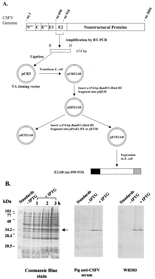
All DNA manipulations were performed essentially according to established procedures (29). The cloning scheme is illustrated (see Fig. 1A). A cDNA coding for the N-terminal portion (aa 690 to 910) of the CSFV Alfort/187 E2 was synthesized from the virus RNA by the reverse transcriptase PCR procedure described previously (11), using primers PA and PB (Table 1). Amplification by PCR was carried out with a GeneAmp PCR system 9700 thermocycler (Perkin-Elmer, Foster City, Calif.). The PCR product of 674 bp was purified using a Wizard PCR Preps DNA purification system (Promega) and ligated into a pCR3 vector followed by transformation into E. coli DH5α. Recombinant plasmids thus constructed were designated pCRE2AB. A 674-bp BamHI-HindIII fragment from pCRE2AB was subcloned into the BamHI and HindIII sites of the pQE30 vector (QIAGEN) to create pQEE2AB. This construct was analyzed with restriction enzymes and sequenced using an ABI model 373 automatic sequencer with an ABI PRISM Dye Terminator Cycle Sequencing Ready Reaction kit (Perkin-Elmer) to ensure that the sequence of E2 gene fragments was correct and in frame at the fusion point. In order to select a vector system suitable for expression of the CSFV E2 gene fragments for epitope mapping, two additional constructs, pHTE2AB and pETE2AB, were made by inserting a 674-bp BamHI-HindIII fragment from pQEE2AB into the BamHI and HindIII sites of pProEx HT and pET30, respectively. For protein expression, pQEE2AB, pHTE2AB, and pETE2AB were transformed into E. coli hosts M15[pREP4], DH5α, and BL21(DE3)/pLysS, respectively.
FIG. 1.
(A) Schematic representation of cloning and expression in E. coli of a cDNA fragment coding for an N-terminal portion (aa 690 to 910) of CSFV Alfort/187 E2 protein, designated E2AB. Amino acids are numbered according to the polyprotein derived from the CSFV Alfort/187 genome (27) (GenBank accession number X87939). The restriction endonuclease sites used to clone the cDNA fragment were depicted as follows: B, BamHI; and H, HindIII. Three expression constructs (pQEE2AB, pHTE2AB, and pETE2AB) were created and tested for their ability to express E2AB from T5, trc, and T7 promoters, respectively. E2AB (□) was well expressed only from pETE2AB, yielding the recombinant product with N-terminal (█) and C-terminal ( ) fusions. A six-histidine tag was placed in the N-terminal fusion for affinity purification of the expressed protein. (B) SDS-PAGE and Western blot analysis of the fusion protein expressed from pETE2AB in the absence (−) or presence (+) of IPTG in E. coli. The cells were lysed with the SDS–β-mercaptoethanol sample buffer and heated at 95°C for 5 min before being loaded onto the gel. In all three panels, each lane except the leftmost lane (molecular mass standards) contained total cell proteins from 1 ml of culture with an A590 of 0.2. Left panel, a 3-h time course experiment showing expression of the E2AB fusion protein (indicated by ←) following induction with 1 mM IPTG; middle panel, reactivity of the E2AB fusion protein with polyclonal IgG antibodies of an experimentally CSFV-infected pig serum; right panel, reactivity of the E2AB fusion protein with MAb WH303.
TABLE 1.
List of primersa used to amplify cDNA fragments coding for targeted regions of the CSFV E2 protein
| Primer | Nucleotide sequence | Position (nucleotides) |
|---|---|---|
| PA | 5′ GGATCCCAGCTAGCCTGCAAGGAAGA 3′ | 2441 to 2460 |
| PB | 5′ AAGCTTGGGTAGTGTGGGAGTCCGTC 3′ | 3083 to 3102 |
| P3 | 5′ GTGGAATTCGAGCTCCTGTTCGACGGGAC 3′ | 2681 to 2700 |
| P4 | 5′ ACAGTCGACTCTGTTCTCAGAGTTGTTGGG 3′ | 2869 to 2889 |
| P5 | 5′ GTGGAATTCGTGGTAAAGACCTTCAGGAG 3′ | 2891 to 2910 |
| P6 | 5′ ACAGTCGACGGGTAGTGTGGGAGTCCGTC 3′ | 3083 to 3102 |
| P7 | 5′ GTGGAATTCTACAATACAACCTTGTTGAAC 3′ | 2783 to 2803 |
| P8 | 5′ ACAGTCGACTTGCCCCCCAACTTACAGTAG 3′ | 2974 to 2994 |
| P9 | 5′ GTGGAATTCCAGCTAGCCTGCAAGGAAGAT 3′ | 2441 to 2461 |
| P10 | 5′ GTGGAATTCGACGGGACCGTTAAGGCCATT 3′ | 2561 to 2581 |
| P11 | 5′ ACAGTCGACCCGTTCAACAAGGTTGTATTG 3′ | 2785 to 2805 |
| P14 | 5′ ACAGAATTCGGGTGGACGGGTGTTATAGAG 3′ | 2834 to 2854 |
| P15 | 5′ GTGGTCGACCAATCCATTCTGTGCGGAAAG 3′ | 2920 to 2940 |
| P33 | 5′ GTGGAATTCAGCCCAACAACTCTGAGAACAG 3′ | 2867 to 2888 |
| P44 | 5′ ACAGAATTCACGGGTGTTATAGAGTGCACAGC 3′ | 2840 to 2862 |
| P45 | 5′ GTGGAATTCGTTATAGAGTGCACAGCAGTG 3′ | 2846 to 2866 |
| P46 | 5′ ACAGAATTCGAGTGCACAGCAGTGAGCCCA 3′ | 2852 to 2872 |
| P47 | 5′ ACAGAATTCACAGCAGTGAGCCCAACAACTC 3′ | 2858 to 2879 |
| P48 | 5′ ACAGTCGACCTCAGAGTTGTTGGGCTCACTGC 3′ | 2861 to 2883 |
| P49 | 5′ ACAGTCGACACCACTTCTGTTCTCAGAGTTG 3′ | 2874 to 2895 |
Deletion constructs.
DNA fragments coding for a targeted region of the CSFV E2 were derived by PCR from pETE2AB with a thermostable DNA polymerase mixture of Taq and Deep Vent as described previously (17) by using a pair of oligonucleotide primers listed in Table 1. EcoRI or SalI restriction sites are incorporated at 5′ ends of the primers. The PCR products were inserted as EcoRI-SalI fragments into the EcoRI and SalI sites of pET30 to produce a series of deletion constructs: pETE2-7/6, pETE2-5/6, pETE2-7/8, pETE2-15/14, pETE2-3/49, pETE2-44/6, pETE2-45/6, pETE2-46/6, pETE2-47/6, pETE2-33/6, pETE2-3/4, pETE2-3/48, pETE2-9/8, pETE2-9/4, pETE2-9/11, and pETE2-10/11. The primer pairs used for PCR amplification are indicated in respective constructs. pETE2SH was constructed by subcloning a 428-bp SacI-HindIII fragment from pETE2AB into the SacI and HindIII sites of pET30, and pETE2BS was obtained by inserting a 252-bp BamHI-SacI fragment from pETE2AB into the BamHI and SacI sites of pET30. All constructs were sequenced as above to ensure that the E2 gene segments were in frame at the fusion point. The constructs express an additional 50-aa fusion at the N terminus, including a six-histidine tag and an additional 37-aa fusion at the C terminus, with the exception that the recombinant product expressed from pETE2BS contains an additional 20-aa fusion at the C terminus.
Expression of the E2 gene segments in E. coli.
BL21(DE3)/pLysS cells harboring an E2 deletion construct derived from pETE2AB were cultured at 37°C in 10 to 20 ml of Luria-Bertani broth supplemented with kanamycin (30 μg/ml). Following growth to an A590 of between 0.5 and 1.0, the culture was induced by addition of 1 mM isopropyl-β-d-thiogalactopyranoside (IPTG) to express the recombinant fusion protein for 3 h. Whole-cell proteins from induced cells were analyzed by Western blotting for reactivity to WH303 or pig polyclonal IgG antibodies. For purification of the minimal antigenic domain of E2 expressed from pETE2-9/11, a culture of 2 liters was grown. After induction with IPTG for 3 h, cells were harvested by centrifugation at 10,000 × g for 20 min. The cell pellets were stored frozen (−80°C) until use.
SDS-PAGE and Western blotting.
SDS-polyacrylamide gel electrophoresis (SDS-PAGE) was carried out by the method described by Laemmli (16), with 4% stacking gels and 12% resolving gels and a Bio-Rad minigel apparatus. The separated proteins were either stained with Coomassie blue or analyzed by use of Western blots (19) probed with MAb WH303 or IgG antibodies (hyperimmune serum) from an experimentally CSFV-infected pig (4). Bound antibodies were detected by using horseradish peroxidase (HRP)-conjugated goat anti-mouse (Jackson ImmunoResearch Laboratories, Inc., West Grove, Pa.) or rabbit anti-swine IgG (ICN Pharmaceuticals Inc., Aurora, Ohio) with a 4-chloro-1-naphthol–H2O2 substrate kit (Bio-Rad, Mississauga, Ontario, Canada) according to the manufacturer's instructions.
Immunological analysis of synthetic peptides.
A 15-mer peptide (pep 415, CTAVSPTTLRTEVVK) containing the WH303 epitope and a 15-mer nonspecific peptide (pep 191, KWGGNWTCVKGEPVT) were synthesized and purified (>95%) using high-pressure liquid chromatography in the Eastern Quebec Peptide Sequencing Facility, Le Centre Hospitalier de l'Université Laval (CHUL) Research Center (Sainte-Foy, Quebec, Canada). Reactions of these synthetic peptides with WH303 were assessed by enzyme-linked immunosorbent assay (ELISA) and dot blots. The ELISA was carried out as described below. The wells of a microtiter plate were coated with purified recombinant E2AB (1 μg/ml in 60 mM carbonate buffer [pH 9.6], 100 μl/well), incubated overnight at room temperature, and frozen at −20°C. After washing with PBS-T (0.05% Tween 20 in PBS [pH 7.2]), various dilutions of a synthetic peptide in PBS-T (50 μl/well) were added to the plate followed by the addition of WH303 (50 μl/well, in PBS-T) previously titrated to give an A414 of between 0.8 and 1.2 in an ELISA. Control wells of buffer and MAb were also prepared. The plate was placed at room temperature for 1 h and washed with PBS-T, followed by incubation with horseradish peroxidase-conjugated goat anti-mouse IgG (1:8,000 dilution in PBS-T, 100 μl/well) for 1 h at room temperature. After a final washing with PBS-T, 100 μl of a substrate solution containing 2,2′-azinobis(3-ethylbenzthiazolinesulfonic acid) (1 mM) and H2O2 (0.015%) in 50 mM sodium citrate buffer (pH 4.5) was added to each well. Following incubation with moderate shaking for 10 min at room temperature, absorbances were determined at 414 nm on a Labsystems Multiskan Bichromatic plate photometer. Dot blotting was performed by spotting a peptide solution onto a nitrocellulose membrane pretreated with 4% (wt/vol) ovalbumin in PBS for 1 min and than 2.5% glutaraldehyde for 10 min. Blots were probed with WH303 followed by immunostaining as described above.
Purification of the minimal antigenic domain of CSFV E2.
Frozen cells from a 2-liter culture were resuspended in about 40 ml of the extraction buffer (6 M guanidine hydrochloride, 0.1 M NaH2PO4, 1 mM phenylmethylsulfonyl fluoride, and 0.01 M Tris [pH 8.0]) with stirring at room temperature for 1 h and then at 4°C overnight. The cell extract was centrifuged at 30,900 × g for 30 min. The supernatant was collected and loaded at 0.5 ml/min onto a column of Ni-NTA agarose (1 by 7.5 cm) that had been preequilibrated with buffer A (8 M urea, 0.1 M NaH2PO4, and 0.01 M Tris [pH 8.0]). The column was washed with 40 ml of buffer A followed by buffer B (8 M urea, 0.1 M NaH2PO4, 0.5 M NaCl, and 0.01 M Tris [pH 6.3]) and buffer C (8 M urea, 0.1 M NaH2PO4, 0.5M NaCl, 5 mM imidazole, and 0.01 M Tris [pH 5.9]). The denatured recombinant product was refolded by washing the column with 40 ml of Tris-buffered saline (TBS; pH 7.4) containing 1 M urea followed by 80 ml of TBS (pH 7.4). Proteins were then eluted from the column with 200 mM imidazole in TBS. Fractions of 2 ml were collected, and the absorbance was monitored at 280 nm. The peak fractions were pooled, mixed with an equal volume of buffer D (10 mM glycine, 5% glycerol, 0.005% Tween 20, and 25 mM Tris-HCl [pH 8.0]), and loaded at 1 ml/min onto a column of Q-Sepharose (1.5 by 11.5 cm) that had been preequilibrated with buffer D. After washing with 50 ml of buffer D, proteins were eluted with a 10 to 500 mM NaCl linear gradient in buffer D (100 ml). Fractions of 2 ml were collected, their A280s were determined, and they were then analyzed by SDS-PAGE. The fractions containing the desired protein were pooled and stored at −20°C. Protein concentration was determined by the method described by Bradford (2), using bovine serum albumin as the standard.
RESULTS AND DISCUSSION
This study was undertaken to define the epitope of WH303 and the minimal region (or domain) of CSFV Alfort/187 E2 required for binding IgG antibodies from an experimentally CSFV-infected pig. This can be achieved by expressing various truncated forms of the protein in E. coli and testing the reactivity of recombinant products with specific antibodies. A structural model for the CSFV E2, based upon the antigenic study with a panel of MAbs (35), predicted that the N-terminal part of E2 was surface exposed with a membrane-spanning region composed of a short stretch of C-terminal residues. Accordingly, the exposed region (aa 690 to 910) of E2, designated E2AB, was expressed and targeted for a series of deletion experiments to determine the binding site(s) recognized by WH303 or pig polyclonal IgG antibodies. It was recognized that a recombinant protein could be expressed at various levels from different vectors (8). Three constructs (pQEE2AB, pHTE2AB, and pETE2AB) were generated to test expression of E2AB by using three different vector systems enabling IPTG-induced expression of the inserted gene fragment from T5, trc, and T7 promoters, respectively (Fig. 1A). All the constructs were designed to express a six-histidine fusion to the N terminus of E2AB. Expression of pQEE2AB in strain M15[pRep4], pHTE2AB in DH5α, and pETE2AB in BL21(DE3)/pLysS was analyzed by SDS-PAGE and Western blotting with MAb WH303 or the hyperimmune serum of an experimentally CSFV-infected pig. pQEE2AB did not appear to yield a measurable level of the recombinant product, as revealed by SDS-PAGE and Western blotting probed with WH303 (data not shown). Although expression of the E2AB fusion protein from pHTE2AB was not evident on SDS-PAGE, the fusion protein was clearly detected by both WH303 and pig IgG antibodies (data not shown), indicating a low level of expression. Expression with pETE2AB in E. coli was prominent after induction with IPTG, as shown by SDS-PAGE analysis of the recombinant product in a 3-h time course experiment (Fig. 1B). Upon induction, a protein band of approximately 34.5 kDa appeared; this was close to the predicted size of the E2AB fusion protein (34.812 kDa). This protein was detected on Western blots probed with anti-histidine tag MAb (data not shown), IgG antibodies in a pig hyperimmue serum, and WH303. The noninduced cells containing pETE2AB (Fig. 1B) and the noninduced or induced cells harboring pET30 (not shown) displayed no protein band in this region. These results exhibited a significant expression of the fusion protein from pETE2AB and indicated that the antigenicity of the expressed protein appears to be independent of postranslational modifications, such as glycosylation. Thus, the pET30 system was used to produce targeted deletions of CSFV Alfort/187 E2 throughout this study.
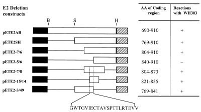
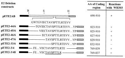
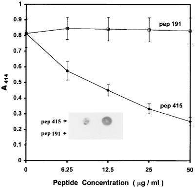
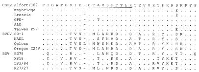
Deletion analysis was first performed to define the epitope recognized by WH303, which was shown to react with all 56 strains of CSFV and none of the BVDV and BDV strains tested in a previous study by Edwards and Sands (10). The finding that WH303 was capable of recognizing the E2AB polypeptide on Western blots (Fig. 1B) indicated that its epitope was located within this region. The fusion polypeptides expressed from a number of deletion constructs (Fig. 2) were analyzed by Western blotting to map this epitope. Deletions of 79 and 114 aa from the N terminus of E2AB, as demonstrated by the constructs pETE2SH and pETE2-7/6, did not affect the binding of polypeptides to WH303. However, the polypeptide expressed from pETE2-5/6, which gave a deletion of 150 aa from the N terminus of E2AB, completely abolished its reactivity with WH303. These results indicated that at least a part of the epitope was located within 36 aa (between aa 804 and 839). Three constructs (pETE2-7/8, pETE2-15/4, and pETE2-3/49) with deletions at both termini of E2AB were generated to express the polypeptides that contain a common 19-mer peptide (GWTGVIECTAVSPTTLRTEVV) between aa 821 and 839. All these products reacted with WH303, indicating that this 19-mer peptide contains the epitope recognized by WH303. In order to further map the WH303-defined epitope, five constructs with serial deletions of 2 or 3 aa from the N terminus of the 19-mer peptide (pETE2-44/6, pETE2-45/6, pETE2-46/6, pETE2-47/6, and pETE2-33/6) were made and tested for the binding of polypeptides to WH303 (Fig. 3). All the recombinant products except the one expressed from pETE2-33/6 reacted with WH303, suggesting that the 3-mer peptide TAV between aa 829 and 831 contained a residue(s) critical for binding WH303. Similarly, two constructs (pETE2-3/4 and pETE2-3/48) with serial deletions of 2 aa from the C terminus of the 19-mer peptide were generated; both polypeptides were capable of binding WH303. These deletion experiments have mapped the epitope recognized by WH303 to a 9-mer peptide TAVSPTTLR between aa 829 and 837, which contains no predicted N-linked glycosylation sequence. To further confirm this, a synthetic peptide (pep 415) containing the 9-mer peptide sequence was evaluated for its ability to inhibit WH303 binding to the recombinant E2AB in a competitive ELISA and for its ability to bind WH303 in a dot blot. As shown in Fig. 4, pep 415 was capable of inhibiting WH303 from binding to E2AB in a concentration-dependent manner. Consistent with the ELISA result was the observation that pep 415 immobilized on a nitrocellulose membrane was detected by WH303 in a dot blot (Fig. 4, inset). In contrast, a nonspecific peptide denoted pep 191 had no inhibitory effect on binding of WH303 to E2AB in the ELISA and showed no reaction with WH303 in dot blotting, indicating that the reaction of pep 415 with WH303 was specific. All these experiments support the conclusion that the epitope recognized by WH303 was a 9-mer peptide TAVSPTTLR between aa 829 and 837. Previously, Yu et al. (40) mapped a linear epitope to a few residues in the C-terminal membrane-spanning region of the CSFV strain Brescia E2, which is highly conserved among different pestiviruses. The present study has defined a different epitope at a high resolution on the CSFV Alfort/187 E2 protein. Comparison of the sequences around the TAVSPTTLR region among the E2 proteins from different strains of pestiviruses indicated that the sequence TAVSPTTLR is strongly conserved among the strains of CSFV and is highly variable among strains of BVDV and BDV (Fig. 5). This provides a structural basis for the finding of Edwards and Sands (10) that WH303 reacted with all 56 strains of CSFV but reacted with none of the BVDV and BDV strains tested. The practical implication of these results is that a synthetic peptide containing the WH303-defined epitope may be useful for detection of the E2 protein in a competitive binding assay for the diagnosis of viral infection and for the identification of CSFV strains. The peptide TAVSPTTLR, in combination with MAb WH303, might be a generally useful fusion tag system for immunoaffinity purification of recombinant proteins. The interaction of pep 415 in both liquid and solid phases with WH303 (Fig. 4) further supports the feasibility of these practical uses.
FIG. 2.
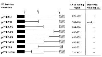
Localization of the WH303-defined epitope within a region of the CSFV Alfort/187 E2 protein. (Left panel) Deletion constructs for expression of targeted regions of E2 were generated as described in Materials and Methods. The restriction endonuclease sites used to construct the deletions are depicted as follows: B, BamHI; H, HindIII; and S, SacI. □, the regions of E2; —, deleted portions of E2; █, the N-terminal fusion; , C-terminal fusion. (Right panel) Reactivity of the fusion polypeptides with WH303 was analyzed by Western blotting. +, specific reaction showing bands of similar intensity; −, no reaction.
FIG. 3.
High-resolution mapping of the WH303-defined epitope. Serial deletions of 2 or 3 aa from the N or C terminus of the 19-mer peptide (GWTGVI ECTAVSPTTLRTEVV) between aa 821 and 839 of the CSFV Alfort/187 E2 protein were constructed as described in Materials and Methods, and the recombinant products were analyzed by Western blots probed with WH303. The regions of E2 are represented by single-letter amino acid codes. Other symbols used here are explained in the Fig. 2 legend.
FIG. 4.
Inhibition of binding of MAb WH303 to the recombinant E2AB by synthetic peptides. The binding of WH303 to the immobilized E2AB was tested by ELISA in the presence of varying amounts of pep 415 or pep 191. Error bars represent standard deviations (n = 3). A dot blot (inset) shows the reaction of immobilized pep 415 or pep 191 (both at 1 and 10 μg) with WH303.
FIG. 5.
Comparison of the CSFV Alfort/187 E2 sequence around the TAVSPTTLR region (underlined) with those from other strains of pestiviruses. The E2 protein sequences selected here for comparison are from CSFV strains Alfort/187 (27) (GenBank accession number X87939), Weybridge (39) (GenBank accession number X71780), Brescia (22) (GenBank accession number M31768), GPE− (14) (GenBank accession number D49533), ALD (14) (GenBank accession number D49532), and Taiwan P97 (30) (GenBank accession number U43924); BVDV strains SD-1 (7) (GenBank accession number M96751), NADL (5), Osloss (6) (GenBank accession number M96687), and Oregon C24V (25) (GenBank accession number L07496); and BDV strains BD78 (31) (GenBank accession number U18330), X818 (1) (GenBank accession number AF037405), L83/84 (1) (GenBank accession number U00890), and R27/27 (1) (GenBank accession number U00891). Amino acid residues that match exactly those of the CSFV Alfort/187 E2 protein are represented as dots. Gaps are indicated by dashes.
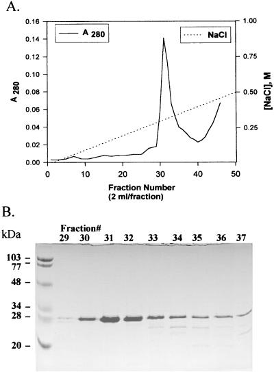
A total of eight constructs of targeted deletions of CSFV Alfort/187 E2 were generated to define the minimal domain essential for binding polyclonal IgG antibodies from an experimentally CSFV-infected pig (Fig. 6). The E2SH fusion protein (aa 769 to 910), representing a deletion of 79 aa from the N terminus of E2AB, showed a weak reaction with pig IgG antibodies. A deletion of 115 aa from the N terminus of E2AB, as shown by the product expressed from pETE2-7/6, eliminated the reactivity with pig IgG antibodies. These results suggested that at least partial IgG-binding sites were confined to the N-terminal region within about 100 aa residues. In order to confirm this, C-terminal deletions of E2AB were accomplished by making three constructs (pETE2-9/8, pETE2-9/4, and pE2-9/11), which deleted 37, 71, and 98 aa from the C terminus, respectively. All three fusion protein fragments were capable of reacting with pig IgG antibodies. A further deletion of 140 aa from the C terminus of E2AB, as demonstrated by the fusion product expressed from pETE2BS, destroyed the binding of the protein segment to pig IgG antibodies. These results support the above conclusion that the pig IgG-binding sites are confined to the N-terminal region within about 100 aa. They also implied that the sequence between aa 771 and 812 could contain the IgG-binding sites or elements critical for binding to pig IgG antibodies. To differentiate the two possibilities, a fusion polypeptide spanning aa 730 to 812 expressed from pETE2-10/11, which deleted 98 aa from the C terminus of E2AB and 40 aa from its N terminus, was tested for binding to pig IgG antibodies; no reactivity was observed. It therefore can be concluded that (i) both sequences (aa 771 to 812 and aa 690 to 730) contain elements critical for binding by pig IgG antibodies, (ii) neither sequence alone contains functional pig IgG-binding sites, and (iii) the minimal antigenic domain essential for binding pig IgG antibodies is located within the N-terminal region of 123 residues between aa 690 and 812, 17 aa upstream from the WH303 epitope. These conclusions are consistent with the Western blot result demonstrating no reactivity of the E2-9/11 polypeptide with WH303 (data not shown), unpublished data (A. Clavijo) showing that a number of sera from CSFV-infected animals were incapable of inhibiting WH303 from binding to E2AB in an ELISA, and the structural model for the CSFV E2 proposed by van Rijn et al. (35). This work is the first to define a minimal antigenic domain required for binding polyclonal antibodies from an infected host. It is also of practical importance in that this minimal antigenic domain may be (i) a potential vaccine and (ii) a more specific diagnostic reagent than the full-length E2 protein because the regions cross-reactive with other pestiviruses may have been removed. Moreover, the smaller size of this domain meets the requirement for development of a particular diagnostic test, such as the fluorescence polarization assay (3, 18, 19, 23, 24), for the detection of anti-CSFV antibodies. Since the minimal antigenic domain was expressed from pETE2-9/11 in E. coli in large amounts and purified to homogeneity by a combination of affinity and ion-exchange chromatography (Fig. 7), evaluation of its diagnostic potential is now possible.
FIG. 6.
Definition of the minimal domain of the CSFV Alfort/187 E2 protein essential for binding by polyclonal IgG antibodies of an experimentally CSFV-infected pig serum. Deletion constructs for expression of targeted regions of E2 were generated as described in Materials and Methods. Reactivity of the fusion products with pig antiserum was analyzed by Western blotting. Symbols used here are explained in the Fig. 2 legend.
FIG. 7.
(A) Q-Sepharose chromatography of the fusion protein containing the minimal antigenic domain of CSFV Alfort/187 E2 protein. The fusion protein, purified first by Ni-NTA agarose affinity chromatography, was applied onto a column of Q-Sepharose (1.5 by 11.5 cm) and eluted with a 10 to 500 mM NaCl linear gradient in 100 ml of buffer D (10 mM glycine, 5% glycerol, 0.005% Tween 20, and 25 mM Tris-HCl [pH 8.0]). (B) SDS-PAGE analysis of the peak fractions (no. 29 to 37) collected from Q-Sepharose chromatography. Each lane except the leftmost lane (molecular mass standards) contained 10 μl of the individual sample.
In summary, the present study has employed recombinant technology to dissect the major glycoprotein E2 of CSFV strain Alfort/187 in order to map its MAb and polyclonal antibody-binding sites. The epitope recognized by WH303 was successfully defined to be a 9-mer peptide (TAVSPTTLR) between aa 829 and 837. In addition, the minimal domain of E2 required for binding pig polyclonal IgG antibodies was reported for the first time to be located in the N-terminal region of E2 within about 100 aa. Successful definition of the WH303 or pig polyclonal IgG-binding sites of the CSFV Alfort/187 E2 has provided useful information for the future development of diagnostic tests. Since synthesis of a peptide containing the WH303-defined epitope and production and purification of the minimal antigenic domain of CSFV Alfort/187 E2 from E. coli are easily achieved, studies are in progress to evaluate these reagents for their potential use in the diagnosis of CSFV infection.
ACKNOWLEDGMENTS
We thank G. Randall and F. Thomas for helpful discussion and suggestions, S. Nadin-Davis and K. Nielsen for critical review of the manuscript, and A. Bergern for determination of DNA sequences.
This work was supported in part by Diachemix Corporation (Grayslake, Ill.).
REFERENCES
- 1.Becher P, Shannon A D, Tautz N, Thiel H-J. Molecular characterization of border disease virus, a pestivirus from sheep. Virology. 1994;198:542–551. doi: 10.1006/viro.1994.1065. [DOI] [PubMed] [Google Scholar]
- 2.Bradford M M. A rapid and sensitive method for the quantitation of microgram quantities of protein utilizing the principle of protein-dye binding. Anal Biochem. 1976;72:248–254. doi: 10.1016/0003-2697(76)90527-3. [DOI] [PubMed] [Google Scholar]
- 3.Bughio N I, Lin M, Surujballi O P. Use of recombinant flagellin protein as a tracer antigen in a fluorescence polarization assay for diagnosis of leptospirosis. Clin Diagn Lab Immunol. 1999;6:599–605. doi: 10.1128/cdli.6.4.599-605.1999. [DOI] [PMC free article] [PubMed] [Google Scholar]
- 4.Clavijo A, Zhou E-M, Vydelingum S, Heckert R. Development and evaluation of a novel antigen capture assay for the detection of classical swine fever virus antigens. Vet Microbiol. 1998;60:155–168. doi: 10.1016/s0378-1135(98)00160-6. [DOI] [PubMed] [Google Scholar]
- 5.Collett M S, Larson R, Gold C, Strick D, Anderson D K, Purchio A F. Molecular cloning and nucleotide sequence of the pestivirus bovine viral diarrhea virus. Virology. 1988;165:191–199. doi: 10.1016/0042-6822(88)90672-1. [DOI] [PubMed] [Google Scholar]
- 6.de Moerlooze L, Lecomte C, Brown-Shimmer S, Schmetz D, Guiot C, Vandenbergh D, Allaer D, Rossius M, Chappuis G, Dina D, Renard A, Martial J A. Nucleotide sequence of the bovine viral diarrhea virus Osloss strain: comparison with related viruses and identification of specific DNA probes in the 5′ untranslated region. J Gen Virol. 1993;74:1433–1438. doi: 10.1099/0022-1317-74-7-1433. [DOI] [PubMed] [Google Scholar]
- 7.Deng R, Brock K V. Molecular cloning and nucleotide sequence of a pestivirus genome, noncytopathic bovine viral diarrhea virus strain SD-1. Virology. 1992;191:867–869. doi: 10.1016/0042-6822(92)90262-n. [DOI] [PubMed] [Google Scholar]
- 8.Ge Y, Charon N W. FlaA, a putative flagellar outer sheath protein, is not an immunodominant antigen associated with Lyme disease. Infect Immun. 1997;65:2992–2995. doi: 10.1128/iai.65.7.2992-2995.1997. [DOI] [PMC free article] [PubMed] [Google Scholar]
- 9.Gough N M. Rapid and quantitative preparation of cytoplasmic RNA from small numbers of cells. Anal Biochem. 1988;173:93–95. doi: 10.1016/0003-2697(88)90164-9. [DOI] [PubMed] [Google Scholar]
- 10.Edwards S, Sands J J. Antigenic comparisons of hog cholera virus isolates from Europe, America and Asia using monoclonal antibodies. DTW (Dtsch Tieraerztl Wochenschr) 1990;97:79–81. [PubMed] [Google Scholar]
- 11.Harding M, Lutze-Wallace C, Prud'homme I, Zhong X, Rola J. Reverse transcriptase-PCR assay for detection of hog cholera virus. J Clin Microbiol. 1994;32:2600–2602. doi: 10.1128/jcm.32.10.2600-2602.1994. [DOI] [PMC free article] [PubMed] [Google Scholar]
- 12.Hulst M M, Westra D F, Wensvoort G, Moormann R J M. Glycoprotein E1 of hog cholera virus expressed in insect cells protects swine from hog cholera. J Virol. 1993;67:5435–5442. doi: 10.1128/jvi.67.9.5435-5442.1993. [DOI] [PMC free article] [PubMed] [Google Scholar]
- 13.Hulst M M, Panoto F E, Hoekman A, van Gennin H G P, Moormann R J M. Inactivation of the RNase activity of glycoprotein Ernsof classical swine fever virus results in a cytopathogenic virus. J Virol. 1998;72:151–157. doi: 10.1128/jvi.72.1.151-157.1998. [DOI] [PMC free article] [PubMed] [Google Scholar]
- 14.Ishikawa K, Nagai H, Katayama K, Tsutsui M, Tanabayashi K, Takeuchi K, Hishiyama M, Saitoh A, Takagi M, Gotoh K, Muramatsu M, Yamada A. Comparison of the entire nucleotide and deduced amino acid sequences of the attenuated hog cholera vaccine strain GPE− and the wild-type parental strain ALD. Arch Virol. 1995;140:1385–1391. doi: 10.1007/BF01322665. [DOI] [PubMed] [Google Scholar]
- 15.Konig M, Lengsfeld T, Pauly T, Stark R, Thiel H-J. Classical swine fever virus: independent induction of protective immunity by two structural glycoproteins. J Virol. 1995;69:6479–6486. doi: 10.1128/jvi.69.10.6479-6486.1995. [DOI] [PMC free article] [PubMed] [Google Scholar]
- 16.Laemmli U K. Cleavage of structural proteins during the assembly of the head of bacteriophage T4. Nature (London) 1970;227:680–685. doi: 10.1038/227680a0. [DOI] [PubMed] [Google Scholar]
- 17.Lin M. Molecular analysis of flaB, a periplasmic flagellar core protein gene in pathogenic leptospires. J Biochem Mol Biol Biophys. 1999;2:181–187. [Google Scholar]
- 18.Lin M, Nielsen K. Binding of the Brucella abortuslipopolysaccharide O-chain fragment to a monoclonal antibody. Quantitative analysis by fluorescence quenching and polarization. J Biol Chem. 1997;272:2821–2827. doi: 10.1074/jbc.272.5.2821. [DOI] [PubMed] [Google Scholar]
- 19.Lin M, Sugden E A, Jolley M E, Stilwell K. Modification of the Mycobacterium bovisextracellular protein MPB70 with fluorescein for rapid detection of specific serum antibodies by fluorescence polarization. Clin Diagn Lab Immunol. 1996;3:438–443. doi: 10.1128/cdli.3.4.438-443.1996. [DOI] [PMC free article] [PubMed] [Google Scholar]
- 20.Moennig V. Characteristics of the virus. In: Liess B, editor. Classical swine fever and related viral infections. Boston, Mass: Martinus Nijhoff Publishers; 1988. pp. 55–88. [Google Scholar]
- 21.Moennig V, Plagemann P G W. The pestiviruses. Adv Virus Res. 1992;41:53–98. doi: 10.1016/s0065-3527(08)60035-4. [DOI] [PubMed] [Google Scholar]
- 22.Moormann R J M, Warmerdam P A M, van der Meer B, Schaper W M M, Wensvoort G, Hulst M M. Molecular cloning and nucleotide sequence of hog cholera virus strain Brescia and location in the genome of the sequence encoding envelope protein E1. Virology. 1990;177:184–198. doi: 10.1016/0042-6822(90)90472-4. [DOI] [PubMed] [Google Scholar]
- 23.Nielsen K, Gall D, Jolley M, Leishman G, Balsevicius S, Smith P, Nicoletti P, Thomas F. Homogeneous fluorescence polarization assay for detection of antibody to Brucella abortus. J Immunol Methods. 1996;195:161–168. doi: 10.1016/0022-1759(96)00116-0. [DOI] [PubMed] [Google Scholar]
- 24.Nielsen K, Gall D, Lin M, Massangill C, Samartino L, Perez B, Coats M, Hennager S, Dajer A, Nicoletti P, Thomas F. Diagnosis of bovine brucellosis using a homogeneous fluorescence polarization assay. Vet Immunol Immunopathol. 1998;66:321–329. doi: 10.1016/s0165-2427(98)00195-0. [DOI] [PubMed] [Google Scholar]
- 25.Paton D J, Lowings J P, Barrett A D. Epitope mapping of the gp53 envelope protein of bovine viral diarrhea virus. Virology. 1992;190:763–772. doi: 10.1016/0042-6822(92)90914-b. [DOI] [PubMed] [Google Scholar]
- 26.Paton D J, Sands J J, Lowings J P, Smith J E, Ibata G, Edwards S. A proposed division of the pestivirus genus using monoclonal antibodies, supported by cross-neutralisation assays and genetic sequencing. Vet Res. 1995;26:92–109. [PubMed] [Google Scholar]
- 27.Ruggli N, Moser C, Mitchell D, Hofmann M, Tratschin J D. Baculovirus expression and affinity purification of protein E2 of classical swine fever virus strain Alfort/187. Virus Genes. 1995;10:115–126. doi: 10.1007/BF01702592. [DOI] [PubMed] [Google Scholar]
- 28.Rumenapf T, Stark R, Meyers G, Thiel H-J. Structural proteins of hog cholera virus expressed by vaccinia virus: further characterization and induction of protective immunity. J Virol. 1991;65:589–597. doi: 10.1128/jvi.65.2.589-597.1991. [DOI] [PMC free article] [PubMed] [Google Scholar]
- 29.Sambrook J, Fritsch E F, Maniatis T. Molecular cloning: a laboratory manual. 2nd ed. Cold Spring Harbor, N.Y: Cold Spring Harbor Laboratory Press; 1989. [Google Scholar]
- 30.Shiu J-S, Chang M-H, Liu S-T, Ho W-C, Lai S-S, Chang T-J, Chang Y-S. Molecular cloning and nucleotide sequence determination of three envelope genes of classical swine fever virus Taiwan isolate p97. Virus Res. 1996;41:173–178. doi: 10.1016/0168-1702(96)01286-5. [DOI] [PubMed] [Google Scholar]
- 31.Sullivan D G, Chang G J, Trent D W, Akkina R K. Nucleotide sequence analysis of the structural gene coding region of the pestivirus border disease virus. Virus Res. 1994;33:219–228. doi: 10.1016/0168-1702(94)90104-x. [DOI] [PubMed] [Google Scholar]
- 32.van Rijn P A, Bossers A, Wensvoort G, Moormann R J M. Classical swine fever virus (CSFV) envelope glycoprotein E2 containing one structural antigenic unit protects pigs from lethal CSFV challenge. J Gen Virol. 1996;77:2737–2745. doi: 10.1099/0022-1317-77-11-2737. [DOI] [PubMed] [Google Scholar]
- 33.van Rijn P A, van Gennip R G P, de Meijer E J, Moormann R J M. A preliminary map of epitopes on envelope glycoprotein E1 of HCV strain Brescia. Vet Microbiol. 1992;33:221–230. doi: 10.1016/0378-1135(92)90050-4. [DOI] [PubMed] [Google Scholar]
- 34.van Rijn P A, van Gennip H G P, de Meijer E J, Moormann R J M. Epitope mapping of envelope glycoprotein E1 of hog cholera virus strain Brescia. J Gen Virol. 1993;74:2053–2060. doi: 10.1099/0022-1317-74-10-2053. [DOI] [PubMed] [Google Scholar]
- 35.van Rijn P A, Miedema G K W, Wensvoort G, van Gennip H G P, Moormann R J M. Antigenic structure of envelope glycoprotein E1 of hog cholera virus. J Virol. 1994;68:3934–3942. doi: 10.1128/jvi.68.6.3934-3942.1994. [DOI] [PMC free article] [PubMed] [Google Scholar]
- 36.Weiland E, Stark R, Hass B, Rumenapf T, Meyers G, Thiel H-J. Pestivirus glycoprotein which induces neutralizing antibodies forms part of a disulfide-linked heterodimer. J Virol. 1990;64:3563–3569. doi: 10.1128/jvi.64.8.3563-3569.1990. [DOI] [PMC free article] [PubMed] [Google Scholar]
- 37.Wengler G. Flaviviridae. In: Francki R I B, Fauquet C M, Knudson D L, Brown F, editors. The classification and nomenclature of viruses: fifth report of the International Committee on Taxonomy of Viruses. New York, N.Y: Springer-Verlag; 1991. pp. 223–233. [Google Scholar]
- 38.Wensvoort G. Topographical and functional mapping of epitopes on hog cholera virus with monoclonal antibodies. J Gen Virol. 1989;70:2865–2876. doi: 10.1099/0022-1317-70-11-2865. [DOI] [PubMed] [Google Scholar]
- 39.Yu M, McColl K A, Gould A R. Cloning and nucleotide sequence determination of the major envelope glycoprotein (gp55) gene of hog cholera virus (Weybridge) Virus Res. 1993;28:203–208. doi: 10.1016/0168-1702(93)90137-c. [DOI] [PubMed] [Google Scholar]
- 40.Yu M, Wang L-F, Shiell B J, Morrissy C J, Westbury H A. Fine mapping of a C-terminal linear epitope highly conserved among the major envelope glycoprotein E2 (gp51 to gp54) of different pestiviruses. Virology. 1996;222:289–292. doi: 10.1006/viro.1996.0423. [DOI] [PubMed] [Google Scholar]


