Abstract
Pollution by heavy metal ions has a serious impact on human health and the environment, which is why the monitoring of heavy metal ions is of great practical importance. In this work, we describe the development of an electrochemical sensor for the detection of cadmium (Cd2+) involving the doping of porous SiO2 spheres with ZnO nanoparticles. Zinc oxide is chosen as the central dopant in the composite material to increase the conductivity and thus improve the electrochemical detection of Cd2+ ions with the SiO2 spheres. The resulting composite is characterized by electrochemical spectroscopic XRD and microscopic methods. As a result, the developed sensor shows good selectivity towards the targeted Cd2+ ions compared to other divalent ions. After optimization of the experimental conditions, the electrochemical sensor shows two different linear ranges between 2.5 × 10⁻¹¹ molL−1 to 1.75 × 10⁻¹⁰ molL−1 and 2 × 10⁻⁹ molL−1 to 1.75 × 10⁻⁹ molL−1, indicating a change from diffusion-controlled to surface-controlled oxidation of Cd2+. A detection limit was reached at 4.4 × 10⁻¹¹ molL−1. In addition, it offers good repeatability and recovery, and can detect accurate trace amounts of Cd2+ ions in real samples such as tap water or seawater by spiking these samples with known Cd2+ concentrations. This setup also provides satisfactory recovery rates in the range of 89–102%.
Keywords: electrochemical sensor, Zinc oxide, Porous silica coating, Cd2+ detection, ZnO@SiO2 NPs
1. Introduction
Cadmium is present in the environment in elemental form or as various salts that enter drinking water sources through natural processes (leaching from the soil), human activities (product refinement or technological applications), or leaching from certain types of pipes and known components [1]. This not only affects the health of humans, animals, and plants, but also the ability of the environment to sustain life. As heavy metals are not biodegradable, they accumulate in the food chain. According to the World Health Organization (WHO) [2], a heavy metal concentration above the permissible limit can be toxic, carcinogenic, and harmful to human health [3]. Their high water solubility facilitates their spread in the environment and leads to environmental pollution [4]. The maximum level of Cd2+ ions in drinking water is 3.0 μgL−1, according to US Environmental Protection Agency (USEPA) standards [5]. It accumulates in humans, reacts with enzymes, and generates free radicals [6], leading to serious health problems. Recent studies have classified Cd2+ as a type I human carcinogen [7]. Therefore, the reliable detection of Cd2+ is of utmost importance. Effective methods for the determination of Cd2+ ions require high selectivity and sensitivity [8]. Various methods are used for the detection of heavy metal ions, which are divided into three classes: electrochemical [9,10,11], gravimetric [12,13], and optical detection methods [14].
However, despite the excellent sensitivity and selectivity achieved with these techniques, sophisticated instrumentation and competent personnel for careful sample preparation are unsuitable for on-site screening [15,16,17]. Among the recently developed technologies for Cd2+ detection, electrochemical sensors are considered economical because the equipment and operating procedures are generally simpler and less expensive than other technologies [18]. In addition, electrochemical sensors have many advantages, including being cheap, portable (in situ real-time detection), simple, and offering higher sensitivity and specificity than the detection methods already mentioned [19,20,21]. It has been found that that nanocomposites are core components and play an important role in the development of sensitive electrochemical Cd2+ sensors. Electrochemical methods are widely used due to their low cost, simple equipment, easy operation, and strong anti-interference capabilities [22,23]. To improve the sensitivity of the electrochemical sensor, various chemically modified electrodes with nanomaterials [24], magnetic substances [25,26], automotive materials [27], and biological composites [28] have been used. However, these methods are limited in their ability to specifically detect Cd2+ ions. Although silica nanoparticles (NP) are an electronic insulator, they are widely used in electrochemical analysis due to their large effective surface area with high selectivity and good sensitivity to heavy metal ions [29,30,31,32].
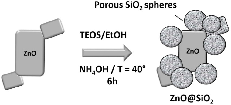
In this work, a sensitive and selective electrochemical sensor based on ZnO@SiO2 was fabricated for the detection of Cd2+ ions in real samples. First, these particles were prepared by a modified sol-gel method in the presence of tetraethyl orthosilicate (TEOS). In this technique, a layer of silicon dioxide spheres is formed around the ZnO particles at a controlled temperature of 40 °C. The electrochemical performance of the fabricated sensors was investigated under optimized experimental conditions. The results showed that these sensors were very sensitive to Cd2+ ions. Furthermore, by comparing the response current of the electrochemical sensor with other metal ions using differential pulse voltammetry (DPV), it was proven that the sensor exhibited increased signal performance for Cd2+ ions. The sensor was also tested on real samples, with deviations ranging from 89 to 102% regarding the spiked Cd2+ concentration as 100%.
2. Materials and Methods
2.1. Materials and Reagents
Zinc oxide nanoparticles (ZnO NPs, nanopowder with a particle size < 100 nm), tetraethyl orthosilicate (TEOS), and ethanol (EtOH) were sourced from Sigma-Aldrich. Sodium hydroxide (NaOH), potassium hexacyanoferrate (II) trihydrate (K4[Fe(CN)6]·3H2O), potassium hexacyanoferrate (III) (K3[Fe(CN)6]), potassium chloride (KCl), copper (II) dichloride (CuCl2), lead nitrate (Pb(NO3)2), cadmium nitrate (Cd(NO3)2·4H2O), nickel (II) dichloride hexahydrate (NiCl2·6H2O), manganese (II) dichloride tetrahydrate (MnCl2·4H2O), and iron (II) dichloride (FeCl2) were acquired from Sigma-Aldrich through Chimisi and Chimie Tunisie companies (Tunisia). Acetate buffer solution (ABS, 0.1 molL−1; pH 5) was formulated by mixing a 0.1 molL−1 acetic acid solution (CH3COOH, HAc) and a 0.1 molL−1 sodium acetate solution (CH3COONa; NaAc) in a 1:1 ratio. This ABS served as the supporting electrolyte, with pH adjustment achieved by incorporating solutions of NaOH or HCl.
Varied concentrations of copper (II), lead (II), nickel (II), manganese (II), iron (II), and cadmium (II) were prepared in an acetate buffer solution (ABS) using distillated water. A stock solution of Cd2⁺was diluted until the desired trace concentrations were obtained. For each dilution, the solutions were stirred until stabilization of the corresponding concentration. The experiments for the detection of Cd2+ concentrations were performed using firstly the lowest concentration followed by the higher concentration.
2.2. Preparation of ZnO@SiO2
In this step, 0.5 g ZnO nanoparticles were dispersed in 50 mL ethanol and 10 mL water for 30 min in an ultrasonic bath. Then 1.6 mL of ammonia solution (1 molL−1) and 8 mL of TEOS were successively added to the Zinc oxide dispersion with vigorous stirring for 6 h at 40 °C. The mixture was washed three times with ethanol and distilled water and finally dried in an oven at 60 °C for 12 h (Figure 1).
Figure 1.
Schematic illustration of the formation of the ZnO@SiO2 nanocomposite.
2.3. Fabrication of ZnO@SiO2/GCE Modified Electrodes
Before each use, glassy carbon electrodes (GCE, Ø = 3 mm) were polished with 0.05 μm aluminum oxide powder using emery paper, rinsed with distilled water, and then ultrasonically cleaned in ethanol for 5 min to obtain a reproducible condition. After cleaning the electrode, 1 mg of ZnO@SiO2 was dispersed in 1 mL of ethanol by sonication for 30 min and then 3 to 20 μL of the suspension was applied to the GCE. The modified electrodes were then dried at room temperature before being used for measurements.
2.4. Characterization of As-Prepared Samples
The morphologies and microstructures of the samples were analyzed by scanning electron microscopy (SEM) using a JEOL JEM-2100 FX microscope (JEOL Ltd., Tokyo, Japan). Transmission electron microscopy (TEM) images were obtained using a JEOL 2100 microscope at 200 kV (JEOL Ltd., Tokyo, Japan). X-ray diffraction patterns for powder samples were obtained with a Philips PanalyticalX’Pert powder diffractometer using CuKα (λ = 1540 Å) radiation in the 2θ range from 5° to 80°. Infrared (IR) spectra were recorded using a PerkinElmer Spectrum IR version 10.6.2 spectrometer and ultraviolet-visible spectroscopy was performed using a Beckman DU640 UV/Vis to further characterize the samples. Electrochemical measurements were performed with a computer-controlled Au-tolab PG Potentiostat/Galvanostat (AUT 83965) and controlled by Autolab’s NOVA 2.1.6 software (Metrohm, Switzerland). The measurements were performed in a traditional three-electrode cell configuration. The working electrode consisted of a modified GCE, while a platinum wire served as the counter electrode and Ag/AgCl (3 molL−1 KCl) as the reference electrode. In brief, Cd2+ ions were detected using differential pulse voltammetry (DPV). For this purpose, the Cd2+ ions were first reduced and accumulated on the sensor surface at −1 V vs. Ag/AgCl for 5 min. Subsequently, a potential sweep from −1 to −0.4 V was applied to detect the target species under optimized conditions (see Supplementary Data).
3. Results
3.1. Material Characterization
3.1.1. Morphology and Structure of ZnO@SiO2
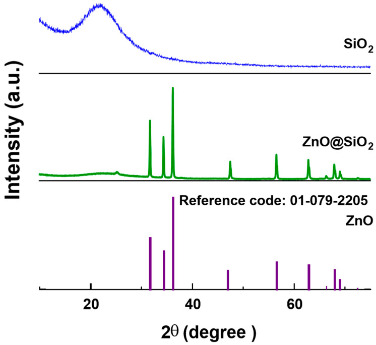
The chemical nature and phase composition of the ZnO@SiO2 nanocomposite were analyzed using the XRD technique. The main peaks in the experimental diffractogram are shown in Figure 2. The characteristic peaks at 2θ = 31.76°, 34.41°, 36.25°, 47.53°, 56.59°, 62.85°, 66.37°, 67.94°, 69.08°, and 76.95° observed in the XRD diffractogram were assigned to planes (100), (002), (101), (102), (110), (103), (200), (112), (201), and (202), respectively. These results confirm the existence of ZnO with a hexagonal wurtzite phase (space group: P63mc, JCPDS Chart No. 01-079-2205). The XRD pattern for SiO2 shows a characteristic broad band centered at about 2θ = 23° associated with the amorphous phase of silica. This broad band is due to the phenomenon of diffuse scattering often observed in amorphous materials. The diffractogram of the ZnO@SiO2 configuration showed that the crystal structure of ZnO was not affected by the presence of SiO2 [33]. This can be attributed to the good dispersion of the ZnO nanoparticles in the SiO2 matrix [34]. These results are in agreement with our previous studies [35,36].
Figure 2.
XRD patterns of SiO2, ZnO@SiO2, and the reference pattern of ZnO (01-079-2205).
3.1.2. Electron Microscopy Analysis
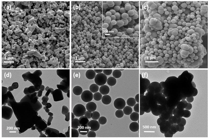
In general, the morphology of almost all metal oxide composites (MO@SiO2) tends to form agglomerates of nanoparticles [37]. Figure 3a shows the scanning electron microscopy (SEM) images of commercial ZnO NPs. The particles have a cubic shape and a rather large size distribution between 100 nm and several micrometers. Figure 3b shows the synthesized SiO2 spheres with an average size of 250 nm. The inset is a magnification of the micrograph to reveal the porous, almost fuzzy, morphology of the NP surface. Figure 3c is a representative SEM image of the ZnO@SiO2 composite. The ZnO particles are mainly covered by the SiO2 spheres and only a few ZnO particles with a SiO2 coating can be seen. Figure 3d–f are TEM images of ZnO particles, SiO2 spheres, and the ZnO@SiO2 composite, respectively. ZnO consists of agglomerates of particles with a broad size distribution, which explains the need for ultrasound to disperse these particles. The SiO2 spheres also form agglomerates, in which the cross-linked SiO2 is clearly visible in the TEM image. The ZnO@SiO2 composite forms denser clusters of SiO2 spheres around the ZnO particle. However, the formation of a SiO2 layer on the ZnO particles is not clearly visible in this microscopic study.
Figure 3.
SEM image of (a) commercial ZnO NPs, (b) synthesized SiO2 NPs with a zoom inset, and (c) ZnO@SiO2.agglomerates. Representative TEM images of (d) commercial ZnO NPs, (e) synthesized SiO2 NPs, and (f) ZnO@SiO2.
3.1.3. Characterization of the Prepared Materials by Infrared Spectroscopy
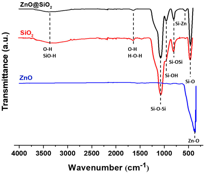
Figure 4 shows the FTIR spectra of ZnO, SiO2, and ZnO@SiO2 samples. Absorption bands at 451, 794, and 1065 cm−1 are observed for the SiO2 nanoparticles. These bands are close to the bands described in the literature [38] (466, 808, and 1100 cm−1) and are consistent with the SiO2 bonding structure. The band corresponds to the symmetric stretching vibration between Si-O bonds of Si-O-Si [39]. Changes in the band characteristics of SiO2 indicate that O-Si-O is perturbed by the presence of oxides in its environment. Studies show that in a mixed crystal, the substitution or filling of vacancies can lead to a shift in the fundamental transverse optical modes. The ZnO spectrum shows only one strong absorption peak at 359 cm−1. This is the characteristic band of the monoclinic phase of pure Zn-O [40]. The band at 3300 cm−1 in the SiO2 and ZnO@SiO2 spectra indicates the presence of water, which is due to moisture absorption from the environment, the -OH stretching vibration of H2O, and the -OH stretching vibration of Si-OH. In addition to the characteristic peaks of ZnO and SiO2, the spectrum of ZnO@SiO2 samples exhibits another small absorption peak at 556 cm−1, which is due to the formation of some Si-O-Zn bonds. These results are in agreement with the results of FTIR studies in the literature [41,42].
Figure 4.
FT-IR spectra of ZnO, SiO2, and ZnO@SiO2 NPs.
3.1.4. UV–Vis Spectra
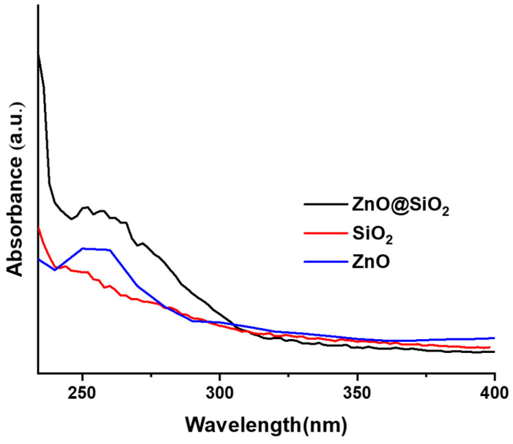
Figure 5 shows the UV-Vis absorption spectra of ZnO, SiO2, and ZnO@SiO2, measured in the wavelength range between 228 nm and 400 nm. In contrast to the spectra of ZnO@SiO2 and ZnO, the spectrum of the pure SiO2 spheres does not show a clearly resolved absorption band, which confirms that the band found in the spectrum of ZnO@SiO2 corresponds to the ZnO in the composite. The spectrum of ZnO@SiO2 shows a clear band between 240 nm and 300 nm, while the band of ZnO@SiO2 appears to be broader, indicating an increase in the transition levels due to the SiO2 spheres bound to the ZnO [43]. In a similar study on a CaO@SiO2 nanocomposite, the maximum absorption wavelength was reported to be 300 nm [44], which is consistent with the optical properties of the present study. The narrow bandwidth at 270 nm is the result of small ZnO nanoparticles remaining in the porous silica structure [38,45].
Figure 5.
UV–Vis spectra for samples of SiO2 and ZnO@SiO2.
3.2. Electrochemical Studies
3.2.1. Optimization of Experimental Variables
To evaluate the effect of different parameters for the detection of Cd2+ with ZnO@SiO2 modified electrodes, we determined the optimal conditions, such as the amount of composite, drying time, pH, and accumulation time. As for the amount, different volumes (3 μL to 20 μL) of ZnO@SiO2 solution (1 mg mL−1 in ethanol) were applied to the GCE surface, and the highest DPV signal was obtained at 7 µL (Figure S1a). Higher amounts obviously passivate the electrode, which is due to the insulating character of SiO2, despite the ZnO doping. We also observed that the drying time of the composite significantly affects the sensor performance. At a drying time of 2 h, the response current reaches its maximum, and at more than 2 h, the response current decreases significantly (Figure S1b). This decrease in current can be explained by the fact that our ZnO@SiO2 layer requires a certain amount of moisture to interact sufficiently with the Cd2+ ions. If it is too dry, the ZnO@SiO2 becomes hydrophobic and the pores are not properly filled by the analyte solutions. To investigate the effects of pH on the response of the electrode, DPV experiments were performed in the pH range from 3 to 8, as shown in Figure S1c. An increase in pH from 3.0 to 5.0 leads to an increase in peak current at −0.78 V vs. Ag/AgCl, with the maximum intensity for Cd2+ ions being reached at pH 5.0. Subsequently, the peak currents decreased with increasing pH values. At a mildly acidic pH value, Cd2+ is completely ionized, while at higher pH values hydroxides are formed which impair the oxidation capacity of the Cd2+ ions. Conversely, at lower pH values (approx. < 4) protons can compete with Cd2+ ions for the binding sites.
Finally, the effect of incubation time on peak current intensity was investigated between 1 and 10 min in a 0.1 molL−1 acetate buffer solution at pH 5.0 containing 10−9 molL−1 Cd2+. Figure S1d shows that the current increases with increasing accumulation time from 1 min to 5 min, which can be attributed to the significant increase in the oxidation of the metal ions on the ZnO@SiO2/GCE surface. These results confirm that the ZnO@SiO2 nanoparticles have a good ability to rapidly accumulate the analyte due to their large active surface area. After 5 min at −1 V vs. Ag/AgCl, the intensity of the current peak decreased. The accumulation time and potential serves to reduce the captured Cd2+ ions to Cd0, which are then re-oxidized to Cd2+ during the DPV measurements. Longer accumulation times had no positive effect on the response, indicating that the electrode surface had reached saturation of the Cd2+ that can be reduced and oxidized. Figure 6 illustrates the capture and adsorption of Cd2+ ions on the surface bound silanol (Si-OH) functions, most likely via dominant polar interactions because at pH 5 these functions can be considered as completely protonated. These ions are reduced to Cd(0) during accumulation and concentration at −1.0 V vs. Ag/AgCl. It is further shown that the pathways of electrons are mediated via the ZnO particles to the SiO2 shell. During reverse potential scanning using DPV, Cd(0) is re-oxidized to Cd2+, giving the electrochemical signal.
Figure 6.
Schematic representation of the adsorption of Cd2+ ion of the SiO2 surface via polar interactions, the formation of Cd(0) during accumulation via ZnO mediated electron transfers, and reformation of Cd2+ during DPV measurements.
3.2.2. Detection of Cd2+
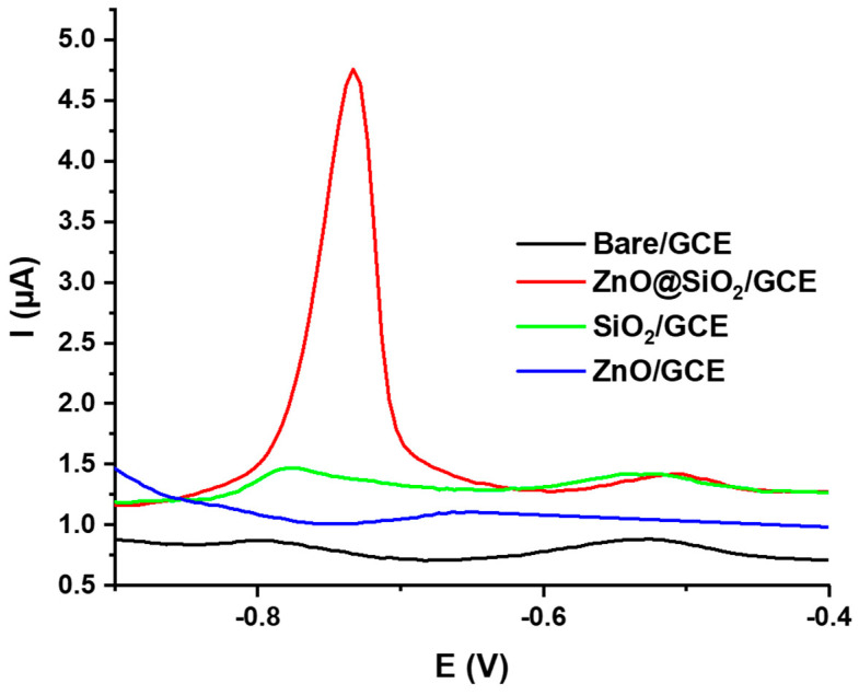
After optimizing the experimental parameters, we investigated the ability of the ZnO@SiO2-modified GCE as an electrochemical sensor for the detection of Cd2+ ions in aqueous solutions. The DPVs of a bare GCE, SiO2/GCE, and ZnO@SiO2/GCE electrodes were recorded in a 0.1 molL−1 ABS (pH = 5:0) solution containing 1.75 10−9 molL−1 Cd2+ (Figure 7). No obvious response was observed for the bare GCE and ZnO-modified electrode when the accumulation time was 5 min. A small peak with an oxidation current of 1.3 µA was observed for the SiO2-modified electrode, which is much lower compared to the ZnO@SiO2-modified electrode, which showed a strong reaction peak at −0.78 V vs. Ag/AgCl with a peak current Ipeak = 4.75 μA. This significant difference can be attributed to the insulating nature of SiO2, which prevents signal trapping for Cd2+ detection. Doping with conductive ZnO particles significantly improves the electron transfer between the electrode and the trapped Cd2+ ions.
Figure 7.
DPV results of bare GCE, SiO2/GCE, ZnO/GCE, and ZnO@SiO2/GCE electrodes measured in 1.75 × 10⁻⁹ molL−1 Cd2+. Data were recorded after drop casting 7 µL of a 1 mg mL−1 ZnO@SiO2 solution in ethanol, a drying time of 2 h, and an accumulation time of 5 min in 0.1 molL−1 in ABS buffer at pH 5.
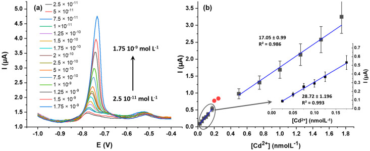
This observation proves the suitability of ZnO@SiO2 as a sensitive layer for the electrochemical detection of Cd2+ ions. Figure 8a shows the DPV responses of ZnO@SiO2/GCE at different Cd2+ concentrations. After baseline correction for each DPV, the resulting calibration curve (Figure 8b) shows two linear regions, with linearity values of R2 = 0.993 for low concentrations and R2 = 0.986 for higher concentrations. These two linearities can be attributed to the change in the electrochemical Cd2+ oxidation from diffusion-controlled domain at lower concentrations (from 2.5 × 10⁻¹¹ molL−1 to 1.75 × 10⁻¹⁰ molL−1) and to surface-controlled processes at higher concentrations (from 2 × 10⁻¹⁰ molL−1 to 1.75 × 10⁻⁹ molL−1). The linearity is not perfect, but satisfying considering the overall low concentration range. At higher concentrations, the electrode surface becomes saturated where no current increase could be measured above 10−6 molL−1. The detection limit is remarkably low, reaching 2.5 × 10⁻¹¹ mol L−1 (S/N = 3).
Figure 8.
(a) Differential pulse voltammograms for Cd2+ at varying concentrations using a ZnO@SiO2/GCE electrode in 0.1 molL−1 ABS buffer pH 5) and (b) calibration curves for Cd2+ detection on ZnO@SiO2/GCE electrodes. The concentration range between 0.2 and 0.5 nmolL−1 (red points) are not be considered due to the change in the electrochemical Cd2+ oxidation process.
This sensor setup was also compared with SiO2/GCE without ZnO dopants (Figure S2). A consistent DPV response could be obtained at much higher concentrations with a linear range between 5 × 10⁻⁷ and 2.5 × 10⁻⁶ molL−1. ZnO doping thus enables monitoring of trace amounts of Cd2+ ions, while SiO2 spheres alone are better suited for quantification of Cd2+ ions in highly polluted environments. By focusing on the detection of trace amounts, the advantageous properties of ZnO doping in this composite can be compared to recently reported sensors [46,47,48,49,50,51,52,53,54,55,56], as summarized in Table 1.
Table 1.
Comparison of our setup with other electrochemical sensors for the determination of Cd2+ by DPV.
| Modified Electrode | Method | Linear Range (molL−1) | Detection Limit (molL−1) | Reference |
|---|---|---|---|---|
| Chitosan-carbon nanotubes/GCE | SWASV | 1.3 × 10⁻⁵–3.9 × 10⁻⁵ | 7.1 × 10⁻⁶ | [46] |
| LAL-AuNPs/GCE | SWASV | 3.0 × 10⁻⁷–1.4 × 10⁻⁶ | 3.0 × 10⁻⁷ | [47] |
| Porous carbon-PdNPs/GCE | DPV | 5.0 × 10⁻⁷–5.5 × 10⁻⁶ | 4.1 × 10⁻⁸ | [48] |
| MIL-53(Fe)/GCE | DPV | 1.5 × 10⁻⁷–4.5 × 10⁻⁷ | 1.6 × 10⁻⁸ | [49] |
| Fe3O4/RGO/GCE | SWASV | 0−8 × 10⁻⁷ | 5.6 × 10⁻⁸ | [2] |
| Nano-PPCPE | DPASV | 10−7–3 × 10⁻⁶ | 7.8 × 10⁻⁸ | [50] |
| PA/PPY/GO/GCE | DPV | 4.4 × 10⁻⁸–1.3 × 10⁻⁶ | 1.9 × 10⁻⁸ | [51] |
| MSK-NPs@GRCPE | DPASV | 5 × 10⁻¹¹–2 × 10⁻⁶ | 5.44 × 10⁻⁹ | [52] |
| NH2@GCE | DPV | 3 × 10⁻⁷–1.5 × 10⁻⁵ | 2 × 10⁻⁷ | [53] |
| GCE/ZSM-5/Pt (5.4%) | DPV | 10−7–7.2 × 10⁻⁶ | 1.2 × 10⁻⁹ | [54] |
| PPh3/MWCNTs/IL/CPE | DPASV | 1 × 10⁻¹⁰–1.5 × 10⁻⁷ | 7.4 × 10⁻⁵ | [55] |
| GCE/SBA-15/ZrO2 (30%) | SWASV | 10−7–5.5 × 10⁻⁶ | 3.14 × 10⁻⁷ | [56] |
| SiO2/GCE | DPV | 5 × 10⁻⁷–2.5 × 10⁻⁶ | 5 × 10⁻⁷ | This work |
| ZnO@SiO2/GCE | DPV | 2.5 × 10⁻¹¹–1.75 × 10⁻¹⁰ and 2 × 10⁻¹⁰–1.75 × 10⁻⁹ | 4.4 × 10⁻¹¹ | This work |
SWASV: square wave anodic stripping voltammetry; DPASV: differential pulse anodic stripping voltammetry; DPV: differential pulse voltammetry; LAL: laser ablation in liquid; Fe3O4/RGO/GCE: magnetite-reduced graphene oxide modified glassy carbon electrode; Nano-PPCPE: nanoporous pseudo carbon paste electrode; PA/PPY/GO/GCE: Phytic acid-functionalized polypyrrole–graphene oxide functionalized glassy carbon electrode.
3.2.3. Appropriateness of ZnO@SiO2 for Specific Cd2+ Detection
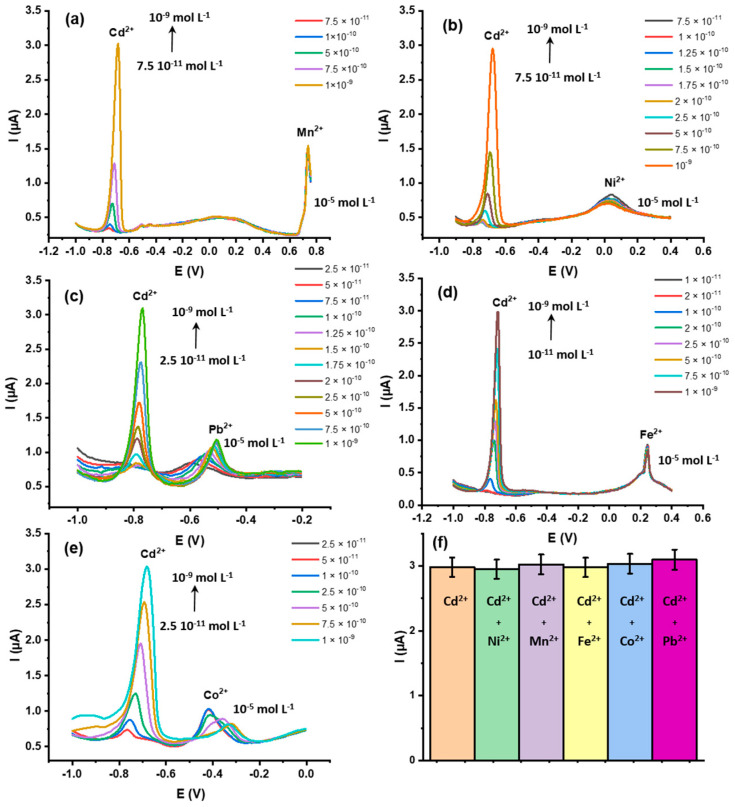
The ZnO@SiO2 nanocomposite was selected for the specific detection of Cd2+ because it has a cavity structure with suitable sizes for the investigated model ion. To evaluate the sensor efficiency, ionic species with redox potentials close to Cd2+, such as Ni2+, Mn2+, Fe2+, Co2+, and Pb2+, were selected and used as interfering ions. As can be seen in Figure 9, the ZnO@SiO2 electrode showed a significant response current for Cd2+ ions at −0.78 V versus Ag/AgCl and significantly lower currents for the other divalent ions within this potential range. Only Mn2+ showed a good response, but the potential (−0.73 V) is clearly separated from the Cd2+ peak potential. In addition, a higher concentration (10−5 molL−1) was used for all tested ions than for Cd2+ (max. 1 10−9). We further investigated possible changes in the peak currents with Cd2+ at different concentrations in the presence of these potentially interfering ions (Figure 9a–e). Within the concentration range between 1 10−9 and 1 10−11 molL−1, the signals are well reproducible, making this setup suitable for the detection of trace Cd2+ in contaminated samples [57,58]. The histogram in Figure 9f confirms the good reproducibility of the sensor formation (n = 3).
Figure 9.
Differential pulse voltammograms of ZnO@SiO2/GCE with different Cd2+ concentrations in the presence of potential interfering ions (a) Mn2+, (b) Ni2+, (c) Pb2+, (d) Fe2+, and (e) Co2+). (f) Histogram of the maximum current peaks for Cd2+ at 10−9 molL−1 in the presence of the different M2+ ions (10−5 molL−1). The electrodes were prepared by drop casting 7 µL of a 1 mg mL−1 ZnO@SiO2 solution in ethanol, with a drying time of 2 h, and the measurements were taken with 5 min of accumulation time in 0.1 mol L−1 ABS buffer at pH 5.
3.2.4. Repeatability and Recovery
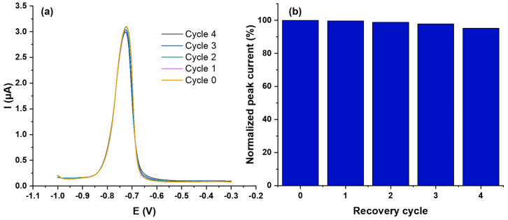
Repeatability and stability are crucial for the reliable performance of electrochemical sensors. Therefore, we investigated these aspects with respect to the proposed sensor. To evaluate the repeatability of ZnO@SiO2/GCE electrodes, six different electrodes (Figure S3) coated with the same amount of the composite material were formed and subjected to DPV analysis at 10−9 molL−1 Cd2+ in 0.1 molL−1 acetate buffer (pH 5). The oxidation peak potential for Cd2+ was −0.78 V and the peak current showed a relative standard deviation (RSD) of 6.28%, which is still acceptable considering the low Cd2+ concentration (10−9 molL−1 = 8 10−3 µgL−1) and shows the good reproducibility of the presented protocol. The repeatability of the sensor was also investigated. After Cd2+ determination using the DPV method (recovery cycle 0), the tested sensor was immersed in 1 molL−1 EDTA solution for 5 min and reconditioned for subsequent Cd2+ determinations (n = 4). After immersion in an aqueous EDTA solution (1 molL−1), the electrode was rinsed three times with distilled water and dried in air at room temperature for 1 h. The efficiency of this cleaning procedure is shown in Figure S4. Figure 10 shows the results of five consecutive recovery cycles using 10−9 molL−1 Cd2+ in a 0.1 molL−1 ABS buffer solution (pH 5). The current intensity decreased only slightly after the first cycle and reached 99.7% of the original peak current. The following cycles showed greater performance losses, but after five recovery cycles 95.2% could be achieved compared to the freshly manufactured electrode. These results underline the ability to reuse these electrodes several times.
Figure 10.
(a) DPV Sensor responses to 10−5 molL−1 of Cd2+ ions using one prepared ZnO@SiO2/GCE modified electrode five times with four recovery cycles. (b) The resulting histogram visualizing the deviations. The measurements were taken in 1 molL−1 EDTA in 0.1 molL−1 ABS buffer at pH 5.
3.2.5. Applications to Real Samples Analysis
To test the reliability of our ZnO@SiO2/GCE sensor, four different water samples were analyzed: tap water from the laboratory, colored water from a local store (laboratory of the textile department), seawater from Skanes, and seawater from Ksibet Elmediouni (Monastir, Tunisia). All water samples were first diluted with ABS tampon solution in a ratio of 1:99 to avoid any possible detrimental effects on the sensor performance by chloride ions. Then the samples were spiked with Cd2+ by diluting 1 mL of a Cd2+ ABS solution (1.75 10−6 molL−1) with the corresponding volume of the real water sample to obtain the final concentration. The measured peak currents were then included in the generated calibration curve (Figure 8b) to obtain the corresponding concentration. The values obtained correlate well with the amount added, with deviations ranging from 89% (tap water) to 102% (seawater from Ksibet Elmediouni). As almost all optimized parameters (quantity, drying, and accumulation time) were met, with the exception of the pH value, these deviations are mainly due to the different pH values of the various samples (see Table 2). These results support the reliability of the proposed Cd2+ sensor for environmental water samples.
Table 2.
Detection of Cd2+ in different environmental water sources.
| Sample | Add (molL−1) | Found (molL−1) | |
|---|---|---|---|
| Tap water | 1.75 × 10⁻⁹ | 1.57 × 10⁻⁹ | 89.71 |
| Tinted water | 1.75 × 10⁻⁹ | 1.6119 × 10⁻⁹ | 92 |
| Sea water (Skanes) | 1.75 × 10⁻⁹ | 1.7411 × 10⁻⁹ | 99.49 |
| Sea water (Ksibet Elmediouni) |
1.5 × 10⁻⁹ 1.75 × 10⁻⁹ |
1.52 × 10⁻⁹ 1.788 × 10⁻⁹ |
100.66 102.2 |
4. Conclusions
In this study, high performance detection of Cd2+ was achieved using glassy carbon electrodes modified with ZnO@SiO2 nanocomposites. DPV analysis showed that the modification of commercial electrodes used in portable devices with this nanocomposite (ZnO@SiO2) is feasible for the detection of trace amounts of Cd2+. The incorporation of ZnO nanoparticles in the core significantly improved the electrochemical performance of the sensor. The experimental results showed that the electrochemical sensor using DPV transduction has good sensitivity and high selectivity. Under optimal experimental conditions, the ZnO@SiO2 sensor exhibits two distinct linear ranges between 2.5 10−11 to 1.75 10−10 molL−1 and 2 10−10 to 1.75 10−9 molL−1, which are related to a change from diffusion-controlled to surface-controlled oxidation of Cd2+. Ultimately, the ZnO@SiO2 composite proved to be effective in detecting Cd2+ in environmental samples using DPV analysis, which enables reliable detection of Cd2+ in the presence of other metal ions.
Acknowledgments
The authors express their gratitude for the assistance provided by the PMIEL platform Chimie NanoBio ICMG FR 2607 (PCN-ICMG) in conducting SEM and TEM studies. Additionally, they acknowledge support from LabEx ARCANE (ANR-11-LABX-0003-01 and CBH-EUR-GS, ANR-17-EURE-0003). Special appreciation is extended to the Institut Carnot PolyNat (CARN 0007-01) for their support.
Supplementary Materials
The following supporting information can be downloaded at: https://www.mdpi.com/article/10.3390/s24134179/s1, Figure S1: Optimization of experimental variables. Figure S2: Detection of Cd2+ with SiO2 nanospheres. Figure S3: Repeatability studies. Figure S4: recovery of the sensor after EDTA treatment.
Author Contributions
A.D.: Investigation, Methodology, Formal Analysis, Writing—Original Draft. M.H.: Methodology, Investigation, Writing—Original Draft, Supervision. P.A.S.-C.: Methodology, Investigation, Writing—Original Draft, Supervision. S.C.: Methodology, Writing—Original Draft, Supervision. H.B.: Methodology, Formal Analysis, Supervision. All authors have read and agreed to the published version of the manuscript.
Institutional Review Board Statement
Not applicable.
Informed Consent Statement
Not applicable.
Data Availability Statement
All experimental data are available and can be shared on request.
Conflicts of Interest
The authors declare no conflicts of interest.
Funding Statement
This research was funded by the Tunisian Ministry of Higher Education and Scientific Research (LR11ES55).
Footnotes
Disclaimer/Publisher’s Note: The statements, opinions and data contained in all publications are solely those of the individual author(s) and contributor(s) and not of MDPI and/or the editor(s). MDPI and/or the editor(s) disclaim responsibility for any injury to people or property resulting from any ideas, methods, instructions or products referred to in the content.
References
- 1.Kubier A., Wilkin R.T., Pichler T. Cadmium in soils and groundwater, A review. Appl. Geochem. 2019;108:104388. doi: 10.1016/j.apgeochem.2019.104388. [DOI] [PMC free article] [PubMed] [Google Scholar]
- 2.Sun Y.F., Chen W.K., Li W.J., Jiang T.J., Liu J.H., Liu Z.G. Selective detection toward Cd2+ using Fe3O4/RGO nanoparticle modified glassy carbon electrode. J. Electroanal. Chem. 2014;714:97–102. doi: 10.1016/j.jelechem.2013.12.030. [DOI] [Google Scholar]
- 3.Fabry G.V.M., Lombaert N., Lison D. Dietary exposure to cadmium and risk of breast cancer in postmenopausal women: A systematic review and meta-analysis. Environ. Int. 2016;86:1–13. doi: 10.1016/j.envint.2015.10.003. [DOI] [PubMed] [Google Scholar]
- 4.Bansod B., Kumar T., Thakur R., Rana S., Singh I. A review on various electrochemical techniques for heavy metal ions detection with different sensing platforms. Biosens. Bioelectron. 2017;94:443–455. doi: 10.1016/j.bios.2017.03.031. [DOI] [PubMed] [Google Scholar]
- 5.Farzin L., Shamsipur M., Sheibani S. A review: Aptamer-based analytical strategies using the nanomaterials for environmental and human monitoring of toxic heavy metals. Talanta. 2017;174:619–627. doi: 10.1016/j.talanta.2017.06.066. [DOI] [PubMed] [Google Scholar]
- 6.Motlagh M.G., Taher M.A. Novel imprinted polymeric nanoparticles prepared by sol–gel technique for electrochemical detection of toxic cadmium(II) ions. Chem. Eng. J. 2017;327:135–141. doi: 10.1016/j.cej.2017.06.091. [DOI] [Google Scholar]
- 7.Aglan R.F., Hamed M.M., Saleh H.M. Selective and sensitive determination of Cd2+ ions in various samples using a novel modified carbon paste electrode. J. Anal. Sci. Technol. 2019;10:7. doi: 10.1186/s40543-019-0166-4. [DOI] [Google Scholar]
- 8.Komarek J., Krasensky P., Balcar J., Rehulka P. Determination of palladium and platinum by electrothermal atomic absorption spectrometry after deposition on a graphite tube. Spectrochim. Acta Part B. 1999;54:739–743. doi: 10.1016/S0584-8547(99)00038-5. [DOI] [Google Scholar]
- 9.Guesmi S., Moulaee K., Bressi V., Kahri H., Khaskhoussi A., Espro C. Houcine Barhoumi and Giovanni Neri, Non-enzymatic amperometric glucose sensing by novel Cu-MOF synthesized at room temperature. Mater. Adv. 2024;5:1160. doi: 10.1039/D3MA00551H. [DOI] [Google Scholar]
- 10.Suturović Z.J., Kravić S.Ž., Stojanović Z.S., Đurović A.D., Brezo-Borjan T.Ž. Potentiometric stripping analysis of cadmium and lead with constant inverse current in the analytical step using an open tubular Mercury-coated glassy carbon electrode. J. Anal. Methods Chem. 2019;8:3579176. doi: 10.1155/2019/3579176. [DOI] [PMC free article] [PubMed] [Google Scholar]
- 11.Liu N., Zhao G., Liu G. Coupling square wave anodic stripping voltammetry with support vector regression to detect the concentration of lead in soil under the interference of copper accurately. Sensors. 2020;20:6792. doi: 10.3390/s20236792. [DOI] [PMC free article] [PubMed] [Google Scholar]
- 12.Schurr S.L., Genske F., Strauss H., Stracke A. A comparison of sulfur isotope measurements of geologic materials by inductively coupled plasma and gas source mass spectrometry. Chem. Geol. 2020;558:119869. doi: 10.1016/j.chemgeo.2020.119869. [DOI] [Google Scholar]
- 13.Fang Y., Cui B., Huang J., Wang L. Ultrasensitive electrochemical sensor for simultaneous determination of cadmium and lead ions based on one-step co-electropolymerization strategy. Sens. Actuators B. 2019;284:414–420. doi: 10.1016/j.snb.2018.12.148. [DOI] [Google Scholar]
- 14.Saha D., Barakat S., Van Bramer S.E., Nelson K.A., Hensley D.K., Chen J. Noncompetitive and competitive adsorption of heavy metals in sulfur-functionalized ordered mesoporous carbon. ACS Appl. Mater. Interfaces. 2016;8:34132. doi: 10.1021/acsami.6b12190. [DOI] [PubMed] [Google Scholar]
- 15.Harrington C.F., Clough R., Drennan-Harris L.R., Hill S.J., Tyson J.F. Atomic spectrometry update. Elemental speciation. J. Anal. At. Spectrom. 2011;26:1561. doi: 10.1039/c1ja90030g. [DOI] [Google Scholar]
- 16.Gawin M., Konefał J., Trzewik B., Walas S., Tobiasz A., Mrowiec H., Witek E. Preparation of a new Cd (II)-imprinted polymer and its application to determination of cadmium (II) via flow-injection-flame atomic absorption spectrometry. Talanta. 2010;80:1305–1310. doi: 10.1016/j.talanta.2009.09.021. [DOI] [PubMed] [Google Scholar]
- 17.Sarpong K.A., Zhang K., Luan Y., Cao Y., Xu W. Development and application of a novel electrochemical sensor based on aunps and difunctional monomer-mips for the selective determination of tetrabromobisphenol-s in water samples. Microchem. J. 2020;154:104526. doi: 10.1016/j.microc.2019.104526. [DOI] [Google Scholar]
- 18.Kreysa G., Ota K.-I., Savinell R.F. Life Cycle Assessment of Sodium-Nickel-Chloride Batteries. Springer; New York, NY, USA: 2014. [Google Scholar]
- 19.Hu J., Mao D., Duan P., Li K., Lin Y., Wang X., Piao Y. Green synthesis of ZnO/BC nanohybrid for fast and sensitive detection of Bisphenol A in water. Chemosensors. 2022;10:163. doi: 10.3390/chemosensors10050163. [DOI] [Google Scholar]
- 20.Walcarius A. Mesoporous materials-based electrochemical sensors. Electroanalysis. 2015;27:1303–1340. doi: 10.1002/elan.201400628. [DOI] [Google Scholar]
- 21.Yasri N.G., Gunasekaran S. Enhancing Cleanup of Environmental Pollutants. Springer; Berlin/Heidelberg, Germany: 2017. Electrochemical technologies for environmental remediation; pp. 5–73. [Google Scholar]
- 22.Zhang K., Kwabena A.S., Wang N., Lu Y., Cao Y., Luan Y., Liu T., Peng H., Gu X., Xu W. Electrochemical assays for the detection of tbbpa in plastic products based on RGO/AgNDs nanocomposites and molecularly imprinted polymers. J. Electroanal. Chem. 2020;862:114022. doi: 10.1016/j.jelechem.2020.114022. [DOI] [Google Scholar]
- 23.Kitte S.A., Lia S., Nsabimana A., Gao W., Lai J., Liu Z., Xu G. Stainless steel electrode for simultaneous stripping analysis of Cd (II), Pb (II), Cu (II) and Hg (II) Talanta. 2019;191:485–490. doi: 10.1016/j.talanta.2018.08.066. [DOI] [PubMed] [Google Scholar]
- 24.Aravind A., Mathew B. Electrochemical sensor based on nanostructured ion imprinted polymer for the sensing and extraction of Cr (III) ions from industrial wastewater. Polym. Int. 2018;67:1595–1604. doi: 10.1002/pi.5683. [DOI] [Google Scholar]
- 25.Xu W. Selective enrichment-release of trace dibutyl phthalate via molecular-imprinting based photo-controlled switching followed by high-performance liquid chromatography analysis. J. Sep. Sci. 2021;44:513–520. doi: 10.1002/jssc.202000950. [DOI] [PubMed] [Google Scholar]
- 26.Xu W., Gao M., Yin X., Zhang L., Cao Y., Zhang Y., Huang W. Photo-stimulated “turn-on/off” molecularly imprinted polymers based on magnetic mesoporous silicon surface for efficient detection of sulfamerazine. J. Sep. Sci. 2020;43:2550–2557. doi: 10.1002/jssc.202000043. [DOI] [PubMed] [Google Scholar]
- 27.Yang W., Qing Y., Cao Y., Luan Y., Lu Y., Liu T., Xu W., Huang W., Li T., Ni X. A stimuli response, core-shell structured and surface molecularly imprinted polymers with specific pH for rapid and selective detection of sulfamethoxazole from milk sample. React. Funct. Polym. 2020;151:104578. doi: 10.1016/j.reactfunctpolym.2020.104578. [DOI] [Google Scholar]
- 28.Aravind A., Mathew B. Tailoring of nanostructured material as an electrochemical sensor and sorbent for toxic Cd (II) ions from various real samples. J. Anal. Sci. Technol. 2018;9:22. doi: 10.1186/s40543-018-0153-1. [DOI] [Google Scholar]
- 29.Dinari M., Tabatabaeian R. Ultra-fast and highly efficient removal of cadmium ions by magnetic layered double hydroxide/guar gum bio nanocomposites. Carbohydr. Polym. 2018;192:317–326. doi: 10.1016/j.carbpol.2018.03.048. [DOI] [PubMed] [Google Scholar]
- 30.Xu W., Zhang Y., Yin X., Zhang L., Cao Y., Ni X., Huang W. Highly sensitive electrochemical BPA sensor based on titanium nitride-reduced graphene oxide composite and core-shell molecular imprinting particles. Anal. Bioanal. Chem. 2021;413:1081–1090. doi: 10.1007/s00216-020-03069-7. [DOI] [PubMed] [Google Scholar]
- 31.Huang W., Zhou X., Luan Y., Cao Y., Wang N., Lu Y., Liu T., Xu W. A sensitive electrochemical sensor modified with multiwalled carbon nanotubes doped molecularly imprinted silica nanospheres for detecting chlorpyrifos. J. Sep. Sci. 2020;43:954–961. doi: 10.1002/jssc.201901036. [DOI] [PubMed] [Google Scholar]
- 32.Walcarius A. Silica-based electrochemical sensors and biosensors: Recent trends. Curr. Opin. Electrochem. 2018;10:88–97. doi: 10.1016/j.coelec.2018.03.017. [DOI] [Google Scholar]
- 33.Cannas C., Mainas M., Musinu A., Piccaluga G. ZnO/SiO2 nanocomposites obtained by impregnation of mesoporous silica. Compos. Sci. Technol. 2003;63:1187–1191. doi: 10.1016/S0266-3538(03)00040-X. [DOI] [Google Scholar]
- 34.Chireh M., Karam Z.M., Naseri M., Jafarinejad-Farsangi S., Ghaedamini H. Synthesis, characterization and cytotoxicity study of graphene/doped ZnO/SiO2 nanocomposites. Appl. Phys. A. 2022;128:107. doi: 10.1007/s00339-022-05367-6. [DOI] [Google Scholar]
- 35.Castillo J., Arcuri M., Vargas V., Piscitelli V. Synthesis of nanocomposites SiO2@Co3O4, SiO2@ZnO, and SiO2@CuO from rice husks: Spectroscopy and optical properties. Appl. Phys. A. 2022;128:107. doi: 10.1007/s00339-021-05247-5. [DOI] [Google Scholar]
- 36.Govindhan P., Pragathiswaran C. Silver Nanoparticle Decorated on ZnO@SiO2 Nanocomposite and Application for Photocatalytic Dye Degradation of Methylene Blue. Natl. Acad. Sci. Lett. 2019;42:323–326. doi: 10.1007/s40009-018-0746-7. [DOI] [Google Scholar]
- 37.Ali A.M., Harraz F.A., Ismail A.A., Al-Sayari S.A., Algarni H., Abdullah G. Al-Sehemi. Synthesis of amorphous ZnO–SiO2 nanocomposite with enhanced chemical sensing properties. Thin Solid Films. 2015;605:277–282. doi: 10.1016/j.tsf.2015.11.044. [DOI] [Google Scholar]
- 38.Kava A.A., Beardsley C., Hofstetter J., Henry C.S. Disposable glassy carbon stencil printed electrodes for trace detection of cadmium and lead. Anal. Chim. Acta. 2020;1103:58–66. doi: 10.1016/j.aca.2019.12.047. [DOI] [PMC free article] [PubMed] [Google Scholar]
- 39.Liu H., Zhou Y., Qi Y., Sun Z., Gong B. Preparation of thiamphenicol magnetic surface molecularly imprinted polymers for its selective recognition of thiamphenicol in milk samples. J. Liq. Chromatogrn Relat. Technol. 2018;41:868–879. doi: 10.1080/10826076.2018.1531294. [DOI] [Google Scholar]
- 40.Paramitha T., Suryadi J., Raissa R.A., Nugraha T.A., Utami N. Preliminary Study of ZnO/SiO2 Synthesis with Silica from Geothermal Solid Waste and Its Performance Test in Methylene Blue Removal. J. Ris. Kim. 2023;9:266–277. [Google Scholar]
- 41.Raevskaya A.E., Panasiuk Y.V., Stroyuk O.L., Kuchmiy S.Y., Dzhagan V.M., Milekhin A.G., Yeryukov N.A., Sveshnikova L.A., Rodyakina E.E., Plyusnin V.F., et al. Spectral and luminescent properties of ZnO–SiO2 core–shell nanoparticles with size-selected ZnO cores. RSC Adv. 2014;4:63393–63401. doi: 10.1039/C4RA07959K. [DOI] [Google Scholar]
- 42.Galedari N.A., Rahmani M., Tasbihi M. Preparation, characterization, and application of ZnO@SiO2 core–shell structured catalyst for photocatalytic degradation of phenol. Environ. Sci. Pollut. Res. 2017;24:12655–12663. doi: 10.1007/s11356-016-7888-2. [DOI] [PubMed] [Google Scholar]
- 43.Abbaa E., Shehub Z., Lamayi D.W., Yoriyo K.P., Dogara R.K., Ayuk N.C. Novel developments of ZnO/SiO2 nanocomposite: A nanotechnological approach towards insect vector control. J. Nig. Soc. Phys. Sci. 2021;3:262–266. doi: 10.46481/jnsps.2021.198. [DOI] [Google Scholar]
- 44.Danbature W.L., Yoro M., Shehu Z., Madugu Y.D. Solvent FreeMechanochemical Synthesis, Characterization and Antibacterial Potency of CaO/SiO2 Nanocomposite. J. Mater. Sci. Res. Rev. 2019;4:1–7. [Google Scholar]
- 45.Fageria P., Gangopadhyay S., Pande S. Synthesis of ZnO/Au and ZnO/Ag nanoparticles and their photocatalytic application using UV and visible light. RSC Adv. 2014;4:24962. doi: 10.1039/C4RA03158J. [DOI] [Google Scholar]
- 46.Wu K.H., Lo H.M., Wang J.C., Yu S.Y., Yan B.D. Electrochemical detection of heavy metal pollutant using crosslinked chitosan/carbon nanotubes thin film electrodes. Mater. Express. 2017;7:15–24. doi: 10.1166/mex.2017.1351. [DOI] [Google Scholar]
- 47.Xu X., Duan G., Li Y. Fabrication of gold nanoparticles by laser ablation in liquid and their application for simultaneous electrochemical detection of Cd2+, Pb2+, Cu2+, Hg2+ ACS Appl. Mater. Interfaces. 2013;6:65–71. doi: 10.1021/am404816e. [DOI] [PubMed] [Google Scholar]
- 48.Veerakumar P., Veeramani V., Chen S.-M., Madhu R., Liu S.-B. Palladium Nanoparticle Incorporated Porous Activated Carbon: Electrochemical Detection of Toxic Metal Ions. ACS Appl. Mater. Interfaces. 2016;8:1061–1554. doi: 10.1021/acsami.5b10050. [DOI] [PubMed] [Google Scholar]
- 49.Tran H.V., Dang H.T.M., Tran L.T., Tran C.V., Huynh C.D. Metal-Organic Framework MIL-53(Fe): Synthesis, Electrochemical Characterization, and Application in Development of a Novel and Sensitive Electrochemical Sensor for Detection of Cadmium Ions in Aqueous Solutions. Adv. Polym. Technol. 2020;2020:6279278. doi: 10.1155/2020/6279278. [DOI] [Google Scholar]
- 50.Liu Y., Li T., Ling C., Chen Z., Deng Y., He N. Electrochemical sensor for Cd2+ and Pb2+ detection based on nano-porous pseudo carbon paste electrode. Chin. Chem. Lett. 2019;30:2211–2215. doi: 10.1016/j.cclet.2019.05.020. [DOI] [Google Scholar]
- 51.Dai H., Wang N., Wang D., Ma H., Lin M. An electrochemical sensor based on phytic acid functionalized polypyrrole/graphene oxide nanocomposites for simultaneous determination of Cd2+ and Pb(II) Chem. Eng. J. 2016;299:150–155. doi: 10.1016/j.cej.2016.04.083. [DOI] [Google Scholar]
- 52.Rahim A.M.A., Mahmoud E.M.M. Recent advances in the modification of electrodes for trace metal analysis: A review. Anal. Sci. 2023;39:179. doi: 10.1007/s44211-022-00214-3. [DOI] [PubMed] [Google Scholar]
- 53.Yu L., Wan J.W., Meng X.Z., Gu H.W., Chen Y., Yi H.C. A simple electrochemical method for Cd(II) determination in real samples based on carbon nanotubes and metal-organic frameworks. Int. J. Environ. Anal. Chem. 2022;102:4757. doi: 10.1080/03067319.2020.1789611. [DOI] [Google Scholar]
- 54.Tamizhdurai P., Rajakumaran R., Sakthinathan S., Chen S.M., Chiu T.-W., Narayanan S. Highly sensitive detection of environmental pollutant cadmium with ultrasonic irradiated Pt-supported ZSM-5 modified electrode. Microporous Mesoporous Mater. 2020;307:110449. doi: 10.1016/j.micromeso.2020.110449. [DOI] [Google Scholar]
- 55.Bagheri H., Afkhami A., Khoshsafar H., Rezaei M., Shirzadmehr A. Simultaneous electrochemical determination of heavy metals using a triphenylphosphine/MWCNTs composite carbon ionic liquid electrode. Sens. Actuators B Chem. 2013;186:451–460. doi: 10.1016/j.snb.2013.06.051. [DOI] [Google Scholar]
- 56.Tamizhdurai P., Sakthinathan S., Krishnan P.S., Ramesh A., Mangesh V.L., Abilarasu A., Narayanan S., Shanthi K., Chiu T.W. Catalytic activity of ratiodependent SBA-15 supported zirconia catalysts for highly selective oxidation of benzyl alcohol to benzaldehyde and environmental pollutant heavy metal ions detection. J. Mol. Struct. 2019;1176:650–661. doi: 10.1016/j.molstruc.2018.09.007. [DOI] [Google Scholar]
- 57.Ghazali N.N., Mohamad Nor N., Abdul Razak K., Lockman Z., Hattori T. Hydrothermal synthesis of bismuth nanosheets for modified APTES-functionalized screen-printed carbon electrode in lead and cadmium detection. J. Nanopart. Res. 2020;22:211. doi: 10.1007/s11051-020-04946-z. [DOI] [Google Scholar]
- 58.Kong X.F., Yang B., Xiong H., Zhou Y., Xue S.G., Xu B.Q., Wang S.X. Selective removal of heavy metal ions from aqueous solutions with surface functionalized silica nanoparticles by different functional groups. J. Cent. South Univ. 2014;21:3575–3579. doi: 10.1007/s11771-014-2338-0. [DOI] [Google Scholar]
Associated Data
This section collects any data citations, data availability statements, or supplementary materials included in this article.
Supplementary Materials
Data Availability Statement
All experimental data are available and can be shared on request.


