Abstract
Background
To interrupt residual malaria transmission and achieve successful elimination of Plasmodium falciparum in low-transmission settings, the World Health Organization (WHO) recommends the administration of a single dose of 0.25 mg/kg (or 15 mg/kg for adults) primaquine (PQ) combined with artemisinin-based combination therapy (ACT), without glucose-6-phosphate dehydrogenase (G6PD) testing. However, due to the risk of haemolysis in patients with G6PD deficiency (G6PDd), PQ use is uncommon. Thus, this study aimed to assess the safety of a single low dose of PQ administered to patients with G6PD deficiency.
Methods
An observational cohort study was conducted with patients treated for uncomplicated P. falciparum malaria with either single-dose PQ (0.25 mg/kg) (SLD PQ) + ACT or ACT alone. Microscopy-confirmed uncomplicated P. falciparum malaria patients visiting public health facilities in Arjo Didessa, Southwest Ethiopia, were enrolled in the study from September 2019 to November 2022. Patients with uncomplicated P. falciparum malaria were followed up for 28 days through clinical and laboratory diagnosis, such as measurements of G6PD levels and haemoglobin (Hb) concentrations. G6PD levels were measured by a quantiative CareSTART™ POCT S1 biosensor machine. Patient interviews were also conducted, and the type and frequency of clinical complaints were recorded. Hb data were taken on days (D) 7, 14, 21, and 28 following treatment with SLD-PQ + ACT or ACT alone.
Results
A total of 249 patients with uncomplicated P. falciparum malaria were enrolled in this study. Of these, 83 (33.3%) patients received ACT alone, and 166 (66.7%) received ACT combined with SLD-PQ treatment. The median age of the patients was 20 (IQR 28–15) years. G6PD deficiency was found in 17 (6.8%) patients, 14 males and 3 females. There were 6 (7.2%) and 11 (6.6%) phenotypic G6PD-deficient patients in the ACT alone and ACT + SLD-PQ arms, respectively. The mean Hb levels in patients treated with ACT + SLD-PQ were reduced by an average of 0.45 g/dl (95% CI = 0.39 to 0.52) in the posttreatment phase (D7) compared to a reduction of 0.30 g/dl (95% CI = 0.14 to − 0.47) in patients treated with ACT alone (P = 0.157). A greater mean Hb reduction was observed on day 7 in the G6PDd ACT + SLD-PQ group (− 0.60 g/dL) than in the G6PDd ACT alone group (− 0.48 g/dL); however, there was no statistically significant difference (P = 0.465). Overall, D14 losses were 0.10 g/dl (95% CI = − 0.00 to 0.20) and 0.05 g/dl (95% CI = − 0.123 to 0.22) in patients with and without SLD-PQ, respectively (P = 0.412).
Conclusions
This study’s findings indicate that using SLD-PQ in combination with ACT is safe for uncomplicated P. falciparum malaria regardless of the patient's G6PD status in Ethiopian settings. Caution should be taken in extrapolating this finding in other settings with diverse G6DP phenotypes.
Keywords: Plasmodium falciparum, Malaria elimination, Primaquine, Artemisinin-based combination therapies, Haemoglobin, G6PD deficiency, Ethiopia
Background
The global decline in malaria incidence due to increased malaria control measures, particularly the use of artemisinin-based combination therapy (ACT), has stimulated efforts to eliminate Plasmodium falciparum and sparked an upsurge in interest in therapies to stop transmission [1]. In many malaria-endemic areas, artemisinin derivatives are very effective against both asexual and young P. falciparum gametocytes, and this has considerably enhanced the global reduction in malaria transmission [2]. However, mature gametocytes continue to exist after ACT at the microscopic or submicroscopic level, and residual transmission is not interrupted after ACT [3, 4]. This is the reason why artemisinin derivatives against mature P. falciparum gametocytes are not effective in preventing transmission [5].
To treat P. falciparum malaria, the World Health Organization (WHO) recommends incorporating a single low dose of primaquine (SLD-PQ, 0.25 mg/kg of body weight) into artemisinin-based combination therapy (ACT), as part of pre-elimination or elimination programmes [6] and as part of artemisinin resistance containment programmes [7]. In low-endemic settings, the combination of this gametocytocidal drug with ACT is effective [8, 9], and this combination may significantly reduce malaria transmission [10]. While PQ is a promising treatment for relapse of Plasmodium ovale and Plasmodium vivax, cautions about its safety should not be overlooked, particularly in patients with glucose-6-phosphate dehydrogenase deficiency (G6PDd) [11]. Compared to single low-dose (SLD) PQ treatment, the dosage regimen used is noticeably higher, which could put G6PDd patients at greater risk of haemolysis [12]. The WHO currently recommends adding a single dose of 0.25 mg/kg to ACT without glucose-6-phosphate dehydrogenase (G6PD) to reduce transmission in low-transmission settings and accelerate the elimination of P. falciparum [8, 13]. However, due to fears of haemolysis in people with G6PD deficiency (G6PDd) [14], the use of PQ is not as common as anticipated. Hence, the broad use of PQ is hampered by safety concerns of haemolysis in individuals with G6PDd, the most prevalent hereditary enzyme defect reported in all malaria-endemic areas [15].
An estimated 400 million people worldwide suffer from G6PDd, which is an X-linked genetic disorder in malaria-endemic countries [16]. The frequency of G6PDd varies significantly from region to region, even within a country, with the highest prevalence reported in Africa. Additionally, migration and resettlement have an impact on the spread of this genetic disorder [17, 18]. Due to safety concerns, especially for those with G6PDd, which affects up to 37.5% of the continent's population [19], the use of PQ is also restricted, particularly in Africa. In Africa, the predominant G6PD deficiency variants are the mild A-type, while in Asia, there is a heterogeneous mix, with the Mediterranean variant being the most severe. These variations underscore the complexity of managing G6PDd and the importance of tailored healthcare approaches based on regional prevalence and genetic diversity [20]. The safety of PQ in relation to haemolysis at the individual level has prevented its use despite the benefit being solely at the population level [21].
With a growing body of evidence on the safety of the administration of SLD-PQ in recent years, there have been indications that SLD-PQ is well tolerated in G6PDd patients [22].Several randomized and controlled clinical trials on the safety and efficacy of ACT versus SLD-PQ have been conducted in South Africa [21], Tanzania [23, 24], Senegal [6], Swaziland [25], Burkina Faso [26], and Uganda [27], supporting the use of the WHO-recommended SLD-PQ without G6PD testing during the preelimination and elimination phases of malaria.
Although previous studies have shown that SLD-PQ is safe even for people with G6PDd, the WHO recommended additional clinical research to ensure the safety of SLD-PQ in G6PDd individuals in different eco-epidemiological settings [20, 28]. Furthermore, due to pragmatic dosing techniques, some patients may receive a dose greater than the recommended dose of 0.25 mg/kg, which may improve gametocyte clearance but increase the risk of haemolysis [29].
Unfortunately, most studies were clinical trials that lacked population-based data and merely provided quantitative data assessing the effect of the SLD-PQ on haemoglobin (Hb) levels. In addition, previous safety studies were often based on carefully chosen study participants and small groups with relatively high pretreatment Hb levels; therefore, they offered limited information at the population level. The risk of anaemia is likely to increase in individuals with lower pretreatment Hb concentrations [30, 31]. Although PQ is linked to a brief reduction in Hb levels in G6PDd patients, baseline Hb levels continue to be the leading cause of anaemia in such patients [9]. Additionally, severe haemolytic events might be rare and unlikely to be observed in small safety studies [26]. Therefore, whether certain groups are still at risk of clinically significant haemolysis when SLD-PQ treatment occurs at the population level needs to be examined. For noncurative treatments that aim to help the community rather than just the dosed individual, this safety profile is particularly crucial [9]. To address this issue, we evaluated the safety of adding a single fixed low dose of PQ (15 mg tablet or 0.25 mg/kg for a person weighing 60 kg) to ACT regimens of artemether-lumefantrine (AL) to treat patients with P. falciparum malaria in an Ethiopian cohort.
Moreover, changes in the Hb concentration over time were also examined, which is helpful information for clinical practice. Furthermore, previous studies have mainly examined one population group—males [26], children [32], adults [6] or asymptomatic people [33]—while excluding patients with G6PDd [3]. In the present study, all groups were included.
Methods
Study area
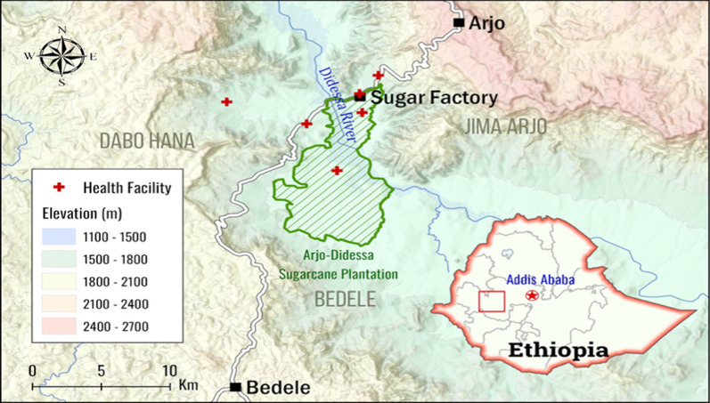
This study was conducted at the Arjo-Didessa Sugar Cane Plantation and surrounding areas of southwestern Ethiopia between September 2018 and November 2022. Arjo-Didessa is located 540 km southwest of Addis Ababa, the nation's capital. The latitude of the study area is 8°41′35.5″N, and the longitude is 36°25′54.9″E. The mean annual rainfall is 1477 mm, which falls over two rainy seasons: one short season between February and March and the other long rainy season ranging between June and September. These rainy seasons correspond to low and high peak transmission seasons, respectively. The two primary malaria parasite species are P. falciparum and P. vivax [34, 35]. Studies in this area have reported P. ovale infections [36]. The rates of malaria transmission are low, unstable, and seasonal. The vast majority of people living in the area are farmers who raise food crops, including corn, sorghum, nuts, and peppers, and a smaller portion of these people work at the Arjo sugar factory and sugarcane plantations. The residents also maintained livestock to help with their living. The study was carried out in health facilities located in three districts: Dabo Hana (Kerka Health Post and Sefera Tabiya Health Center), Jimma Arjo (Arjo-Didessa Sugar Factory Clinic, Abote Didessa Health Center), and Bedele (Command 2 and Command 5 Health Posts). The selection of health facilities was based partly on how close they were to the follow-up study. Fig. 1 (Map of the study sites).
Fig. 1.
Map of the study sites
Study design
This was a prospective cohort study on the impact of a single low dose of primaquine on the prevention and control of P. falciparum malaria. As part of this study, patients treated for uncomplicated P. falciparum malaria with either ACT + SLD-PQ at 0.25 mg/kg or ACT alone were enrolled and assessed for the safety of the addition of SLDPQ. Since the study started in September 2018 and SLD-PQ was not fully adopted nationwide, regions that started utilizing SLD-PQ were targeted for the study. As Study participants from other health facilities that did not enrollSLD-PQ use served as a natural control group. By comparing patients receiving ACT alone with those receiving ACT + SLDPQ, this method aids in evaluating the safety of including SLDPQ in the treatment plan. Then, clinical and laboratory evaluations were conducted on days (D) 0, 7, 14, 21, and 28 or on any day of recurrent illness [37]. Patients who satisfied the study's eligibility criteria, such as uncomplicated P. falciparum mono-infection verified microscopically, provided consent to adhere to the study protocol and were included in the study.
The study excluded pregnant women, lactating mothers, and children under one year old due to contraindications to PQ in these populations [38]. Any history of severe malaria or warning signs, a recent history of blood transfusion, febrile conditions unrelated to malaria, or a known chronic or serious underlying illness were among the exclusion criteria.
Sample size calculation
This study took the primary objective—the mean Hb reduction—into consideration when calculating the proper sample size. The Hb reduction calculation in this study was based on a study in Senegal by Tine et al., which showed that adding primaquine (0·25 mg/kg) to ACT reduced the mean Hb concentration from 13.4 to 11.9 g/dl (1.47) [6]. With a 10% loss to follow-up and a clinically meaningful noninferiority margin of 0.3 g/dl, the intended sample size for the effect of SLD-PQ on Hb was 131 participants per group. This made it possible to determine if the study group and the reference group were not significantly different by 80% at the two-tailed 5% significance level.
Sampling procedure
Following the detection of P. falciparum on D0, artemether + lumefantrine (AL) tablets twice daily for 3 days (Coartem [20 mg of artemether and 120 mg of lumefantrine]; Ipca Laboratories Ltd., India) were given for all positive uncomplicated P. falciparum patient malaria cases as per the National Malaria Treatment Guidelines. In addition, SLD-PQ + ACT was also administered on D0 in selected health clinics and posts in the study area. Following guidelines from the FMoH, Primaquine phosphate dose: of 0.25 mg base per kg was prescribed according to an age-based, non-weight-based treatment schedule. The fixed dose for adults was 15 mg, which was taken as two tablets containing 7.5 mg each. The drug was divided in accordance with the guidelines for children [39]. Researchers directly observed participants taking the primaquine dose at the time of enrollment in the study. Adherence to the ACT regimen was assessed through self-reported adherence during the second and seventh days of the follow-up.
Laboratory procedure
Blood samples for malaria diagnosis using microscopy and dried blood spots for PCR analysis were taken prior to treatment. In addition, capillary blood samples (300 microns) were collected for Hb concentration measurement and determination of G6PD enzyme activity. G6PD was measured once on day 0, and the patient was followed up via scheduled appointments on D7, D14, D21, and D28 for Hb status and microscopy examination. Clinical data, including patient sociodemographic information, malaria symptoms, and history of treatment with anti-malarial drugs, were collected during enrollment.
Microscopy of malaria parasites
According to the WHO malaria microscopy procedure [40], thick and thin smears were taken at patient enrollment and during follow-up, and they were quickly stained with 10% Giemsa. Two different microscopists examined each microscope slide. When there was disagreement about the presence of parasitaemia or whether there was a difference in parasite density of more than 25%, a third independent reading was performed [41]. Accordingly, microscopic examination was carried out on D 0, 7, 14, 21, and 28 using Giemsa-stained blood films at a magnification of 1000x.
G6PD enzyme and Hb measurements
Blood samples from each study participant were tested for G6PD enzyme and Hb levels in accordance with the manufacturer’s instructions. Hb and G6PD levels were measured using CareSTART™ POCT S1 (Access Bio, Inc., New Jersey, USA). As recommended by the manufacturer, Hb and G6PD activity were checked daily prior to sample collection. Every sample was run twice. International units per decilitre, or IU/dl, are used to express G6PD enzyme activity, which was measured using a G6PD strip. The enzyme activity was normalized using Hb values taken concurrently with the G6PD enzyme test and expressed as IU/g Hb. Quantitative POCTs were performed using “biosensors,” portable electronic devices where disposable test strips were inserted. Within five minutes after blood was added, the device's screen displayed the results as a quantitative readout. An unused test strip was inserted into the biosensor, and an aliquot from each blood sample (5 µL) was then placed on the exposed end of the test strip until the device displayed complete automated sample intake. The running times for the G6PD and Hb tests were 4 min and 10 s, respectively. Using a previously described methodology [18], the adjusted male median (AMM) G6PD activity (100% G6PD activity) for males was used to determine the cutoff values for G6PD deficiency. After excluding males who were severely G6PD-deficient (i.e., had less than 10% of the median G6PD enzyme activity), as advised by an expert panel, the percentage of G6PD activity was determined based on their relative proportion to the adjusted median value of G6PD enzyme activity among the male population [42]. For male study participants, class I was G6PD deficient with less than 30% AMM activity, while class II was normal with more than 30% AMM activity. Individuals with G6PD activity < 30%, 30–80%, or > 80% of the AMM activity in females are regarded as G6PD deficient, intermediate, or normal, respectively [43–46].
Clinical and safety assessments
Safety assessments in the present study were performed via haematological analysis and recording of all adverse events [47]. All patients were asked without probing about their health after taking their last dose of anti-malarial medication, and the responses were categorized as none, mild, moderate, severe, or life-threatening. The following adverse events (AEs) were recorded: abdominal pain, nausea, vomiting, haemolysis(if dark urine was encountered using a clean container), itching, rash, and any drug-related severe adverse events (SAEs). AEs were graded using the Primaquine Roll Out Monitoring Pharmacovigilance Tool (PROMPT), a tool that is helpful for assessing the safety of AEs and SAEs [25]. Mild, moderate, severe, and life-threatening AEs were further divided into four categories: mild, easily tolerable; no or little interference with daily activities; moderate, low level of inconvenience; more than little interference with daily activities; severe, interrupted regular daily activities, typically incapacitating; and life-threatening, life-threatening consequences, indicating the need for immediate intervention or death events.
Outcomes
The main goal of the study was to determine how adding SLD-PQ to ACT affects patients’ mean change in Hb levels [48]. In particular, we examined the changes in Hb from Day 0 of baseline to Day 7 after the start of treatment. The mean absolute fall in Hb by Day 7, the mean fractional decline in Hb from Day 0 to Day 7, and the degree of Hb recovery by Day 28 to baseline values were all evaluated in this analysis [22, 49]. Because Day 7 could be the longest day for PQ-induced haemolysis and because earlier observations showed that Hb levels peaked by this day, Day 7 was chosen for assessment [19]. Either an absolute drop of more than 3 g/dL or a fractional reduction of more than 25% was considered a clinically significant Hb decrease [49].
The secondary outcomes of this study were the occurrence of adverse events (AEs), including blood transfusions, AEs that occurred within 7 and 28 days after the administration of primaquine, and other AEs related to the administration of PQ, such as gastrointestinal reactions within 7 days and risk of haemolysis.
Data management and analysis
The data were entered into a Microsoft Excel data sheet and analyzed using Stata (v17, StataCorp LLC, Texas) and IBM SPSS Statistics 27 software. The paired t-test was employed to assess the significance of mean Hb within groups, while the unpaired t-test was used to evaluate their significance between groups. Absolute and relative changes in Hb concentration from the baseline value were reported during the follow-up. A linear mixed-effects model was used to assess the impact of treatment on Hb drop. This model yielded the treatment difference at each time point and a 95% confidence interval. Primaquine’s effect in G6PD normal and G6PD deficient patients was estimated in an intended subgroup analysis using a model fitted with baseline Hb as a covariate, patient as a random effect, and G6PD status, the interaction between time and G6PD status, and the treatment group and G6PD status at each time point as fixed effects. The significance of all P values was assessed using a two-tailed approach, with a threshold set at P < 0.05 for statistical significance. Multiple linear regression model was employed,adjusted for age, sex, and baseline Hb concentration to assess whether there was a significant difference in the mean Hb change between G6PD-deficient patients and normal patients following therapy. The reported complaints and adverse events (AEs) in the ACT alone and ACT + SLD-PQ treatment arms were also compared using Fisher’s exact test.
Results
Overview of the study
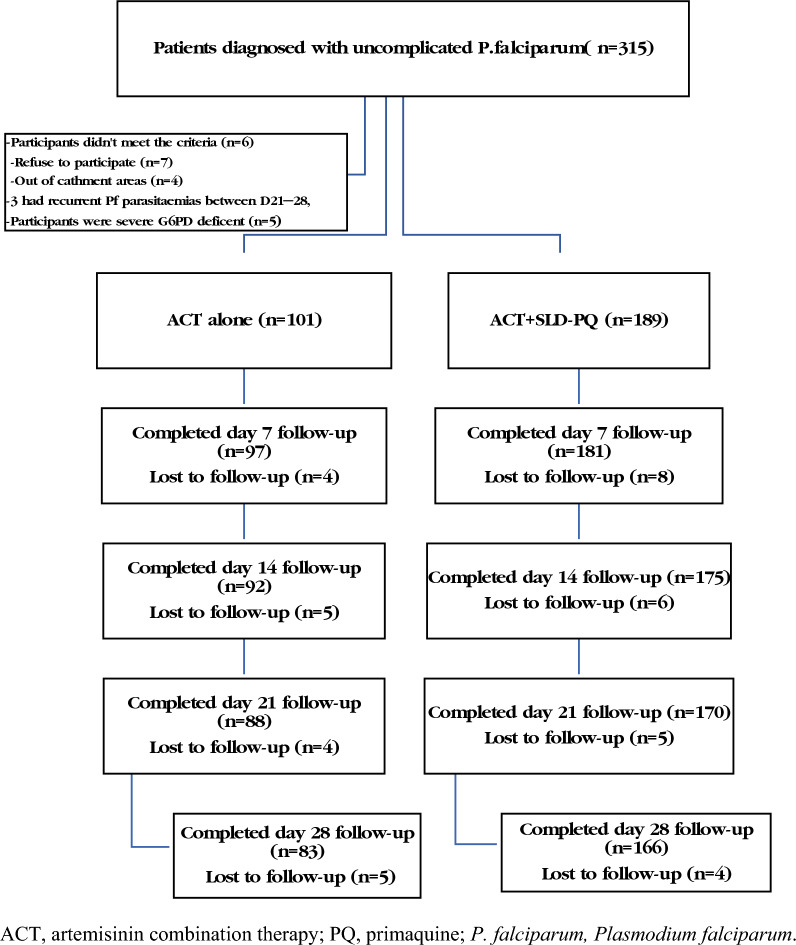
A total of 315 febrile patients with confirmed falciparum malaria were followed for two seasons during high malaria transmission. The study excluded 66 individuals who were not within the catchment area, did not match the inclusion criteria, had severe G6PDd, or declined to participate (Fig. 2). The remaining 249 (79%) patients were eligible and completed the 28 day follow-up. The initial characteristics of the two treatment groups were comparable (Table 1). Patient sociodemographic characteristics included the total number of patients who received ACT alone (n = 83) or ACT + SLD-PQ (n = 166) for uncomplicated falciparum malaria. Adults (aged > 15 years) accounted for the majority of the study population, with a total of 197 (79.12%) individuals and a sex ratio of males to females of 1.8 (Table 1).
Fig. 2.
Flow chart of uncomplicated P. falaciparum malaria patients enrolled for ACT and SLD PQ + ACT in Arjo Didessa, Southwest Region, Ethiopia. ACT: artemisinin combination therapy, PQ: primaquine, P. falciparum: Plasmodium falciparum
Table 1.
Baseline sociodemographic, clinical,parasitological profiles and prognostic profiles of the study participants (n = 249) by treatment group in Arjo-Didessa, southern Ethiopia; 2019–2022
| Characteristics | Treatment arms | P value | |
|---|---|---|---|
| ACT alone 83 (33.33%) |
ACT + SLD-PQ 166 (66.67%) |
||
| Gender (% male no. of males/total no. of individuals) | 65.06(54/83) | 64.46(107/166) | 0.9257 |
| Age(years), median (IQR) | 20(13) | 20 (15) | 0.9276 |
|
Age groups ≤ 5 years, no. (%) 6–15 years,no.(%) 16–30 years,no.(%) ≥ 31 years,no.(%) |
2(2.6) 10(12.8) 49(62.8) 17(21.8) |
4(2.5) 32(20.4) 91(58.0) 30(19.1) |
0.560 |
| Axillary temperature (oC), mean (SD) | 38.13(0.54) | 37.98(.63) | 0.0759 |
| Fever (≥ 37.5 °C) at present, no. (%) | 63(75.90) | 119(71.69) | |
| Asexual parasite density/microlitre, mean (SD) | 13815.67(19655.76) | 17430.92(30420.42) | 0.3259 |
|
G6PD activity**, no. (%) -Normal -Deficient |
77(92.77) 6(7.23) |
155(93.37) 11(6.63) |
0.859 |
| Hb concentration at enrollment (g/dl), mean (SD) | 12.80(1.18) | 12.75(0.89) | 0.736 |
|
G6PDd Hb(g/dl), mean (SD) G6PDn Hb(g/dl), mean (SD) |
12.70(0.88) 12.80(1.21) |
12.7(0.53) 12.76(.91) |
1.0000 0.730 |
Number [Note: means and standard deviation are presented for temperature and haemoglobin; age is presented as median and interquartile range]
IQR: interquartile range, °C: degree Celsius
** as determined by a POCT analyzer indicating G6PD enzyme activity (IU/g Hb)]
Phenotypic G6PD status
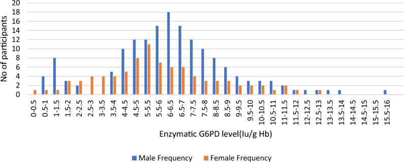
At the time of enrollment, phenotypic point-of-care testing was utilized to screen patients for G6PD deficiency. The adjusted median G6PD enzyme activity was determined for the male participants by excluding 5 patients whose enzyme activity was less than 10% (less than 0.627 U/g Hb) of the median value found for all male participants. The median G6PD activity was 6.27 U/g Hb for all study participants (the range was 0.4–15.22 U/g Hb). The AMM (range) enzyme activity was 6.32 IU/gHb (0.67–15.219), which was 100%. The distribution of enzymatic activity by sex is illustrated in Fig. 3. There was a distinct bimodal distribution in males and a unimodal distribution in females. When the participants’ enzyme activity was between 0.4 and 1.896 U/g Hb (less than 30% of the adjusted male median), they were classified as deficient. Phenotypic G6PD data were recorded for 249 patients; 17 (6.83%) had G6PDd—14 males and 3 females. In the ACT alone arm, there were 6 (2.41%) patients with phenotypic G6PDd, whereas there were 11 (4.42%) patients in the ACT + SLD-PQ arm (Table 1).
Fig. 3.
Distribution of G6PD enzyme levels by sex at Arjo-Didessa, southern Ethiopia; 2019–2022
Haemoglobin profiling
Data from 415 Hb measurements in 83 patients treated with ACT alone and 830 Hb measurements in 166 patients treated with ACT + SLD-PQ were used for the assessment of the changes in the mean Hb level between baseline and days 7, 14, 21, and 28. During enrollment, the mean (SD) Hb level was 12.80 (1.18) g/dL for patients receiving ACT alone and 12.75 (0.89) g/dL for those receiving ACT + SLD-PQ (Table 1). There was no significant difference in the baseline Hb levels between the two treatment arms (p = 0.7357). However, males had greater mean (SD) Hb concentrations at baseline (12.78 (1.03) [95% CI 12.62, 12.94]) than females did (12.75 (0.93) [95% CI 12.55–12.94]). There were no significant differences in the mean Hb concentration between males and females in the two treatment arms (p = 0.8281). However, as illustrated in Table 3, in both groups receiving ACT + SLD-PQ, the mean Hb concentrations, shown as the mean change, decreased in the first week following treatment and reverted to baseline values during follow-up. Similarly, paired analysis of Hb concentrations relative to baseline demonstrated a reduction in the first week after receiving 0.25 mg/kg PQ + ACT or ACT alone. These reductions were significant on day 7 following SLD-PQ (0.25 mg/kg PQ) (P < 0.01) or ACT alone (P < 0.01; Table 2). However, there was no significant difference between the treatment groups, ACT alone and ACT + SLD-PQ (P = 0.157; Table 3).
Table 3.
The mean Haemoglobin concentration in G6PD deficient and normal patients at each time point, the mean and the change from baseline, and the adjusted effect of a single low dose primaquine on the change from baseline estimated from the linear mixed effects model
| G6PD | Mean Hb concentration in g/dL (SD) | Change from baseline mean (SD) | Adjusted difference in change from baseline (95% CI) | ||||
|---|---|---|---|---|---|---|---|
| Days | status | ACT alone | ACT + SLD-PQ | ACT alone | ACT + SLD-PQ | (ACT + SLD-PQ) − (ACT alone) | P-Value** |
| 0 | Normal | 12.80 (1.21) | 12.76 (0.91) | * | * | * | * |
| Deficient | 12.70 (0.88) | 12.70 (0.53) | * | * | * | * | |
| 7 | Normal | 12.51 (1.18) | 12.31 (0.97) | − 0.29 (0.79) | − 0.44 (0.43) | 0.15 (− 00, 0.31) | 0.056 |
| Deficient | 12.22 (0.86) | 12.10 (0.56) | − 0.48 (0.21) | − 0.60 (0.34) | 0.12 (− 0.22, 0.45) | 0.465 | |
| 14 | Normal | 12.77 (1.03) | 12.67 (0.79) | − 0.04 (0.81) | − 0.09 (0.68) | 0.05 (− 0.15, 0.25) | 0.599 |
| Deficient | 12.47 (0.89) | 12.44 (0.72) | − 0.23 (0.40) | − 0.26 (0.66) | 0.03 (− 0.60, 0.67) | 0.9203 | |
| 21 | Normal | 12.86 (0.97) | 12.72 (0.83) | 0.06 (0.93) | − 0.03 (0.61) | 0.09 (− 0.11, 0.29) | 0.383 |
| Deficient | 12.57 (0.94) | 12.53 (0.72) | − 0.13 (0.52) | − 0.17 (0.62) | 0.04 (− 0.60, 0.68) | 0.8970 | |
| 28 | Normal | 12.93 (0.94) | 12.76 (0.86) | 0.13 (0.97) | 0.01 (0.54) | 0.12 (− 0.07, 0.32) | 0.2126 |
| Deficient | 12.68 (0.93) | 12.59 (0.74) | − 0.02 (0.66) | − 0.11 (0.69) | 0.09 (− 0.64, 0.83) | 0.7919 | |
*Baseline Hb values only
**The P-value is from a test of interaction between G6PD status and treatment
Table 2.
Mean change and mean haemoglobin concentration at baseline and day 7 in the treatment group in Arjo-Didessa, southern Ethiopia; 2019–2022
| Treatment groups | N | Mean (baseline) | Mean (on day 7) | Mean diff. | t-value | P value |
|---|---|---|---|---|---|---|
| ACT alone | 83 | 12.80 | 12.49 | 0.30 | 3.6 | 0.0005* |
| ACT + SLD-PQ | 166 | 12.75 | 12.30 | 0.45 | 13.75 | 0.0000* |
[Note: n, number of observations; mean baseline, mean of baseline haemoglobin concentration in g/dl; mean on day 7, mean of day 7 follow-up time haemoglobin concentration in g/dl; Diff; mean difference (95% CI) estimated using a paired Student t test comparing the mean concentration of haemoglobin on day 7 in each group]
ACT: artemisinin combination therapy, PQ: primaquine
After posttreatment (D7), Hb levels decreased on average by 0.45 g/dl (95% CI = 0.39 to 0.52) in patients receiving ACT + SLD-PQ compared to 0.30 g/dl (95% CI = 0.14 to 0.47) in patients receiving ACT alone (P = 0.157; Table 3). Patients with and without SLD-PQ experienced overall (D0–14) Hb losses of 0.10 g/dl (95% CI = − 0.00 to 0.20) and 0.05 g/dl (95% CI = − 0.123 to 0.22), respectively (P = 0.412; Table 3). At the overall follow-up (D0-28), the mean Hb levels were marginally lower in patients treated with ACT + SLD-PQ than in those not treated with ACT + SLD-PQ. Patients in the ACT alone and ACT + SLD-PQ arms recovered their Hb to or above the baseline values by day 28.
Mean Hb (SD) changes following treatment
The overall pattern showed an initial rapid drop in Hb levels followed by a lengthy recovery period; however, the difference between the two arms was not statistically significant (p = 0.157). On day 7, the mean Hb reduction was comparable between the groups.
Hb reduction by G6PD phenotype and treatment arm
To address concerns about haemolysis associated with SLD-PQ use in G6PDd individuals, Hb concentrations were assessed at enrollment and throughout follow-up. When all the G6PDd patients were phenotypically combined, compared with those in G6PDn patients, the mean Hb reduction was − 0.24 g/dl (P = 0.359). Overall, the baseline mean (± SD) Hb concentrations were similar between G6PDd patients and G6PDn patients [12.77 (1.01) g/dL vs. 12.70 (0.64) g/dL] and during each follow-up period [D7 12.14 (0.66) g/dL vs. 12.38 (1.05) g/dL; D14 12.45 (0.75) g/dL vs. 12.70 (0.87) g/dL; D21 12.54 (0.78) g/dl vs. 12.77 (0.88) g/dL; D28 12.62 (0.79) g/dl].
Reduction in Hb levels from Day 0 to Day 7 based on phenotypic G6PD status and treatment arm
The mean Hb concentrations, expressed as an absolute or relative change, decreased in the first week following treatment in all groups receiving ACT + SLD-PQ and recovered to baseline levels at follow-up were presented (Tables 4 and 5). Collectively, patients identified as phenotypic G6PDd did not exhibit any statistically significant difference in Hb reduction compared to G6PDn patients. A further breakdown of the absolute mean Hb reduction by phenotypic G6PD status is presented in Table 4. For patients treated with ACT alone, the absolute mean Hb reduction among G6PDn and G6PDd individuals was [0.26 g/dL (95% CI: − 0.73, 0.21), P = 0.279]. In the ACT + SLD-PQ arm, corresponding results were 0.34 g/dL (95% CI: − 0.70, 0.01, P = 0.058) and 0.16 g/dL (95% CI: − 0.31, − 0.00, P = 0.05), respectively. No statistically significant differences was observed in absolute mean Hb reduction compared to G6PDn patients treated with ACT alone.
Table 4.
Mean absolute Haemoglobin reduction between days 0,7,14,21 and 28 by phenotypic G6PD status & treatment arm
| Treatment group | ||||
|---|---|---|---|---|
| G6PD normal | G6PD deficient | |||
| Mean absolute Hb reduction (g/dL) (95% CI) | ACT alone (n = 77) |
ACT + SLD-PQ (n = 155) | ACT alone (n = 6) |
ACT + SLD-PQ(n = 11) |
|
Day 7, Hb change, g/dL, mean (SD) Compared to G6PD normal ACT alone, g/dL, mean difference (95% CI) *P value |
− 0.29 (0.79) |
− 0.44 (0.43) − 0.16 (− 0.31, − 0.00) 0.047 |
− 0.48 (0.21) − 0.26 (− 0.73, 0.21) 0.279 |
− 0.60 (0.34) − 0.34 (− 0.70, 0.01) 0.058 |
|
Day 14, Hb change, g/dL, mean (SD) Compared to G6PD normal ACT alone, g/dL, mean difference (95% CI) *P value |
− 0.04 (0.81) |
− 0.09 (0.68) − 0.05 (− 0.25, 0.14) 0.588 |
− 0.23 (0.40) − 0.24 (− 0.84, 0.37) 0.437 |
− 0.26 (0.66) − 0.25 (− 0.71, 0.21) 0.284 |
|
Day 21, Hb change, g/dL, mean (SD) Compared to G6PD normal ACT alone, g/dL, mean difference (95% CI) *P value |
0.06 (0.93) |
− 0.03 (0.61) − 0.09 (− 0.29, 0.11) 0.366 |
− 0.13 (0.52) − 0.26 (− 0.86, 0.34) 0.398 |
− 0.17 (0.62) − 0.27 (− 0.72 to 0.19) 0.255 |
|
Day 28, Hb change, g/dL, mean (SD) Compared to G6PD normal, g/dL, mean difference (95% CI) *P value |
0.13 (0.97) |
0.01 (0.54) − 0.13 (− 0.32, 0.07) 0.199 |
− 0.02 (0.66) − 0.23 (− 0.83, 0.36) 0.436 |
− 0.11 (0.69) − 0.28 (− 0.73, 0.16) 0.212 |
*The P-value is from a test of interaction between G6PD status and treatment from the Linear mixed effect model
Table 5.
Mean relative Haemoglobin reduction between days 0,7,14,21 and 28 by phenotypic G6PD status & treatment arm
| Treatment Group | ||||
|---|---|---|---|---|
| G6PD Normal | G6PD Deficient | |||
| Mean fractional Hb reduction (%) (95% CI) | ACT alone (n = 77) | ACT + SLD-PQ (n = 155) | ACT alone (n = 6) | ACT + SLD-PQ(n = 11) |
|
Day 7, Hb change, %, mean (SD) Compared to G6PD normal, g/dL, mean difference (95% CI) P value |
− 2.07 (6.28) |
− 3.48 (3.34) − 1.42 (− 2.62, − 0.21) 0.021 |
− 3.80 (1.63) − 2.25 (− 5.94,1.44) 0.231 |
− 4.72 (2.59) − 2.91 (− 5.70, − 0.11) 0.041 |
|
Day 14, Hb change, %, mean (SD) Compared to G6PD normal, g/dL, mean difference (95% CI) P value |
0.09 (7.51) |
− 0.48 (5.51) − 0.58 (− 2.27, 1.11) 0.502 |
− 1.81 (3.07) − 2.24 (− 7.42, 2.94) 0.395 |
− 2.02 (5.37) − 2.29 (− 6.21, 1.63) 0.252 |
|
Day 21, Hb change, %, mean (SD) Compared to G6PD normal, g/dL, mean difference (95% CI) P value |
0.89 (8.33) |
− 0.10 (4.93) − 1.00 (− 2.69, 0.69) 0.243 |
− 1.02 (3.99) − 2.50 (− 7.67, 2.67) 0.342 |
− 1.32 (5.05) − 2.52 (− 6.44, 1.39) 0.206 |
|
Day 28, Hb change, %, mean (SD) Compared to G6PD normal, g/dL, mean difference (95% CI) P value |
1.54 (8.68) |
0.16 (4.31) − 1.39 (− 3.04, 0.26) 0.098 |
− 0.04 (5.15) − 2.33 (− 7.38, 2.72) 0.365 |
− 0.80 (5.58) − 2.73 (− 6.55, 1.10) 0.162 |
G6PDdd: glucose-6-phosphate dehydrogenase deficiency, G6PDn: normal glucose-6-phosphate dehydrogenase
Hb recovery
By Day 28, 38 out of 83 patients (45.8%) in the ACT alone arm and 71 out of 166 patients (42.8%) in the ACT + SLD-PQ arm had recovered their Hb levels. No statistically significant difference was found in the proportion of patients with Hb recovery by day 28 compared to G6PDn patients treated with ACT alone (P = 0. 0.212). Again, there was no significant difference in mean Hb recovery between the G6PDd ACT alone and the G6PDd ACT + SLD-PQ arms at day 28, even though there was no documented Hb concentration recovery in either treatment arm (P = 0.792).
Relative Hb reduction
Table 5 presents the percentage relative mean Hb reductions based on phenotypic G6PD status. For patients treated with ACT alone, the relative mean Hb reduction in G6PDd individuals was 2.25% (95% CI − 5.94, 1.44, p = 0.231). In contrast, significant differences were observed in the relative mean Hb reduction between G6PDd and G6PDn patients treated with ACT + SLD-PQ compared to G6PDn patients treated with ACT alone (2.91% [95% CI − 5.70, − 0.11], p = 0.041 and 1.42% [95% CI − 2.62, − 0.21], p = 0.021, respectively). After adjusting for baseline parasitaemia, Hb levels, age, and sex, Table 5 shows a significant reduction in relative Hb for G6PDd and G6PDn patients treated with ACT + SLD-PQ.To potentially better capture haemolysis, maximum decreases in Hb concentration(the largest reduction in Hb compared to baseline at any time point during follow-up were assessed on day 7). after adjustment for baseline Hb concentration. Participants with G6PDd exhibited larger maximum drops in Hb concentration compared to G6PDn participants, both in absolute (mean difference, − 0.60 g/dL; 95% CI − 0.70, − 0.01, P = 0.058) and relative (mean difference, − 4.72 g/dL; 95% CI − 5.70, − 0.11, P = 0.041) terms. Moreover, none of the patients experienced acute haemolysis during follow-up, which is defined as a Hb drop of absolute (> 3 g/dl) or fractional(> 25%) Hb concentration reductions were seen in any of the treatment arms.
Mean Hb concentration reduction by G6PD phenotype
A paired t-test model showed a negative association between D7-D0, D14-D0, D21-D0, and D28-D0 decreases in the G6PDd and G6PDn treatment groups, but G6PDn, in comparison to G6PDd, was linked to a positive change in D28-D0 Hb, with a mean increase of 0.05 g/dl (95% CI − 0.14, P = 0.316). The decrease in the mean Hb concentration after D 7 was greater in the G6PDd group than in the G6PDn group, but the difference was not significant (P = 0.359): − 0.56 g/dl vs. − 0.39 g/dL, respectively; ΔHb concentration = − 0.24 (− 0.75, 0.27) g/dL. Table 3 shows the reduction in the mean Hb concentration according to the G6PD phenotype. On day 14, the mean decreases in Hb in the G6PDn and G6PDd groups were nearly equal to their values from the previous day, which were − 0.07 g/dl and − 0.25 g/dl, respectively. On day 21, however, there was no mean Hb decrease in G6PDn individuals, but there was a mean Hb decrease of − 0.16 g/dl in G6PDd individuals. On day 28, the mean Hb reduction in G6PDd patients was − 0.102 g/dl; however, the mean increase in Hb in G6PDn patients was 0.05 g/dl. This difference was not significant (P = 0.337).
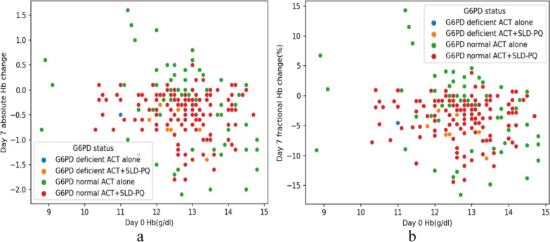
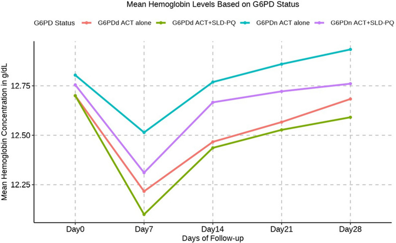
In comparison to those with G6PDn ACT alone, both the ACT alone and the ACT + SLD-PQ G6PDd cohorts experienced a lower mean Hb concentration, with mean changes ranging from 0.45 g/dl [95% CI − 0.486 to 0.079] to 0.54 g/dl [95% CI − 1.067 to 0.238] (Table 5 and Fig. 4). In addition, on day 7 following ACT + SLD-PQ treatment, the Hb concentration in these G6PDd participants ranged from 12.70 to 12.10 g/dL, and that in the ACT-alone G6PDd participants ranged from 12.70 to 12.22. The distribution of patients between treatment arms was unaffected by sex or phenotypic G6PD phenotype. Although the mean changes in Hb in these groups were greater than those in the G6PDn ACT treatment group, a significant difference in Hb levels was not detected on Day 7 posttreatment for G6PDd ACT + SLD-PQ vs. G6PDn ACT alone (P = 0.109, P = 0.304; Table 5).
Fig. 4.
Change in absolute Hb concentration by day 7(Hb day 7-day 0) (a) and change in fractional Hb concentration by day 7[100*(absolute Hb /baseline Hb)] (b) plotted against the concentration of Hb at baseline, for each group
However, the absolute mean reduction in Hb levels on day 7 was not significantly lower in either G6PDd (− 0.54 g/dl 95% CI − 1.19, 0.12; P = 0.109) or G6PDn (− 0.20 g/dl 95% CI − 0.48, 0.08; P = 0.154) ACT + SLD-PQ individuals (Table 5). There were consistently lower Hb concentrations in G6PDd participants treated with ACT + SLD-PQ than in G6PDn participants, although these differences were not clinically significant Fig. 5.
Fig. 5.
Mean haemoglobin concentrations in G6PDd and G6PDn study participants who received ACT + SLD-PQ or ACT alone
Adverse events in the treatment groups
In the study, abdominal pain, appetite loss, fatigue, and nausea were the major AEs, followed by skin rash, cough, headache, diarrhoea, and vomiting. Out of the 249 study participants who completed the follow-up, 94 AEs were recorded—51 (30.7%) from the ACT + SLD-PQ cohort and 43 (51.8%) from the ACT alone group (Table 6). All of the reported AEs were rated as mild. The difference in the incidence of adverse events (AEs) between the treatment groups was not significant (Table 6). There were no SAEs in either treatment group. None of the participants required blood transfusions. In addition, during the study follow-up, every adverse event (AE) resolved, and none of the adverse events stopped participating.
Table 6.
Adverse events (AEs) among study participants by G6PD status and treatment group, Arjo Didessa, Ethiopia
| Treatment arms | |||||
|---|---|---|---|---|---|
| Adverse events | ACT only | ACT + SLD-PQ | P value | ||
| G6PDn n (%) | G6PDd n (%) | G6PDn n (%) | G6PDd n (%) | ||
| Head ache | 4 (11.43%) | 0 | 1 (2.38%) | 1 (11.11%) | 0.097 |
| Nausea | 6 (17.14%) | 1 (12.5%) | 4 (9.52%) | 1 (11.11%) | 0.062 |
| Vomiting | 1 (2.86%) | 0 | 1 (2.38%) | 1 (11.11%) | 0.705 |
| Abdominal pain | 5 (17.14%) | 2 (25%) | 12 (28.57%) | 2 (22.22%) | 0.460 |
| loss of appetite | 5 (14.29%) | 2 (25%) | 9 (21.43%) | 1 (11.11%) | 0.321 |
| Fatigue | 6 (17.14%) | 1 (12.5%) | 5 (11.91%) | 1 (11.11%) | 0.098 |
| Skin rashes | 2 (5.71%) | 1 (12.5%) | 5 (11.91%) | 1 (11.11%) | 0.626 |
| Cough | 4 (11.43) | 1 (12.5%) | 3 (7.14%) | 0 | 0.084 |
| Diarrhea | 1 (2.86%) | 0 | 2 (4.76%) | 1 (11.11%) | 0.593 |
| Total | 35 (100%) | 8 (100%) | 42 (100%) | 9 (100%) | |
Discussion
This observational cohort study's findings provide significant new insights into the safety profile of ACT + SLD-PQ combination therapy for managing uncomplicated falciparum malaria in individuals with G6PDd from Ethiopia. This study is the first of its kind in which patients with confirmed enzymatic G6PDd were included in an effort to more accurately reflect real-world circumstances. Further proof of the haematological effects of this treatment regimen in the PQ cohort is provided by the study using longitudinal panel measurements from the Arjo-Didessa. Interestingly, a week after starting treatment, there was a noticeable drop in the mean Hb concentration, suggesting a temporary haematological effect. However, it was also observed that Hb levels returned to baseline values by the 28th day of treatment, indicating a recuperative trend during treatment. It is remarkable, nonetheless, that the G6PD deficit did not exhibit the same pattern.
Various studies examining the relationship between G6PDd and posttreatment haemolysis induced by oxidant anti-malarial drugs have used Hb concentration (mean decrease) as the primary outcome measure because changes in Hb levels are objectively measurable [20, 24, 26, 27]. According to the findings in this study, there was no statistically significant difference (P = 0.157) between the treatment groups that got ACT + SLD-PQ and ACT-alone. Results from an earlier study conducted in Burkina Faso are similar, supporting the idea that there is a consistent pattern in this context [29]. Further investigation is necessary in light of the noted decrease in Hb levels among individuals who were only given ACT. There are various reasons related to the pathophysiology of malaria and its treatment that could be responsible for this reduction. The loss of red blood cells carrying parasites during schizont rupture may be the reason for the decrease in Hb levels in the ACT alone group. Furthermore, haemolysis is caused by systemic inflammation and oxidative stress brought on by the malaria infection, which leads to the loss of both parasitized and non-parasitized red blood cells [50–53].
Similarly, the mean changes in Hb concentration concerning baseline values and the mean variations in Hb concentration concerning the status of G6PD were further analysed. The findings showed that in all groups treated with ACT + SLD-PQ, mean Hb concentrations, whether expressed as absolute values or relative changes, decreased during the first week of therapy and recovered to baseline levels during follow-up. A week after the administration of ACT + SLD-PQ, G6PDd participants showed a decrease in their mean Hb concentration compared to their baseline values. On day 7, there was no significant difference in the mean Hb levels between the patients treated with ACT-alone and those treated with ACT + SLD-PQ (coefficient, − 0.45; 95% CI − 1.01, 0.10; P = 0.108). According to a previous study that assessed the safety of this SLD-PQ as a transmission-blocking therapy showed comparable decreases in Hb levels, Hb levels in G6PDd patients temporarily decreased after they were treated with ACT + SLD-PQ [29]. The findings of this study were consistent with those of previous studies indicating low safety concerns associated with SLD-PQ in G6PDd malaria patients [24, 26, 54, 55]. Additionally, SLD-PQ treatment was linked to a temporary drop in Hb levels in G6PDd individuals [54]. Similarly, a recent systematic review [56] reported that the haemolytic effects of SLD-PQ (0.1 and 0.25 mg/kg) were less likely in individuals who received G6PDd than in those who received a previous dose of 0.75 mg/kg. Similarly, a recent systematic review [56] reported that the haemolytic effects of SLD-PQ (0.1 and 0.25 mg/kg) were less likely in individuals who received G6PDd than in those who received a previous dose of 0.75 mg/kg. Overall, these findings imply that reduced G6PD enzyme activity may not lead to a considerable reduction in Hb levels following day 7 after ACT + SLD-PQ therapy when only given at the recommended dose. Contrasting results are reported in a study in Senegal [6], where patients with G6PDd on Day 7 post-treatment had considerably lower Hb levels than G6PDn persons. This variation highlights the variety in haematological responses to antimalarial medication among various populations and settings, implying the possibility of regional or population-specific variables affecting the dynamics of Hb after treatment.
In this study, there were no significant differences seen in the mean absolute reduction in Hb levels by day 7 after treatment initiation based on G6PD status. G6PDd patients treated with ACT alone showed an absolute mean Hb decrease of 0.26 g/dL (95% CI − 0.73, 0.21, P = 0.279). On the other hand, the similar results for G6PDn and G6PDd patients in the ACT + SLD-PQ arm were [0.34 g/dL (95% CI − 0.70, 0.01, P = 0.058) and 0.16 g/dL (95% CI − 0.31, − 0.00, P = 0.05)], respectively. Comparing the absolute mean Hb reduction to G6PDn individuals treated with ACT alone, no statistically significant changes were found. The results of this investigation are consistent with earlier studies that looked at the safety of SLD-PQ with G6PD level [23, 26]. Other research, however, has demonstrated a notable drop in absolute mean Hb levels between the treatment arms although they did not compare the treatment outcomes by G6PD status [25]. This shows that early in the course of treatment, there is a consistent haematological response to antimalarial therapy. Comparing G6PDd patients with normal G6PD activity and those who received ACT alone or in combination with SLD-PQ, similar trends were found [26]. Moreover, the absence of acute haemolysis, which is characterized by a drop in fractional Hb levels of > 25% or a decrease in 3 g/dl or above during the follow-up, as evidenced by the absence of significant drops in Hb levels, indicates the safety of both treatment arms in terms of haemolytic risk.
This study also provides significant inverse data in that most of the available studies did not investigate the relative mean Hb concentration decline in G6PDn patients who were treated with primaquine in particular. These results demonstrate that, when treated with SLD-PQ, individuals with different G6PD statuses had significantly different relative mean Hb levels. There was not a significant distinction in the relative mean Hb reduction between G6PDd and G6PDn individuals receiving ACT alone. In contrast to G6PDn patients treated with ACT alone, both G6PDd and G6PDn patients treated with SLD-PQ and ACT showed a significant relative mean Hb reduction (2.91% [95% CI − 5.70, − 0.11], P = 0.041, and 1.42% [95% CI − 2.62, − 0.21], P = 0.021, respectively). The precise reasons behind the observed decrease in Hb levels in patients receiving combination therapy remain unclear.
The study also revealed that by Day 28, a comparable proportion of patients in both the ACT alone arm and ACT + SLD-PQ arm had recovered their Hb levels. This lack of statistically significant difference (p = 0. 0.212) suggests that the addition of SLD-PQ to the treatment regimen did not significantly affect Hb recovery rates in G6PDn patients. These results align with earlier research on Tanzanian individuals with uncomplicated P. falciparum infections. These earlier studies likewise reported a decrease in Hb levels on Day 7 after the start of treatment, followed by a period of recovery [23]. Mean changes in Hb concentration provided a broad comparison between treatment groups; maximal reductions might more accurately represent extreme haemolysis [57]. The baseline Hb concentration was taken into account when comparing the results, and the maximum reductions in Hb concentration (the largest reduction in Hb concentration compared to baseline at any time point during follow-up) were greater in both the ACT alone and ACT + SLD-PQ groups on Day 7 posttreatment, with a relative mean difference of − 0.20 g/dL (95% CI − 0.08, 0.47; P = 0.157). The study did not show any significant differences between the two groups in terms of the maximum reductions in Hb concentration, which was predicted [58]. There were no statistically significant differences in the absolute mean maximal reduction in Hb concentration or the relative mean maximal reduction in Hb concentration between G6PDd patients receiving ACT + SLD-PQ and ACT alone (P = 0.058 and 0.066, respectively). The results of the current study align with what has been observed in similar research studies before [22]. It is important to keep in mind, nevertheless, that data from other research have suggested that G6PDd patients showed larger Hb falls compared to G6PDn in terms of their mean fractional Hb changes [26, 59]. Overall, these findings imply that reduced G6PD enzyme activity may not lead to a considerable reduction in Hb levels following day 7 after ACT + SLD-PQ therapy when only given at the recommended dose.
The biological plausibility of haemolysis caused by primaquine in patients with G6PDd demonstrates complex relationships between drug metabolism and red blood cell function. The liver metabolizes primaquine, one of the most important anti-malarial medications, producing reactive metabolites that cause oxidative stress in red blood cells [60]. Red blood cells with G6PDd are more susceptible to oxidative damage because their defense systems are compromised, resulting in insufficient enzymatic activity. Due to this increased susceptibility, those with G6PD deficiency are more likely to have haemolysis from primaquine because the delicate balance between antioxidants and oxidants is tilted in favour of cellular damage [61]. This possibility is supported by clinical data, which includes reports of haemolytic responses in G6PDd individuals after primaquine treatment. Nonetheless, the suggestion by the World Health Organization that a single low dose of primaquine be used in the fight against malaria is part of a larger public health strategy that aims to eliminate malaria [62]. However, recent research—including the one that was mentioned—supports the safety of primaquine in those who lack G6PD and offers a detailed analysis of the drug's risk–benefit ratio [27].
One limitation of this study was the small number of G6PD-deficient study participants, and an unequal number of study participants were followed from the two study arms. Furthermore, determining Hb levels on Day 3 would have yielded a more thorough comprehension of the nadir Hb. Moreover, future studies employing sizable population-based cohorts are recommended to strengthen the robustness of the conclusions about the safety of SLD-PQ in G6PD-deficient individuals.
Conclusion
This study's findings indicate that using SLD-PQ in combination with ACT is safe for uncomplicated falciparum malaria regardless of the patient’s G6PD status in Ethiopian settings. Caution should be taken in extrapolating this finding in other settings with diverse G6DP phenotypes.
Acknowledgements
We thank the parents, legal guardians, and study participants for their patience and dedication; all of the study team members for their contributions; the International Centre of Excellence for Malaria Research (ICEMR-Ethiopia) collaboration, which provided logistical and laboratory support for this study; the local physicians at the Arjo Sugar Factory Health Centre, who guided the evaluation of the potential adverse effects of primaquine; and the Tropical and Infectious Diseases Research Centre (TIDRC) of Jimma University for providing logistics.
Abbreviations
- ACT
Artemisinin-based combination therapy
- AMM
Adjusted male median (AMM)
- CI
Confidence interval
- FMoH
Federal Ministry of Health
- G6PD
Glucose-6-phosphate dehydrogenase
- G6PDd
Glucose-6-phosphate dehydrogenase deficient
- Hb
Haemoglobin
- ICEMR
International Center of Excellence for Malaria Research
- IQR
Interquartile Range
- NIH
National Institute of Health
- SD
Standard deviation
- SLD-PQ
Single low-dose (0.25 mg/kg) primaquine
- WHO
World Health Organization
Appendix
Table 7.
Results of multiple linear regression showing the mean Hb concentration and range of change from baseline for patients with and without G6PD deficiency on day 7
| Day 7 | Coef. | t value | [95% CI] | P value |
|---|---|---|---|---|
| Sex: base male | 0 | |||
| Female | − 0.23 | − 1.64 | − 0.50–0.05 | 0.102 |
| Age | 0.01 | 1.71 | − 0.00–0.02 | 0.089 |
| G6PD status | ||||
| G6PDd ACT alone | − 0.45 | − 1.03 | − 1.31–0.41 | 0.304 |
| G6PDn ACT + SLD-PQ | − 0.20 | − 1.43 | − 0.48–0.08 | 0.154 |
| G6PDd ACT + SLD-PQ | − 0.54 | − 1.61 | − 1.19–0.12 | 0.109 |
Coef is the mean Hb coefficient of variation estimated using a multiple linear regression model comparing the mean reduction in Hb concentration on day 7 in each group
G6PD-d: glucose-6-phosphate dehydrogenase deficiency, G6PD-n: normal glucose-6-phosphate dehydrogenase
Table 8.
The mean haemoglobin concentration and range of change from baseline to day 14 for patients with and without G6PD deficiency
| Day 14 | Coef | t value | [95% CI] | p value |
|---|---|---|---|---|
| Sex: base male | 0 | |||
| Female | − 0.016 | − 1.40 | − 0.39–0.07 | 0.163 |
| Age | 0.012 | 2.29 | 0.00–0.02 | 0.023 |
| G6PD status | ||||
| G6PDd ACT alone | − 0.44 | − 1.19 | − 1.17–0.29 | 0.234 |
| G6PDn ACT + SLD-PQ | − 0.10 | − 0.85 | − 0.34–0.13 | 0.397 |
| G6PDd ACT + SLD-PQ | − 0.45 | − 1.61 | − 1.01–0.10 | 0.108 |
Coef, mean Hb coefficient of variation estimated using a multiple linear regression model comparing the mean reduction in Hb concentration on day 14 in each group
G6PD-d: glucose-6-phosphate dehydrogenase deficiency, G6PD-n: normal glucose-6-phosphate dehydrogenase
Author contributions
Conceptualization: KH, BP, DY, and GY. Data curation: KH, HG, AA, and AT. Data analysis: KH, ZG, HG. Funding acquisition: GY. Investigation: KH, HG, AA, AT, and AD. Methodology: KH, HG, DY, BP, and GY. Project administration: DY, TD, GY, DY. Supervision: KH, TD, DY, BP, and GY. Writing – original draft: KH. Writing: Review and editing: KH, HG, AA, AD, AT,DY, ML, CK, JK, GZ, XW, TD, SK, BP, GY.
Funding
The National Institutes of Health (NIH) (D43 TW001505, R01 A1050243, and U19 AI129326) funded this research. The study’s design, the gathering, analysis, and reporting of the data, the interpretation of the findings, the choice to publish, and the writing of the paper were all performed without the sponsors’ input.
Data availability
The data that support the findings of this study are available from the corresponding author, [Kassahun Habtamu], upon reasonable request.
Declarations
Ethics approval and consent to participate
This study was approved by the Institutional Review Board at the University of Addis Ababa's Natural & Computational Sciences (IRB) (CNSDO/20 1 111/2018). There were no unanticipated problems involving risks to volunteers or others, and no significant deviations were noted. Before the study was screened, all willing participants older than eighteen years were asked to provide written informed consent; for those younger, approval was obtained from parents or legal guardians. A witness who could read was utilized for patients who could not provide their consent, and children's consent was also sought.
Competing interests
The authors declare no competing interests.
Footnotes
Publisher's Note
Springer Nature remains neutral with regard to jurisdictional claims in published maps and institutional affiliations.
References
- 1.Pousibet-Puerto J, Salas-Coronas J, Sánchez-Crespo A, Molina-Arrebola MA, Soriano-Pérez MJ, Giménez-López MJ, et al. Impact of using artemisinin-based combination therapy (ACT) in the treatment of uncomplicated malaria from Plasmodium falciparum in a non-endemic zone. Malar J; 2016;15:339. [DOI] [PMC free article] [PubMed]
- 2.WWARN Gametocyte Study Group Gametocyte carriage in uncomplicated Plasmodium falciparum malaria following treatment with artemisinin combination therapy: a systematic review and meta-analysis of individual patient data. BMC Med. 2016;14:79. doi: 10.1186/s12916-016-0621-7. [DOI] [PMC free article] [PubMed] [Google Scholar]
- 3.Gonçalves BP, Tiono AB, Ouédraogo A, Guelbéogo WM, Bradley J, Nebie I, et al. Single low dose primaquine to reduce gametocyte carriage and Plasmodium falciparum transmission after artemether-lumefantrine in children with asymptomatic infection: a randomised, double-blind, placebo-controlled trial. BMC Med. 2016;14:40. doi: 10.1186/s12916-016-0581-y. [DOI] [PMC free article] [PubMed] [Google Scholar]
- 4.Roth JM, Sawa P, Omweri G, Osoti V, Makio N, Bradley J, et al. Plasmodium falciparum gametocyte dynamics after pyronaridine-artesunate or artemether-lumefantrine treatment. Malar J. 2018;17:223. doi: 10.1186/s12936-018-2373-7. [DOI] [PMC free article] [PubMed] [Google Scholar]
- 5.Munro BA, McMorran BJ. Antimalarial drug strategies to target Plasmodium gametocytes. Parasitologia. 2022;2:101–124. doi: 10.3390/parasitologia2020011. [DOI] [Google Scholar]
- 6.Tine RC, Sylla K, Faye BT, Poirot E, Fall FB, Sow D, et al. Safety and efficacy of adding a single low dose of primaquine to the treatment of adult patients with Plasmodium falciparum malaria in Senegal, to reduce gametocyte carriage: a randomized controlled trial. Clin Infect Dis. 2017;65:535–543. doi: 10.1093/cid/cix355. [DOI] [PMC free article] [PubMed] [Google Scholar]
- 7.White NJ. Primaquine to prevent transmission of falciparum malaria. Lancet Infect Dis. 2013;13:175–181. doi: 10.1016/S1473-3099(12)70198-6. [DOI] [PubMed] [Google Scholar]
- 8.Taylor WR, Naw HK, Maitland K, Williams TN, Kapulu M, D’Alessandro U, et al. Single low-dose primaquine for blocking transmission of Plasmodium falciparum malaria–a proposed model-derived age-based regimen for sub-Saharan Africa. BMC Med. 2018;16:11. doi: 10.1186/s12916-017-0990-6. [DOI] [PMC free article] [PubMed] [Google Scholar]
- 9.Stepniewska K, Allen EN, Humphreys GS, Poirot E, Craig E, Kennon K, Yilma D, et al. Safety of single-dose primaquine as a Plasmodium falciparum gametocytocide: a systematic review and meta-analysis of individual patient data. BMC Med. 2022;20:350. doi: 10.1186/s12916-022-02504-z. [DOI] [PMC free article] [PubMed] [Google Scholar]
- 10.Dicko A, Brown JM, Diawara H, Baber I, Mahamar A, Soumare HM, et al. Primaquine to reduce transmission of Plasmodium falciparum malaria in Mali: a single-blind, dose-ranging, adaptive randomised phase 2 trial. Lancet Infect Dis. 2016;16:674–684. doi: 10.1016/S1473-3099(15)00479-X. [DOI] [PMC free article] [PubMed] [Google Scholar]
- 11.Liu H, Zeng W, Malla P, Wang C, Lakshmi S, Kim K, et al. Risk of hemolysis in Plasmodium vivax malaria patients receiving standard primaquine treatment in a population with high prevalence of G6PD deficiency. Infection. 2023;51:213–222. doi: 10.1007/s15010-022-01905-9. [DOI] [PMC free article] [PubMed] [Google Scholar]
- 12.Avalos S, Mejia RE, Banegas E, Salinas C, Gutierrez L, Fajardo M, et al. G6PD deficiency, primaquine treatment, and risk of haemolysis in malaria-infected patients. Malar J. 2018;17:415. doi: 10.1186/s12936-018-2564-2. [DOI] [PMC free article] [PubMed] [Google Scholar]
- 13.WHO. Guidelines for Malaria, 16 February 2021. Geneva, World Health Organization; 2021.
- 14.Recht J, Ashley EA, White NJ. Use of primaquine and glucose-6-phosphate dehydrogenase deficiency testing: divergent policies and practices in malaria endemic countries. PLoS Negl Trop Dis. 2018;12:e0006230. doi: 10.1371/journal.pntd.0006230. [DOI] [PMC free article] [PubMed] [Google Scholar]
- 15.Chu CS, Bancone G, Nosten F, White NJ, Luzzatto L. Primaquine-induced haemolysis in females heterozygous for G6PD deficiency. Malar J. 2018;17:101. doi: 10.1186/s12936-018-2248-y. [DOI] [PMC free article] [PubMed] [Google Scholar]
- 16.Howes RE, Battle KE, Satyagraha AW, Baird JK, Hay SI. G6PD deficiency: global distribution, genetic variants and primaquine therapy. Adv Parasitol. 2013;81:133–201. doi: 10.1016/B978-0-12-407826-0.00004-7. [DOI] [PubMed] [Google Scholar]
- 17.He Y, Zhang Y, Chen X, Wang Q, Ling L, Xu Y. Glucose-6-phosphate dehydrogenase deficiency in the Han Chinese population: molecular characterization and genotype–phenotype association throughout an activity distribution. Sci Rep. 2020;10:17106. doi: 10.1038/s41598-020-74200-y. [DOI] [PMC free article] [PubMed] [Google Scholar]
- 18.Sathupak S, Leecharoenkiat K, Kampuansai J. Prevalence and molecular characterization of glucose-6-phosphate dehydrogenase deficiency in the Lue ethnic group of northern Thailand. Sci Rep. 2021;11:2956. doi: 10.1038/s41598-021-82477-w. [DOI] [PMC free article] [PubMed] [Google Scholar]
- 19.Awandu SS, Raman J, Makhanthisa TI, Kruger P, Frean J, Bousema T, et al. Understanding human genetic factors influencing primaquine safety and efficacy to guide primaquine roll-out in a pre-elimination setting in southern Africa. Malar J. 2018;17:120. doi: 10.1186/s12936-018-2271-z. [DOI] [PMC free article] [PubMed] [Google Scholar]
- 20.Chen I, Diawara H, Mahamar A, Sanogo K, Keita S, Kone D, et al. Safety of single-dose primaquine in G6PD-deficient and G6PD-normal males in Mali without malaria: an open-label, phase 1, dose-adjustment trial. J Infect Dis. 2018;217:1298–1308. doi: 10.1093/infdis/jiy014. [DOI] [PMC free article] [PubMed] [Google Scholar]
- 21.Raman J, Allen E, Workman L, Mabuza A, Swanepoel H, Malatje G, et al. Safety and tolerability of single low-dose primaquine in a low-intensity transmission area in South Africa: an open-label, randomized controlled trial. Malar J. 2019;18:209. doi: 10.1186/s12936-019-2841-8. [DOI] [PMC free article] [PubMed] [Google Scholar]
- 22.Dysoley L, Kim S, Lopes S, Khim N, Bjorges S, Top S, et al. The tolerability of single low dose primaquine in glucose-6-phosphate deficient and normal falciparum-infected Cambodians. BMC Infect Dis. 2019;19:250. doi: 10.1186/s12879-019-3862-1. [DOI] [PMC free article] [PubMed] [Google Scholar]
- 23.Mwaiswelo R, Ngasala BE, Jovel I, Gosling R, Premji Z, Poirot E, et al. Safety of a single low-dose of primaquine in addition to standard artemether-lumefantrine regimen for treatment of acute uncomplicated Plasmodium falciparum malaria in Tanzania. Malar J. 2016;15:316. doi: 10.1186/s12936-016-1341-3. [DOI] [PMC free article] [PubMed] [Google Scholar]
- 24.Mwaiswelo RO, Ngasala B, Msolo D, Kweka E, Mmbando BP, Mårtensson A. A single low dose of primaquine is safe and sufficient to reduce transmission of Plasmodium falciparum gametocytes regardless of cytochrome P450 2D6 enzyme activity in Bagamoyo district. Tanzania Malar J. 2022;21:84. doi: 10.1186/s12936-022-04100-1. [DOI] [PMC free article] [PubMed] [Google Scholar]
- 25.Poirot E, Soble A, Ntshalintshali N, Mwandemele A, Mkhonta N, Malambe C, et al. Development of a pharmacovigilance safety monitoring tool for the rollout of single low-dose primaquine and artemether-lumefantrine to treat Plasmodium falciparum infections in Swaziland: a pilot study. Malar J. 2016;15:384. doi: 10.1186/s12936-016-1410-7. [DOI] [PMC free article] [PubMed] [Google Scholar]
- 26.Bastiaens GJ, Tiono AB, Okebe J, Pett HE, Coulibaly SA, Goncalves BP, et al. Safety of single low-dose primaquine in glucose-6-phosphate dehydrogenase deficient falciparum-infected African males: two open-label, randomized, safety trials. PLoS ONE. 2018;13:e0190272. doi: 10.1371/journal.pone.0190272. [DOI] [PMC free article] [PubMed] [Google Scholar]
- 27.Taylor WR, Olupot-Olupot P, Onyamboko MA, Peerawaranun P, Weere W, Namayanja C, et al. Safety of age-dosed, single low-dose primaquine in children with glucose-6-phosphate dehydrogenase deficiency who are infected with Plasmodium falciparum in Uganda and the Democratic Republic of the Congo: a randomised, double-blind, placebo-controlled, non-inferiority trial. Lancet Infect Dis. 2023;23:471–483. doi: 10.1016/S1473-3099(22)00658-2. [DOI] [PubMed] [Google Scholar]
- 28.WHO. Evidence review group: the safety and effectiveness of single dose primaquine as a P. falciparum gametocytocide. Bangkok, Thailand 2012:13–15.
- 29.van Beek SW, Svensson EM, Tiono AB, Okebe J, D’Alessandro U, Gonçalves BP, et al. Model-based assessment of the safety of community interventions with primaquine in sub-Saharan Africa. Parasit Vectors. 2021;14:524. doi: 10.1186/s13071-021-05034-4. [DOI] [PMC free article] [PubMed] [Google Scholar]
- 30.Shekalaghe S, Drakeley C, Gosling R, Ndaro A, Van Meegeren M, Enevold A, et al. Primaquine clears submicroscopic Plasmodium falciparum gametocytes that persist after treatment with sulphadoxine-pyrimethamine and artesunate. PLoS ONE. 2007;2:e1023. doi: 10.1371/journal.pone.0001023. [DOI] [PMC free article] [PubMed] [Google Scholar]
- 31.Stepniewska K, Humphreys GS, Gonçalves BP, Craig E, Gosling R, Guerin PJ, et al. Efficacy of single-dose primaquine with artemisinin combination therapy on Plasmodium falciparum gametocytes and transmission: an individual patient meta-analysis. J Infect Dis. 2022;225:1215–1226. doi: 10.1093/infdis/jiaa498. [DOI] [PMC free article] [PubMed] [Google Scholar]
- 32.Mukaka M, Onyamboko MA, Olupot-Olupot P, Peerawaranun P, Suwannasin K, Pagornrat W, et al. Pharmacokinetics of single low dose primaquine in Ugandan and Congolese children with falciparum malaria. eBioMedicine. 2023;96:104805. [DOI] [PMC free article] [PubMed]
- 33.Dicko A, Roh ME, Diawara H, Mahamar A, Soumare HM, Lanke K, et al. Efficacy and safety of primaquine and methylene blue for prevention of Plasmodium falciparum transmission in Mali: a phase 2, single-blind, randomised controlled trial. Lancet Infect Dis. 2018;18:627–639. doi: 10.1016/S1473-3099(18)30044-6. [DOI] [PMC free article] [PubMed] [Google Scholar]
- 34.Demissew A, Hawaria D, Kibret S, Animut A, Tsegaye A, Lee MC, et al. Impact of sugarcane irrigation on malaria vector Anopheles mosquito fauna, abundance and seasonality in Arjo-Didessa. Ethiopia Malar J. 2020;19:344. doi: 10.1186/s12936-020-03416-0. [DOI] [PMC free article] [PubMed] [Google Scholar]
- 35.Tsegaye A, Demissew A, Hawaria D, Abossie A, Getachew H, Habtamu K, et al. Anopheles larval habitats seasonality and environmental factors affecting larval abundance and distribution in Arjo-Didessa sugar cane plantation. Ethiopia Malar J. 2023;22:350. doi: 10.1186/s12936-023-04782-1. [DOI] [PMC free article] [PubMed] [Google Scholar]
- 36.Getachew H, Demissew A, Abossie A, Habtamu K, Wang X, Zhong D, et al. Asymptomatic and submicroscopic malaria infections in sugar cane and rice development areas of Ethiopia. Malar J. 2023;22:341. doi: 10.1186/s12936-023-04762-5. [DOI] [PMC free article] [PubMed] [Google Scholar]
- 37.WHO. Methods for surveillance of antimalarial drug efficacy. Geneva, World Health Organization; 2015.
- 38.WHO. Guidelines for malaria. Geneva, World Health Organization; 2023.
- 39.Ethiopia FMoH. National malaria guidelines. Addis Ababa, 2018.
- 40.WHO. Giemsa staining of malaria blood films. Geneva, World Health Organization; 2016.
- 41.WHO. Basic malaria microscopy. Geneva, World Health Organization; 2010.
- 42.Domingo GJ, Satyagraha AW, Anvikar A, Baird K, Bancone G, Bansil P, et al. G6PD testing in support of treatment and elimination of malaria: recommendations for evaluation of G6PD tests. Malar J. 2013;12:391. doi: 10.1186/1475-2875-12-391. [DOI] [PMC free article] [PubMed] [Google Scholar]
- 43.Shenkutie TT, Nega D, Hailu A, Kepple D, Witherspoon L, Lo E, et al. Prevalence of G6PD deficiency and distribution of its genetic variants among malaria-suspected patients visiting Metehara health centre. Eastern Ethiopia Malar J. 2022;21:260. doi: 10.1186/s12936-022-04269-5. [DOI] [PMC free article] [PubMed] [Google Scholar]
- 44.Lo E, Zhong D, Raya B, Pestana K, Koepfli C, Lee M-C, et al. Prevalence and distribution of G6PD deficiency: implication for the use of primaquine in malaria treatment in Ethiopia. Malar J. 2019;18:340. doi: 10.1186/s12936-019-2981-x. [DOI] [PMC free article] [PubMed] [Google Scholar]
- 45.Leung-Pineda V, Weinzierl EP, Rogers BB. Preliminary investigation into the prevalence of G6PD deficiency in a pediatric African American population using a near-patient diagnostic platform. Diagnostics. 2023;13:3647. doi: 10.3390/diagnostics13243647. [DOI] [PMC free article] [PubMed] [Google Scholar]
- 46.Domingo GJ, Advani N, Satyagraha AW, Sibley CH, Rowley E, Kalnoky M, et al. Addressing the gender-knowledge gap in glucose-6-phosphate dehydrogenase deficiency: challenges and opportunities. Int Health. 2019;11:7–14. doi: 10.1093/inthealth/ihy060. [DOI] [PMC free article] [PubMed] [Google Scholar]
- 47.Thriemer K, Degaga TS, Christian M, Alam MS, Rajasekhar M, Ley B, et al. Primaquine radical cure in patients with Plasmodium falciparum malaria in areas co-endemic for P. falciparum and Plasmodium vivax (PRIMA): a multicentre, open-label, superiority randomised controlled trial. Lancet. 2023;402:2101–10. [DOI] [PMC free article] [PubMed]
- 48.Shekalaghe S, Mosha D, Hamad A, Mbaga TA, Mihayo M, Bousema T, et al. Optimal timing of primaquine to reduce Plasmodium falciparum gametocyte carriage when co-administered with artemether–lumefantrine. Malar J. 2020;19:34. doi: 10.1186/s12936-020-3121-3. [DOI] [PMC free article] [PubMed] [Google Scholar]
- 49.Taylor WR, Kheng S, Muth S, Tor P, Kim S, Bjorge S, et al. Hemolytic dynamics of weekly primaquine antirelapse therapy among Cambodians with acute Plasmodium vivax malaria with or without glucose-6-phosphate dehydrogenase deficiency. J Infect Dis. 2019;220:1750–1760. doi: 10.1093/infdis/jiz313. [DOI] [PMC free article] [PubMed] [Google Scholar]
- 50.Kyeremeh R, Antwi-Baffour S, Annani-Akollor M, Adjei JK, Addai-Mensah O, Frempong M. Comediation of erythrocyte haemolysis by erythrocyte-derived microparticles and complement during malaria infection. Adv Hematol. 2020;2020:1640480. doi: 10.1155/2020/1640480. [DOI] [PMC free article] [PubMed] [Google Scholar]
- 51.Mosha D, Kakolwa MA, Mahende MK, Masanja H, Abdulla S, Drakeley C, et al. Safety monitoring experience of single-low dose primaquine co-administered with artemether–lumefantrine among providers and patients in routine healthcare practice: a qualitative study in Eastern Tanzania. Malar J. 20:392. [DOI] [PMC free article] [PubMed]
- 52.White NJ. Anaemia and malaria. Malar J. 2018;17:371. doi: 10.1186/s12936-018-2509-9. [DOI] [PMC free article] [PubMed] [Google Scholar]
- 53.White NJ. What causes malaria anemia? Blood. 2022;139:2268–2269. doi: 10.1182/blood.2021015055. [DOI] [PubMed] [Google Scholar]
- 54.Nelwan EJ, Shakinah S, Pasaribu A. Association of G6PD status and haemolytic anaemia in patients receiving anti-malarial agents: a systematic review and meta-analysis. Malar J. 2023;22:77. doi: 10.1186/s12936-023-04493-7. [DOI] [PMC free article] [PubMed] [Google Scholar]
- 55.Mwaiswelo RO, Kabuga H, Kweka EJ, Baraka V. Is it time for Africa to adopt primaquine in the era of malaria control and elimination? Trop Med Health. 2022;50:17. doi: 10.1186/s41182-022-00408-5. [DOI] [PMC free article] [PubMed] [Google Scholar]
- 56.Uthman OA, Graves PM, Saunders R, Gelband H, Richardson M, Garner P. Safety of primaquine given to people with G6PD deficiency: systematic review of prospective studies. Malar J. 2017;16:346. doi: 10.1186/s12936-017-1989-3. [DOI] [PMC free article] [PubMed] [Google Scholar]
- 57.Barcellini W, Fattizzo B: Clinical applications of hemolytic markers in the differential diagnosis and management of hemolytic anemia.Disease markers 2015, 2015. [DOI] [PMC free article] [PubMed]
- 58.Price RN, White NJ. Drugs that reduce transmission of falciparum malaria. Lancet Infect Dis. 2018;18:585–586. doi: 10.1016/S1473-3099(18)30070-7. [DOI] [PMC free article] [PubMed] [Google Scholar]
- 59.Bancone G, Chowwiwat N, Somsakchaicharoen R, Poodpanya L, Moo PK, Gornsawun G, et al. Single low dose primaquine (0.25 mg/kg) does not cause clinically significant haemolysis in G6PD deficient subjects. PLoS One 2016;11:e0151898. [DOI] [PMC free article] [PubMed]
- 60.Ganesan S, Chaurasiya ND, Sahu R, Walker LA, Tekwani BL. Understanding the mechanisms for metabolism-linked hemolytic toxicity of primaquine against glucose 6-phosphate dehydrogenase deficient human erythrocytes: evaluation of eryptotic pathway. Toxicology. 2012;294:54–60. doi: 10.1016/j.tox.2012.01.015. [DOI] [PubMed] [Google Scholar]
- 61.Luzzatto L, Ally M, Notaro R. Glucose-6-phosphate dehydrogenase deficiency. Blood. 2020;136:1225–1240. doi: 10.1182/blood.2019000944. [DOI] [PubMed] [Google Scholar]
- 62.WHO. Policy brief on single-dose primaquine as a gametocytocide in Plasmodium falciparum malaria. Geneva, World Health Organization; 2015.
Associated Data
This section collects any data citations, data availability statements, or supplementary materials included in this article.
Data Availability Statement
The data that support the findings of this study are available from the corresponding author, [Kassahun Habtamu], upon reasonable request.


