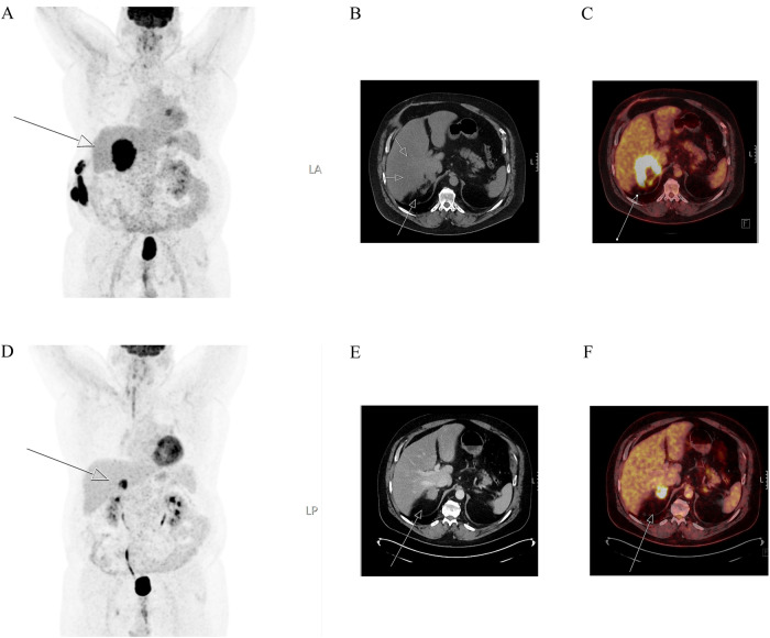
Fig. 3.
18F-FDG-PET/CT imaging of the same patient histologically and immunohistochemically demonstrated in Fig. 2. Maximum intensity projection (MIP) image (left A, and D), CT images (middle B and E) and fused images (right C and F) of 18F-FDG-PET/CT show the adrenal gland metastasis (white arrow) in the right adrenal. The metastasis increased in size from 4 (E, F) to 9 (A−C) centimeters between examinations, which were carried out at an 8-month interval

