Abstract
Background
While the urogenital microbiota has recently been characterized in healthy male and female dogs, the influence of sex hormones on the urogenital microbiome of bitches is still unknown. A deeper understanding of the cyclic changes in urinary and vaginal microbiota would allow us to compare the bacterial populations in healthy dogs and assess the impact of the microbiome on various urogenital diseases. Therefore, the aim of this study was to characterize and compare the urogenital microbiota during different phases of the estrous cycle in healthy female dogs. DNA extraction, 16 S rDNA library preparation, sequencing and informatic analysis were performed to determine the vaginal and urinary microbiota in 10 healthy beagle dogs at each phase of the estrous cycle.
Results
There were no significant differences in alpha and beta diversity of the urinary microbiota across the different cycle phases. Similarly, alpha diversity, richness and evenness of vaginal bacterial populations were not significantly different across the cycle phases. However, there were significant differences in vaginal beta diversity between the different cycle phases, except for between anestrus and diestrus.
Conclusion
This study strongly suggests that estrogen influences the abundance of the vaginal microbiota in healthy female dogs, but does not appear to affect the urinary microbiome. Furthermore, our data facilitate a deeper understanding of the native urinary and vaginal microbiota in healthy female dogs.
Supplementary Information
The online version contains supplementary material available at 10.1186/s12917-024-04104-w.
Keywords: Vaginal microbiota, Urinary microbiota, Hormonal influence, Dogs, Estrous cycle
Background
In recent years, there has been a growing interest in the role of the microbiome in health and disease. Early studies of the urogenital microbiome primarily relied on culture-based techniques. Unfortunately, these techniques often failed to detect most of the resident microflora [1]. In human medicine, more recent studies have used 16 S rDNA gene sequencing to show that urine is not sterile [2]. In dogs, studies established that samples collected via cystocentesis differ from those collected via midstream voiding [3]. Genome phylogenetic analysis of bacterial strains isolated from the vagina and bladder of women suggest that the female urogenital microbiota is interconnected, comprising various health-associated commensals, such as Lactobacillus, Corynebacterium, Streptococcus, Actinomyces, Gardnerella and Bifidobacterium species [2–10]. Furthermore, sex hormones contribute to the regulation of the vaginal microbiota in women, which may modify mucosal estrogen levels [11–13]. However, while the vaginal microbiota varies between prepubertal, pubertal and post-menopausal women [14, 15], in most women, it remains relatively stable throughout the menstrual cycle, with little variation in diversity and only modest fluctuations in species richness [16].
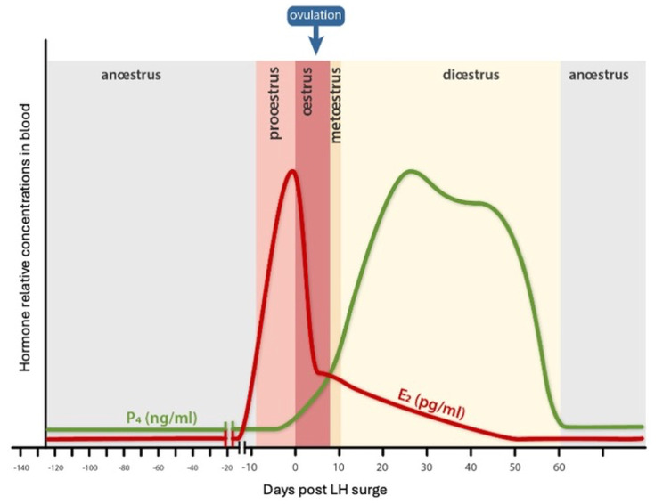
In veterinary medicine, 16 S rDNA gene sequencing has recently been used to characterize the urogenital microbiome in healthy dogs. Four taxa, belonging to the Pseudomonadota (previously Proteobacteria) phylum: Pseudomonas spp, Acinetobacter spp, Sphingobium spp and Bradyrhizobiaceae, dominated the urinary microbiota in dogs of both sexes. Moreover, considerable overlap was observed between the vaginal and bladder microbiota where Pseudomonas and Acinetobacter were the most abundant taxa [6]. Another recent study showed that Hydrotalea, Ralstonia, Mycoplasma, Fusobacterium and Streptoccocus were the predominant species in the vagina of female dogs [17]. The vaginal microbiota of bitches was most diverse, with the highest richness, during the estrous phase of the estrous cycle. However, these differences were only statistically significant between estrous and the prepubertal stage [17]. These results may be related to the age of the dogs in the study as the diversity of the vaginal microbiome continuously changes with age [18]. For the good understanding of the paper, the main hormonal changes and phases of the cycle are illustrated in Fig. 1.
Fig. 1.
Estrogen and Progesterone modifications during female dog cycle
The urinary system is also affected by sex hormones and the estrous cycle [19]. Urodynamic studies in dogs have shown that urethral pressure, an indicator of urinary continence, decreased during estrous and early diestrus [20]. However, the influence of sex hormones on the urinary microbiota of female dogs is still unknown. Only, difference in urinary microbiota between pre- and post-menopausal women was demonstrated [21, 22].
Therefore, the purpose of our research was to characterize and compare the urogenital microbiota in the different phases of the estrous cycle in healthy bitches. Since the bladder and the vaginal microbiota are closely connected, we hypothesized that sex hormones and the estrous cycle influence both microbiotas.
Methods
Study population
This prospective study was conducted on 10 healthy intact adult female laboratory beagle dogs owned by the Veterinary Faculty of the University of Liège (ULiège ethical approval number: 20-2250; laboratory approval number: LA161012). Dogs were included in the study if they had no signs of systemic, vaginal or lower urinary tract disease. Dogs that had received antibiotics, probiotics and/or anti-inflammatory drugs within 30 days prior to enrolment were excluded. Dogs were housed in a kennel with wood shavings as bedding.
Study design
Samples were obtained from adult dogs at the following phases of the estrous cycle: proestrus, estrous, diestrus and anestrus. Phases of the estrous cycle were identified via visual examination of the vulva, cytologic examination of a vaginal smear and measurement of plasma progesterone concentration (Automated Immunoassay analyzer 360, TOSOH). Urine was collected from all dogs via prepubic cystocentesis after skin disinfection with chlorhexidine soap and alcohol to minimize dermal microbiota contamination. Around 10 ml of urine was aliquoted for routine analysis (specific gravity, dipstick with pH, and microscopic evaluation of the sediment), routine culture and 16 S rRNA amplicon sequencing at each cycle phase. The culture has been performed by mass spectrophotometry (VITEK MS biomérieux) and conventional biochemistry (VITEK 2 Biomérieux). After placing a sterile (UV treatment for 30 min in a BSL2 Biohazard cabinet) otoscope cone beyond the vestibule, a swab moistened with sterile saline solution was passed through the speculum into the anterior vagina that was swabbed for 10 s to collect a genital sample. Negative controls consisted of saline moistened vaginal swabs passed through a sterile speculum. Urine and vaginal samples were stored at -80 °C until DNA extraction.
Total bacterial DNA extraction and amplicon sequencing
16 S rDNA library preparation, sequencing and informatic analysis were performed as previously described [23]. Briefly, total bacterial DNA were extracted from the vaginal swabs and urine with the DNEasy Blood and Tissue kit (QIAGEN Benelux BV; Antwerp, Belgium) with an added bead beating step during lysis. Amplicon sequencing targeting V1V3 hypervariable regions of the 16 S rDNA was performed using a MiSeq sequencer (Illumina; San Diego, California, USA). Sequence reads were cleaned and processed using MOTHUR software package v1.47 and the SILVA v1.38_1 16 S rDNA reference database. The urinary and vaginal microbiota were analyzed separately following the same protocol. Analyses were performed at the genus taxonomic level.
The identification of putative bacterial contaminant population in vaginal swab involves the molecular quantification of the bacterial load in samples. This protocol is described elsewhere and is based upon the quantitative amplification of the V2V3 hypervariable region of the 16 S rDNA by real time PCR [24].
Statistical analysis
Good’s coverage index and ecological indicators, including the α-diversity (inverse Simpson’s index and Shannon index), bacterial richness (Chao1 index) and evenness (Simpson index-based measure) were calculated with MOTHUR v1.47. Differences between groups were assessed using non parametric Friedman ANOVA test for repeated measures, followed by paired post-hoc tests corrected with a two-stage linear step-up Benjamini Hochberg procedure (q threshold = 0.05) with PRISM 9.0.
Beta diversity was visualized with a Bray–Curtis dissimilarity matrix-based non-parametric dimensional scaling (NMDS) model using vegan and vegan3d packages in R. Differences in beta diversity between cycle phases was assessed with analysis of molecular variance (AMOVA) and homogeneity of molecular variance (HOMOVA) tests using MOTHUR (using 10,000 iterations on the subsampled table) on a Bray–Curtis dissimilarity matrix. A p-value < 0.05 was considered significant.
Differential population abundance between cycle phases was evaluated using a negative binomial Wald test in the DESeq2 R package. Differences were considered significant if the corrected p-value was < 0.05 (Benjamini-Hochberg False Discovery Rate multi-testing correction).
To identify putative bacterial contaminants in vaginal swabs sample, a multiple non-parametric Spearman Rho correlation test was performed between genus abundance and total bacteria load, determined by real time PCR. The correlation was considered as significant for Rho value above 0.5 or below − 0.5, with a p-value < 0.05. We also use Decontam package [25] in R to detect putative contaminants in the bacterial profiles, following authors protocols for the prevalence and frequency strategies. For the first strategy, sample library DNA quantification was used. For the second strategy, specific negative controls for vaginal swabs and sequencing run negative controls for the urine samples were used.
A Matrix correlation Mantel test [26] was performed with the Pearson, Spearman and Kendall test to evaluate the correlation between vaginal and urinary microbiota.
Results
Study population characteristics
The mean weight of the dogs included in this study was 14.25 kg (range 12–17 kg). The mean age was 6.5 years (range 5–9 years). Dogs were housed together in a kennel with wood shavings as bedding. They were fed with a strict diet of adult maintenance or light dry food.
Urine analysis
Urine pH was between 4.5 and 8 and urine specific gravity was between 1.005 and 1.048. Mild proteinuria was frequently observed in five dogs and consistently observed in two dogs (Table 1).
Table 1.
Urinalysis and urinary culture
| Beagle | Phase | Dipstick | Specific gravity | Sediment | Urinary culture |
|---|---|---|---|---|---|
| 1 | Proestrus | pH 7 | 1,016 | negative | negative |
| 2 | pH 7, proteins + | 1,005 | negative | negative | |
| 3 | pH 6 | 1,014 | negative | ||
| 4 | pH5 | 1,018 | negative | negative | |
| 5 | pH 7 | 1, 024 | negative | negative | |
| 6 | pH5, proteins + | 1,04 | negative | negative | |
| 7 | pH 8 | 1,015 | negative | negative | |
| 8 | pH 6,5, blood +, bilirubin + | 1,024 | negative | negative | |
| 9 | pH5, blood ++, bilirubine +, proteins + | 1,024 | Epithelial cells and Erythrocyts | negative | |
| 10 | pH 6, proteins + | 1,03 | Epithelial cells | negative | |
| 1 | Estrous | pH 6 | 1,016 | negative | negative |
| 2 | pH 7, proteins++ | 1,027 | negative | 80% Streptococcus infantarius, 20% E. Coli | |
| 3 | pH 7, blood + | 1,024 | Epithelial cells and extracellular bacteria | 100% Lactobacillus gasseri | |
| 4 | pH 5 | 1, 022 | negative | negative | |
| 5 | pH 7 | 1,014 | Epithelial cells | negative | |
| 6 | pH5 +, proteins + | 1,027 | negative | negative | |
| 7 | pH 5 | 1,024 | negative | negative | |
| 8 | pH 8 | 1,008 | negative | negative | |
| 9 | pH 5, blood +, proteins ++ | 1,02 | Erythrocyts | negative | |
| 10 | pH 7 proteins + | 1,032 | negative | negative | |
| 1 | Diestrus | pH7, blood+, bilirubin +, proteins + | 1,026 | negative | negative |
| 2 | pH 6, bilirubin +, proteins + | 1,027 | negative | 100% Enterococcus hirae | |
| 3 | pH 5 | 1,028 | negative | negative | |
| 4 | pH 5 | 1, 022 | negative | negative | |
| 5 | pH 8 | 1,012 | negative | negative | |
| 6 | - | - | - | negative | |
| 7 | pH 6,5, proteins + | 1, 024 | negative | negative | |
| 8 | - | - | - | negative | |
| 9 | pH6, proteins + | 1,014 | negative | negative | |
| 10 | pH 6 | 1,034 | Epithelial cells | negative | |
| 1 | Anestrus | pH 7 | 1,018 | negative | negative |
| 2 | pH 8, proteins ++ | 1,01 | negative | negative | |
| 3 | pH 6, proteins+ | 1,032 | negative | negative | |
| 4 | pH 5, proteins ++ | 1, 022 | negative | negative | |
| 5 | pH 7 | 1,017 | negative | negative | |
| 6 | pH 4,5, proteins + | 1, 020 | negative | negative | |
| 7 | pH 6,5, proteins + | 1,028 | Erythrocyts | negative | |
| 8 | pH 5 | 1,02 | negative | negative | |
| 9 | pH 5 | 1,02 | negative | negative | |
| 10 | pH 6,5 Proteins + | 1,048 | negative | negative |
Urine bacterial cultures
Urine cultures were mostly negative, with the exception of Streptococcus infantarius and Escherichia coli in one estrous sample, Enterococcus hirae in the diestrus sample from one dog and Lactobacillus gasse in the estrous sample from one dog.
Vaginal microbiota
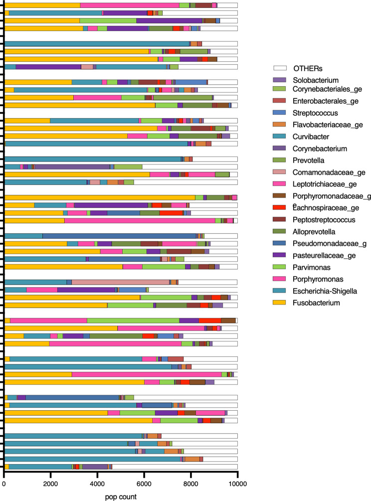
A total of 315 genera were identified in the vaginal samples during the different phases of the estrous cycle. The most abundant bacterial populations belonged to the genera Fusobacterium, Porphyromas, Parvimonas and Escherichia-Shigella, which represented 33.1%, 11.5%, 7.1%, 7% and 5.8%, respectively, of the total bacterial population (Fig. 2). To investigate whether the identified bacterial genera were contaminants, a non-parametric Spearman correlation test was performed between the abundance of each identified genus and the total bacterial population. The negative result (Rho − 0,7972; p < 0.0001) for Escherichia-Shigella suggested that it was a contaminant.
Fig. 2.
Vaginal genus
The vaginal bacterial ecosystem was assessed at the genus level. Alpha diversity, richness and evenness of the bacterial populations did not change significantly throughout the different cycle phases (Fig. 3). AMOVA-based cluster analysis showed significant differences between samples from different cycle phases (anestrus, diestrus, estrous and proestrus, p < 0.0001). Paired analysis showed significant differences between anestrus and estrous (p < 0.0001), anestrus and proestrus (p < 0.0001), diestrus and estrous (p < 0.0001), diestrus and proestrus (p < 0.0001), and estrous and proestrus (p < 0.0002). There was no significant difference between anestrus and diestrus. HOMOVA testing was statistically significant (p = 0.0035). However, paired analysis did not yield statistically different results.
Fig. 3.
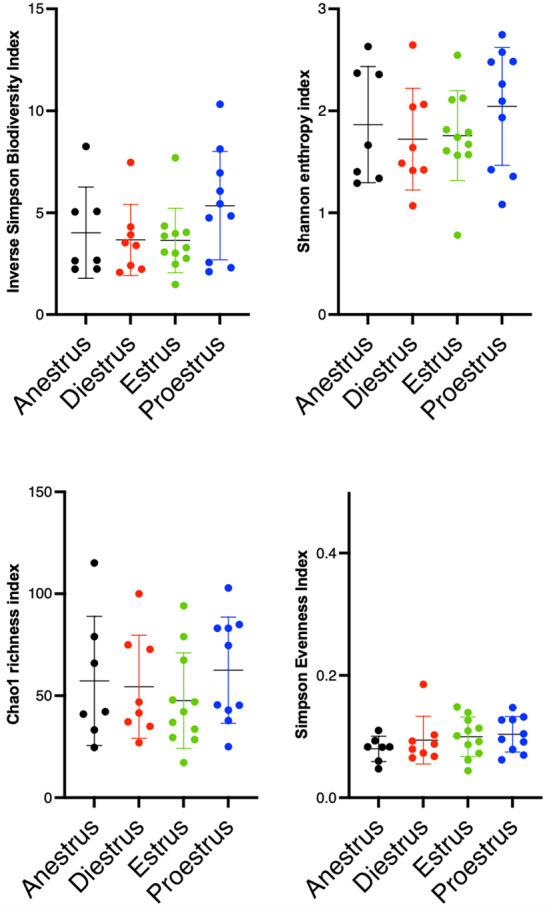
Scatterplots depicting alpha-diversity indices of vaginal microbiota at each phase of the estrous cycle. Each dot represents a subsample. Bacterial intrinsic diversity was calculated using the reciprocal Simpson Biodiversity index, bacterial genus richness was calculated using the Chao1 index and bacterial genus evenness was calculated using the Simpson index. No significant differences were found between the groups based on a Friedman test. Horizontal lines represent the mean, and error bars indicate the 95% CIs for each group and time point
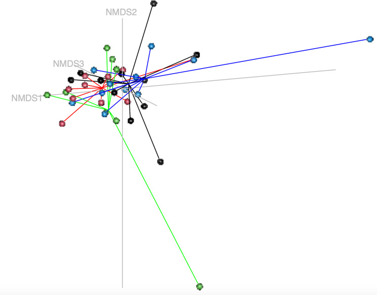
The beta-diversity of the vaginal microbial profile was visualized using a non-metric dimensional scaling (NMDS) model based on a Bray-Curtis dissimilarity matrix including samples from the different cycle phases (anestrus, diestrus, estrous and proestrus, Fig. 4).
Fig. 4.
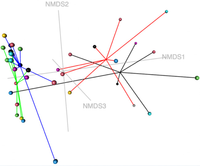
Non-metric multidimensional scaling model (k = 3, stress = 0.0836) based on a Bray-Curtis dissimilarity matrix of the vaginal microbial profiles during estrous cycle phases. Colored dots represent subsamples in the different cycle phases (black: anestrus, red: diestrus, green: estrous, blue: proestrus). AMOVA-based cluster analysis showed significant differences in variance between all estrous cycle phases, except between anestrus and diestrus (p-value = 0.0001). HOMOVA testing showed statistically significant differences (p = 0.0035)
Differential population abundance in cycle phases was evaluated using a negative binomial Wald test in the DESeq2. The abundance of bacterial populations was not significantly different in anestrus and diestrus samples. In contrast, the abundance of five genera (Prevotella, Variovorax and Porphyromonas and two contaminants—Rheinheimera and Corynebacteriale) were significantly different in proestrus and estrous samples. Notably, there were significant differences in the abundance of 32 genera, including Parvimonas, S5.A14, Peptostreptococcae, Anaerovoracacaea genus, Solobacterium, Porphyromonas, Fusobacterium, Alloprevotella, Peptococcus, Fusobacteriales, Porphyromonadacae, Johnsonella and six contaminants (Flavobacteriaceae genus, Corynebacteriales genus, Rheinheimera, Pelomonas, Pseudomonadaceae genus and Escherichia-Shigella), between anestrus and estrous samples. There were also significant differences in the abundance of 28 genera, including S5.A14a, Parvimonas, Porphyromonadacae, Anaerovoracaceae, Peptostreptococcaceae, Solobacterium, Alloprevotella and seven contaminants, between anestrus and proestrus samples. Similarly, there were significant differences in the abundance of 27 genera, including S5.A14a, Porphyromonadacae, Fusobacterium and 10 contaminants, between diestrus and proestrus samples. There were also significant differences in the abundance of 33 genera, including S5.A14a, Porphyromonadacae, Fusobacterium, Bacteroides, Peptostreptococcus, Johnsella, Parvimonas, Peptostreptococcaceae, Prophromonadaceae and 11 contaminants between diestrus and estrous samples. These results are summarized in Table 2.
Table 2.
Differential bacterial population abundance in estrous cycle phases is represented by the adjusted p-value (padj) with DESEQ2 test values. The higher abundance in the first group compared to the second is represented by a log2fold change positive. The possible contaminants according to the correlation test are in bold. The possible contaminants according to the Decontam test are underlined
| Anestrus-Estrous | log2FoldChange | padj | Estrous-Proestrus | log2FoldChange | padj | Anestrus-Proestrus | log2FoldChange | padj | Diestrus-Proestrus | log2FoldChange | padj | Diestrus-Estrus | log2FoldChange | padj |
|---|---|---|---|---|---|---|---|---|---|---|---|---|---|---|
| Flavobacteriaceae genus | 20.50 | 1.05926E-22 | Prevotella | 31.43 | 4.38087E-33 | Flavobacteriaceae genus | 23.03 | 1.84734E-27 | Flavobacteriaceae genus | 28.25 | 1.11844E-44 | Flavobacteriaceae genus | 25.73 | 5.90348E-39 |
| Corynebacteriales genus | 25.36 | 1.41072E-22 | Rheinheimera | -16.12 | 1.49931E-13 | Pelomonas | 22.21 | 9.15592E-19 | Pelomonas | 27.92 | 3.43701E-32 | Rheinheimera | 25.88 | 1.10481E-35 |
| Rheinheimera | 20.14 | 6.14841E-20 | Variovorax | 17.21 | 4.1406E-07 | Prevotella | 21.63 | 8.26798E-13 | Prevotella | 23.67 | 2.15437E-16 | Pelomonas | 24.90 | 4.05461E-27 |
| Pelomonas | 19.19 | 6.68813E-15 | Corynebacteriales genus | -13.67 | 5.20347E-07 | Rhodococcus | 25.16 | 2.37102E-12 | Rhodococcus | 24.90 | 6.33797E-13 | Corynebacteriales genus | 25.69 | 2.50813E-25 |
| Rhodococcus | 23.96 | 8.61807E-12 | Porphyromonas | 5.46 | 0.001965367 | Variovorax | 21.50 | 4.94071E-10 | Variovorax | 23.29 | 1.55098E-12 | Escherichia Shigella | 10.94 | 3.96533E-14 |
| Pseudomonadaceae genus | 12.56 | 1.39975E-09 | Pseudomonadaceae genus | 12.89 | 1.07674E-09 | Escherichia Shigella | 10.13 | 1.92812E-12 | Rhodococcus | 23.70 | 1.42357E-12 | |||
| Conchiformibius | 22.19 | 2.0297E-06 | Corynebacteriales genus | 11.69 | 6.26809E-05 | Pseudomonadaceae genus | 12.78 | 3.3036E-10 | Conchiformibius | 27.57 | 1.16139E-10 | |||
| Parvimonas | -6.42 | 5.23916E-06 | Conchiformibius | 19.43 | 7.23703E-05 | Conchiformibius | 24.81 | 2.62996E-08 | Pseudomonadaceae genus | 12.44 | 2.63062E-10 | |||
| S5 A14a | -11.32 | 2.62116E-05 | Abiotrophia | 19.14 | 7.23703E-05 | Abiotrophia | 23.75 | 6.16636E-08 | S5 A14a | -13.89 | 2.74823E-08 | |||
| Escherichia Shigella | 6.55 | 4.4847E-05 | S5 A14a | -10.23 | 0.000292341 | Pseudomonas | 12.23 | 4.85539E-07 | Porphyromonas | -7.97 | 9.7955E-08 |
Differences in bacterial abundance were evaluated using a negative binomial Wald test in the DESeq2 R package. Differences were considered significant if the corrected p-value < 0.05 (Benjamini-Hochberg False Discovery Rate multi-testing correction). Bacteria were suspected to be a contaminant when the correlation coefficient (r) between the microbiota abundance and presence of the bacteria in the sample was < -0.5. Bacteria suspected to be contaminants are shown in bold and bacteria present at a level lower than 1% of the total population are highlighted in dark grey.
Urinary microbiota
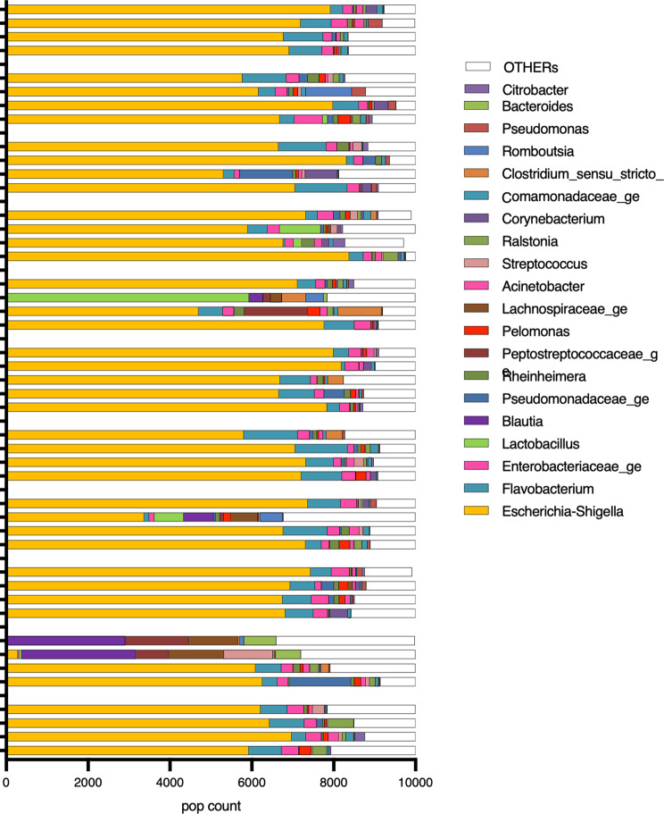
A total of 351 genera were identified in the urinary samples during the different estrous cycle phases. The most abundant bacterial populations belonged to the genera Escherichia-Shigella, Flavobacterium, Enterobacteriaceae genus, Pseudomonadaceae and Rheinheimera, which represented 67%, 6.5%, 3%, 1.3% and 1%, respectively, of the total bacterial population (Fig. 5).
Fig. 5.
Urinary genus
The urinary microbial ecosystem was assessed at the genus level. There were no significant differences in alpha diversity, richness and evenness of the bacterial populations throughout the different cycle phases (Fig. 6).
Fig. 6.
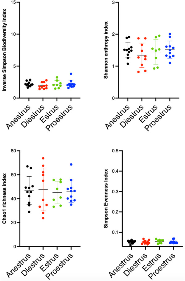
Scatterplots depicting alpha-diversity indices of urinary microbiota at each phase of the estrous cycle. Each dot represents a subsample. Bacterial intrinsic diversity was calculated using the reciprocal Simpson Biodiversity index, bacterial genus richness was calculated using the Chao1 index and bacterial genus evenness was calculated using the Simpson index. No significant differences were found between the groups based on a Friedman test. Horizontal lines represent the mean, and error bars indicate the 95% CIs for each group and time point
AMOVA-based cluster analysis and HOMOVA analysis found no significant differences between samples from the different cycle phases (anestrus, diestrus, estrous and proestrus, Fig. 7).
Fig. 7.
Non-metric multidimensional scaling model (k = 4, stress = 0.084) based on a Bray-Curtis dissimilarity matrix of the urinary microbial profiles during estrous cycle phases. Colored dots represent subsamples in the different cycle phases (black: anestrus, red: diestrus, green: estrous, blue: proestrus). AMOVA- and HOMOVA analysis showed no significant differences between estrous cycle phases
Urinary and vaginal microbiota were compared and no statistically significant correlation was observed.
Discussion
To the authors’ knowledge, this is the first study investigating the changes in both urinary and vaginal microbiomes during the estrous cycle in healthy female dogs. We found significant differences between the most prevalent bacteria present in the vagina and those in the urine. Our data also showed significant changes in the prevalence of various bacterial genera in the vagina during the different phases of the estrous cycle. In contrast, estrous cycle phases did not affect bacterial prevalence in urine samples.
The vaginal microbiome includes bacteria, fungi, viruses, archaea, and candidate phyla radiation bacteria. Within this mix, bacteria are the most prevalent microorganisms in the vagina. Characterization of tissue-specific microbiomes can help identify pathologic microbial changes in various disease states. In this study, we chose to describe the bacterial population in urinary and vaginal samples at the genus level. This allowed precision while avoiding noise associated with identification by species. This best suited the purpose of our research, which was to characterize and compare the urogenital microbiota in the different phases of the estrous cycle in healthy bitches.
Early studies characterizing vaginal microbiota by aerobic and anaerobic culture methods found that E. coli and S. pseudintermedius were the most common isolates in bitches [6, 27]. These results are likely due to the limitations of routine culture. More recent studies using 16 S rDNA gene sequencing to identify bacterial populations have reported a more diverse vaginal microbiome in female dogs. Burton et al. reported Hydrotalea, Ralstonia, Mycoplasma, Fusobacterium and Streptoccocus to be the predominant genera in the vagina of female dogs. [6] Rota et al. identified Mycoplasma, Pasteurellaceae family and Salmonella in healthy bitches of various breeds [28] and Hu et al. found Fusobacteria, Firmicutes, Proteobacteria, Tenericutes and Bacteroidetes in beagles. [19] The results of our study are partly consistent with data from Burton et al. [6] and Hu et al. [19] In our population, Fusobacterium, Porphyromas, Parvimonas and Escherichia-Shigella were the predominant genera in vaginal samples. However, our correlation data suggest that Escherichia-Shigella was a contaminant. The data from our study, and from previous studies, suggest that the vaginal microbiome in dogs is significantly different to that of women; in women, the vaginal microbiota is mostly dominated by Lactobacillus crispatus, Lactobacillus iners, Lactobacillus gasseri or Lactobacillus jensenii [29].
Early studies using aerobic and anaerobic culture characterized urine as sterile [30]. However, a more recent study using 16 S rDNA gene sequencing reported bacteria from the phylum Pseudomonadota, Pseudomonas spp, Acinetobacter spp, Sphingobium spp and Bradyrhizobiaceae to dominate the urinary microbiota in dogs [7]. The results of our study partly overlap with those of the previous report. In our population, Escherichia-Shigella, Flavobacterium, Enterobacteria, Pseudomonadaceae and Rheinheimera were dominant. Moreover, Lactobacillus gasse was identified in the urine of one dog but was not identified in her vaginal microbiota. Escherichia-Shigella, Enterobacteriaceae genus, Pseudomonadaceae and Rheinheimera all belong to the Pseudomonadota phylum. In women, Lactobacillus, Corynebacterium, Streptococcus, Actinomyces, Gardnerella and Bifidobacterium have been observed in urine [3, 4]. These data suggest that the urinary microbiome could be species-specific, as reported for the intestinal microbiota [31]. However, the difference between different study could be due to bias as contamination and analytic and collection method. Mrofchak et al. 2022 suggested in one study that dominant taxa could be shared between humans and dogs [10].
To identify potential contaminants, we performed a correlation test between the presence of each genus and the abundance of the total bacterial population in the vagina. Our results suggest that Escherichia-Shigella, Comamonadaceae, Pseudomonas, Flavobacteriaceae, Enterobacterales, Flavobacterium, Rheinheimera, Pelomonas, Acinetobacter, Saccharimonadales, Chryseobacterium, Parcubactera, Aeromonas, Burkholderiales and Paracoccus are contaminants. In our study, Escherichia-Shigella, Flavobacterium, Enterobacteriaceae genus and Rheinheimera were the dominant genera in urinary microbiota, and we found no correlation between vaginal and urinary microbiota. However, these bacteria have been found in the vaginal microbiota in previous studies. Furthermore, a previous study in dogs found that the genital microbiome was similar to the urinary microbiota [6]. This discrepancy may be due to cross-contamination of the vaginal and urine samples during collection in the study of Burton et al. 2017. As they did not use a sterile speculum for the vaginal swab like we did. Alternatively, this difference may be explained by the difference in methodology sequencing and taxonomic data base that was used (V4).
Sex hormones contribute to the regulation of the vaginal microbiota in women [11–13, 16, 32]. Alpha and beta diversity of the vaginal microbiome varies across the menstrual cycle of women [32]. Moreover, the vaginal microbiome covaries with estradiol level [32]. However, the influence of progesterone is not well known. The results of our study are partially consistent with the results of the human studies. We found that the vaginal microbiota in female dogs also varies across estrous cycle phases. While alpha diversity, which corresponds to the number of taxonomic groups coexisting in the vagina and their distribution of abundances, was not significantly different across cycle phases, beta diversity was significantly different all comparisons, except between diestrus and anestrus. The fact that we observed more stability during the cycle in dogs than has been observed in humans may be due to the fact that we analyzed the microbiota once per cyclic phase, compared with the human study that analyzed vaginal microbiota daily. Additional larger studies with more frequent sampling may help understand this. Our results may also reflect the species-related differences between dogs and humans.
Similar to the results of human studies, our data didn’t demonstrated that progesterone affect the vaginal microbiota, as beta diversity did not differ between diestrus, when progesterone is highest, and anestrus, when there are no significant levels of circulating sex hormones [33]. Furthermore, the variation of beta diversity during estrogenic phases (proestrus and estrous), compared with diestrus and anestrus, suggests that estrogen influences the vaginal microbiome in dogs, as described in women [16].
In women, estrogens stimulate vaginal epithelial cell proliferation, with a mid-cycle peak in intracellular glycogen levels in the vaginal mucosa and a subsequent increase in lactic acid-producing microbes, such as Lactobacillus, in the vaginal milieu [34]. While estrogens have been shown to similarly effect vaginal cell proliferation in the dog [35], the mechanisms underlying the changes in the microbiome remain to be explored. Moreover, Lactobacillus was not identified in the vaginal microbiota in our study population, which suggests a significantly different vaginal environment between women and dogs. However, our results conflict with an earlier study showing that Lactobacillus was present in the vagina of dogs and increased as the dogs aged [19]. The difference in vaginal environment between dogs and women, with a pH of 7 and 4.5 respectively, could explain why the microbiota composition is significantly different in the two species [28]. The presence of blood in the vaginal environment during proestrus may also contribute to the changes in beta-diversity due to the presence of iron or the increased pH [29]. Blood may also influence the vaginal microbiota by providing a substrate for growth and proliferation or flushing out bacteria.
Urodynamic studies have shown that the estrous cycle and sex hormones also affect the urinary system by decreasing urethral pressure during estrous and early diestrus [19, 36]. Estrogens induce an increase in the number of alpha-adrenergic receptors and responsiveness of these receptors to sympathetic stimulation [19]. Estrogens also induce an increase in blood flow to urethral tissues [36], which causes an increase in urethral sphincter tone [37]. In contrast, progesterone potentiates beta-adrenergic activity in the urethra of female dogs, leading to a decrease in urethral smooth muscle tone and, therefore, relaxation [37–40]. We hypothesized that sex hormones may affect the urinary microbiome in female dogs. However, we found no differences in the alpha and beta diversity of the urinary microbiota across the estrous cycle phases. This result may be because, although sex hormones influence urethral function, estrogens and progesterone have not been reported to induce changes in the urothelium, as they do in the vagina.
This study has two main limitations. The first is the possible presence of contaminants in the samples despite the aseptic methods used for collection. The second is the small study population that comprised a homogenous group of dogs in terms of breed, age, housing conditions and diet. Therefore, our study population may not accurately reflect a population of pet dogs. Furthermore, in human medicine, diet has been shown to influence urinary and vaginal microbiota [32, 41]. Our study population was fed a strict diet of adult maintenance or light dry food, which, again, is different to healthy pet dogs, which tend to have a more varied diet, including dry food, wet food and table scraps. Therefore, further studies are need to confirm our data in pet dogs and to explore the effect of diet on the urogenital microbiome of dogs.
Conclusion
This study strongly suggests that estrogen influences the abundance of bacteria in the vaginal microbiota of healthy adult female dogs and provides data comparing the urinary and vaginal microbiota in healthy bitches.
Electronic supplementary material
Below is the link to the electronic supplementary material.
Author contributions
V.G. manuscript preparation and data analysis F.B., S.E., C.P. data collection A.H. and G.D. provided materials B.T. data analysis M-L.VdW. sample analysis S.D. and S.N. author of the project, protocol redaction, data collection and manuscript review.
Funding
The authors have no funding to declare.
Data availability
All sequencing librairies have been deposited to the Genbank SRA database under bioproject PRJNA1082225.
Declarations
Ethics approval and consent to participate
This research was approved by the Ethics Committee of the University of Liège (Ethical approval number: 20-2250).
Consent for publication
Not applicable as study participants were laboratory dogs for which participation was authorized by the Ethics Committee of the University of Liège (Ethical approval number: 20-2250).
Competing interests
The authors report there are no competing interests to declare.
Footnotes
Publisher’s Note
Springer Nature remains neutral with regard to jurisdictional claims in published maps and institutional affiliations.
Stefan Deleuze and Stéphanie Noel contributed equally to this work.
References
- 1.Theron J, Cloete TE. Molecular techniques for determining microbial diversity and community structure in natural environments. Crit Rev Microbiol. 2000;26(1):37–57. doi: 10.1080/10408410091154174. [DOI] [PubMed] [Google Scholar]
- 2.Hilt EE, McKinley K, Pearce MM, Rosenfeld AB, Zilliox MJ, Mueller ER, et al. Urine is not sterile: use of enhanced urine culture techniques to detect resident bacterial flora in the adult female bladder. J Clin Microb. 2014;52:871–6. doi: 10.1128/JCM.02876-13. [DOI] [PMC free article] [PubMed] [Google Scholar]
- 3.Coffey EL, Gomez AM, Ericsson AC, Burton EN, Granick JL, Lulich JP, et al. The impact of urine collection method on canine urinary microbiota detection: a cross-sectional study. BMC Microbiol. 2023;23(1):101. doi: 10.1186/s12866-023-02815-y. [DOI] [PMC free article] [PubMed] [Google Scholar]
- 4.Pearce MM, Hilt EE, Rosenfeld AB, Zilliox MJ, Thomas-White K, Fok C, et al. The female urinary microbiome: a comparison of women with and without urgency urinary incontinence. mBio. 2014;e01283–14. 10.1128/mBio.01283-14. [DOI] [PMC free article] [PubMed]
- 5.Thomas-White K, Forster SC, Kumar N, Van Kuiken M, Putonti C, Stares MD, et al. Culturing of female bladder bacteria reveals an interconnected urogenital microbiota. Nat Commun. 2018;9(1):1557. doi: 10.1038/s41467-018-03968-5. [DOI] [PMC free article] [PubMed] [Google Scholar]
- 6.Burton EN, Cohn LA, Reinero CN, Rindt H, Moore SG, Ericsson A. Characterization of the urinary microbiome in healthy dogs. PLoS ONE. 2017;12(5):e0177783. doi: 10.1371/journal.pone.0177783. [DOI] [PMC free article] [PubMed] [Google Scholar]
- 7.Mrofchak R, Madden C, Evans MV, et Hale VL. Evaluating extraction methods to study canine urine microbiota. PLoS ONE. 2021;16(7):e0253989. doi: 10.1371/journal.pone.0253989. [DOI] [PMC free article] [PubMed] [Google Scholar]
- 8.Melgarejo T, Oakley BB, Krumbeck JA, Tang S, Krantz A, Linde A. Assessment of bacterial and fungal populations in urine from clinically healthy dogs using next-generation sequencing. J Vet Intern Med. 2021;35:1416–26. doi: 10.1111/jvim.16104. [DOI] [PMC free article] [PubMed] [Google Scholar]
- 9.Coffey EL, Gomez AM, Burton EN, Granick JL, Lulich JP, Furrow E. Characterization of the urogenital microbiome in miniature schnauzers with and without calcium oxalate urolithiasis. J Vet Intern Med. 2022;36:1341–52. doi: 10.1111/jvim.16482. [DOI] [PMC free article] [PubMed] [Google Scholar]
- 10.Mrofchak R, Madden C, Evans MV, Kisseberth WC, Dhawan D, Knapp DW, et al. Urine and fecal microbiota in a canine model of bladder cancer and comparison of canine and human urine microbiota. All Life. 2022;15:1245–63. doi: 10.1080/26895293.2022.2154858. [DOI] [Google Scholar]
- 11.Shen J, Song N, Williams CJ, Brown CJ, Yan Z, Xu C, et al. Effects of low dose estrogen therapy on the vaginal microbiomes of women with atrophic vaginitis. Sci Rep. 2016;29(6):34119. doi: 10.1038/srep34119. [DOI] [PMC free article] [PubMed] [Google Scholar]
- 12.Chen KL, Madak-Erdogan Z. Estrogen and Microbiota Crosstalk: should we pay attention? Trends Endocrinol Metab. 2016;27(11):752–5. doi: 10.1016/j.tem.2016.08.001. [DOI] [PubMed] [Google Scholar]
- 13.Romero R, Hassan SS, Gajer P, Tarca AL, Fadrosh DW, Nikita L, et al. The composition and stability of the vaginal microbiota of normal pregnant women is different from that of non-pregnant women. Microbiome. 2014;2(1):4. doi: 10.1186/2049-2618-2-4. [DOI] [PMC free article] [PubMed] [Google Scholar]
- 14.Kaur H, Merchant M, Haque MM, Mande SS. Crosstalk between Female Gonadal Hormones and vaginal microbiota across various phases of women’s gynecological lifecycle. Front Microbiol. 2020;11:551. doi: 10.3389/fmicb.2020.00551. [DOI] [PMC free article] [PubMed] [Google Scholar]
- 15.Smith SB, Ravel J. The vaginal microbiota, host defence and reproductive physiology. J Physiol. 2017;595(2):451–63. doi: 10.1113/JP271694. [DOI] [PMC free article] [PubMed] [Google Scholar]
- 16.Chaban B, Links MG, Jayaprakash TP, Wagner EC, Bourque DK, Lohn Z, et al. Characterization of the vaginal microbiota of healthy Canadian women through the menstrual cycle. Microbiome. 2014;2:23. doi: 10.1186/2049-2618-2-23. [DOI] [PMC free article] [PubMed] [Google Scholar]
- 17.Lyman CC, Holyoak GR, Meinkoth K, Wieneke X, Chillemi KA, DeSilva U. Canine endometrial and vaginal microbiomes reveal distinct and complex ecosystems. PLoS ONE. 2019;14(1):e0210157. doi: 10.1371/journal.pone.0210157. [DOI] [PMC free article] [PubMed] [Google Scholar]
- 18.Golinskan E, Sowinska N, Tomusiak-Plebanek A, Szydlo M, Witka N, Lenarczyk J et al. The vaginal microflora changes in various stages of the estrous cycle of healthy female dogs and the ones with genital tract infections. BMC Vet Res, 17(1):8. 10.1186/s12917-020-02710-y. [DOI] [PMC free article] [PubMed]
- 19.Hu J, Cui L, Wang X, Gao X, Qiu S, Qi H, et al. Dynamics of vaginal microbiome in female beagles at different ages. Res Vet Sci. 2022;149:128–35. doi: 10.1016/j.rvsc.2022.05.006. [DOI] [PubMed] [Google Scholar]
- 20.Hamaide AJ, Verstegen JP, Snaps FR, Onclin KJ, Balligand MH, Ammitzbøll N, Bau BPJ, Bundgaard-Nielsen C, Villadsen AB, Jensen AM et al. Influence of the estrous cycle on urodynamic and morphometric measurements of the lower portion of the urogenital tract in dogs. Am J Vet Res. Pre- and postmenopausal women have different core urinary microbiota. Scientific Reports. 2021;11: Art 2212. https://doi.org/10.1038/s41598-021-81790-8.
- 21.Ammitzb?ll N, Bau,BPJ, Bundgaard-Nielsen C, Villadsen AB, Jensen AM, et al. Pre- and postmenopausal women have different core urinary microbiota. Scientific Reports. 2021;11: Art 2212. [DOI] [PMC free article] [PubMed]
- 22.Park MG, Cho S, Oh MM. Menopausal changes in the Microbiome-A Review focused on the Genitourinary Microbiome. Diagnostics. 2023;13(6):1193. doi: 10.3390/diagnostics13061193. [DOI] [PMC free article] [PubMed] [Google Scholar]
- 23.Daube G, Fontaine J. Ear canal microbiota – a comparison between healthy dogs and atopic dogs without clinical signs of otitis externa. Vet Dermatol. 2018;29(5):425–e140. doi: 10.1038/s42003-019-0540-1. [DOI] [PubMed] [Google Scholar]
- 24.Fastrès A, Taminiau B, Vangrinsven E, Tutunaru AC, Moyse E, Farnir F, Daube G, Clercx C. Effect of an antimicrobial drug on lung microbiota in healthy dogs. Heliyon. 2019;14(11):e02802. doi: 10.1016/j.heliyon.2019.e02802. [DOI] [PMC free article] [PubMed] [Google Scholar]
- 25.Davis NM, Proctor D, Holmes SP, Relman DA, Callahan BJ. Simple statistical identification and removal of contaminant sequences in marker-gene and metagenomics data. bioRxiv. 2017;221499. 10.1101/221499. [DOI] [PMC free article] [PubMed]
- 26.Mantel NA. The detection of disease clustering and a generalized regression approach. Cancer Res. 1967;27:209–20. [PubMed] [Google Scholar]
- 27.Hutchins RG, Vaden SL, Jacob ME, Bowles KD, Wood MW, Bailey CS, et al. Vaginal microbiota of spayed dogs with or without recurrent urinary tract infections. J Vet Intern Med. 2014;28:300–4. doi: 10.1111/jvim.12299. [DOI] [PMC free article] [PubMed] [Google Scholar]
- 28.Rota A, Corrò M, Patuzzi I, Milani C, Masia S, Mastrorilli E, et al. Effect of sterilization on the canine vaginal microbiota: a pilot study. BMC Vet Res. 2020;16(1):455. doi: 10.1186/s12917-020-02670-3. [DOI] [PMC free article] [PubMed] [Google Scholar]
- 29.Ravel J, Gajer P, Abdo Z, et al. Vaginal microbiome of reproductive-age women. Proc Natl Acad Sci. 2011;108:4680–7. doi: 10.1073/pnas.1002611107. [DOI] [PMC free article] [PubMed] [Google Scholar]
- 30.Wolfe AJ, Toh E, Shibata N, Rong R, Kenton K, et al. Evidence of uncultivated bacteria in the adult female bladder. J Clin Microbiol. 2012;50:1376–83. doi: 10.1128/jcm.02876-13. [DOI] [PMC free article] [PubMed] [Google Scholar]
- 31.Deng P, Swanson KS. Gut microbiota of humans, dogs and cats: current knowledge and future opportunities and challenges. Br J Nutr. 2015;113. 10.1017/S0007114514002943. Suppl:S6-17. [DOI] [PubMed]
- 32.Song SD, Acharya KD, Zhu JE. Daily Vaginal Microbiota fluctuations Associated with Natural Hormonal cycle, contraceptives, Diet, and Exercise. mSphere. 2020;5(4):e00593–20. doi: 10.1128/mSphere.00593-20. [DOI] [PMC free article] [PubMed] [Google Scholar]
- 33.Olson PN, Bowen RA, Behrendt MD, Olson JD, Nett TM. Concentrations of reproductive hormones in canine serum throughout late anestrus, proestrus and estrus. Biol Reprod. 1982;27(5):1196–206. doi: 10.1095/biolreprod27.5.1196. [DOI] [PubMed] [Google Scholar]
- 34.Farage MA, Miller KW, Sobe JD. Dynamics of the vaginal ecosystem—hormonal influences. Infect Diseases: Res Treat. 2010;3:1–15. doi: 10.4137/IDRT.S3903. [DOI] [Google Scholar]
- 35.Johnston SD, Root Kustritz MV, Olson PN. Vaginal cytology. In: Kersey R, LeMelledo D, editors. Canine and feline theriogenology. Philadelphia, PA: Saunders; 2001. pp. 32–40. [Google Scholar]
- 36.Noël SM, Farnir F, Hamaide AJ. Urodynamic and morphometric characteristics of the lower urogenital tracts of female Beagle littermates during the sexually immature period and first and second estrous cycles. Am J Vet Res. 2012;73:1657–64. doi: 10.2460/ajvr.73.10.1657. [DOI] [PubMed] [Google Scholar]
- 37.Raz S, Caine M, Zeigler M. The vascular component in the production of intraurethral pressure. J Urol. 1972;108(1):93–6. doi: 10.1016/s0022-5347(17)60650-5. [DOI] [PubMed] [Google Scholar]
- 38.Raz S, Zeigler M, Caine M, Br The effect of progesterone on the adrenergic receptors of the urethra. J Urol. 1972;45(2):131–5. doi: 10.1111/j.1464-410x.1973.tb12129.x. [DOI] [PubMed] [Google Scholar]
- 39.Caine M, Raz S. Some clinical implications of adrenergic receptors in the urinary tract. Arch Surg. 1975;110(3):247–50. doi: 10.1001/archsurg.1975.01360090017003. [DOI] [PubMed] [Google Scholar]
- 40.Holt PE. Urinary incontinence in the bitch due to sphincter mechanism incompetence: prevalence in referred dogs and retrospective analysis of sixty cases. Small Anim Pract. 1985;26:181–90. doi: 10.1111/j.1748-5827.1985.tb02099.x. [DOI] [Google Scholar]
- 41.Fengping L, Zongxin L, Chulei T, Yi F, Chen YQ. Moderation effects of food intake on the relationship between urinary microbiota and urinary interleukin-8 in female type 2 diabetic patients. PeerJ. 2020;8:e8481. doi: 10.7717/peerj.8481. [DOI] [PMC free article] [PubMed] [Google Scholar]
Associated Data
This section collects any data citations, data availability statements, or supplementary materials included in this article.
Supplementary Materials
Data Availability Statement
All sequencing librairies have been deposited to the Genbank SRA database under bioproject PRJNA1082225.


