Abstract
The nonsegmented negative-strand RNA viruses (order Mononegavirales) include many important human pathogens. The order of their genes, which is highly conserved, is the major determinant of the relative levels of gene expression, since genes that are close to the single promoter site at the 3′ end of the viral genome are transcribed at higher levels than those that occupy more distal positions. We manipulated an infectious cDNA clone of the prototypic vesicular stomatitis virus (VSV) to rearrange three of the five viral genes, using an approach which left the viral nucleotide sequence otherwise unaltered. The central three genes in the gene order, which encode the phosphoprotein P, the matrix protein M, and the glycoprotein G, were rearranged into all six possible orders. Viable viruses were recovered from each of the rearranged cDNAs. The recovered viruses were examined for their levels of gene expression, growth potential in cell culture, and virulence in mice. Gene rearrangement changed the expression levels of the encoded proteins in concordance with their distance from the 3′ promoter. Some of the viruses with rearranged genomes replicated as well or slightly better than wild-type virus in cultured cells, while others showed decreased replication. All of the viruses were lethal for mice, although the time to symptoms and death following inoculation varied. These data show that despite the highly conserved gene order of the Mononegavirales, gene rearrangement is not lethal or necessarily even detrimental to the virus. These findings suggest that the conservation of the gene order observed among the Mononegavirales may result from immobilization of the ancestral gene order due to the lack of a mechanism for homologous recombination in this group of viruses. As a consequence, gene rearrangement should be irreversible and provide an approach for constructing viruses with novel phenotypes.
Vesicular stomatitis virus (VSV) is the prototype virus of the order Mononegavirales, the nonsegmented negative-strand RNA viruses that include the causative agents of many serious infectious diseases (32). The negative-sense RNA genomes of these viruses contain five to 10 genes whose expression is controlled at the level of transcription by the viral RNA-dependent RNA polymerase. The polymerase initiates transcription at a single promoter site near the 3′ end of the viral RNA, and transcription is obligatorily sequential (1, 3), but attenuation at each intergenic junction results in a progressive decrease in the transcription of genes that are further from the promoter (18, 41). It has been suggested that this simple method of regulation probably accounts in part for the observation that the gene order within the Mononegavirales is highly conserved (32); proteins that are required in large amounts, such as the nucleocapsid protein (N), are encoded in promoter-proximal genes, whereas those that are needed only in catalytic amounts, such as the large subunit (L) of the RNA polymerase, are encoded more distally. The gene order of VSV, as follows, is typical: 3′ - N - P - M - G - L - 5′.
The replication of VSV can be modulated by changing the relative levels of the individual viral proteins. For example, cells that express the VSV L protein from a vector can complement an L gene mutant virus only if their levels of L expression are low (23), a result that is consistent with the fact that the L gene is furthest from the promoter in VSV and in every other member of the Mononegavirales (32). Furthermore, when the replication of VSV defective interfering (DI) RNAs or genome analogs is supported by the viral N, P, and L proteins expressed in trans from individual vectors, precise molar ratios of the three proteins are needed to achieve optimal RNA replication (29). Since the L and P proteins are subunits of the viral RNA polymerase (12), and N and P form complexes that are involved in encapsidation of the RNA template (16, 19), the sensitivity of RNA replication to the N:P:L ratio probably reflects the formation of several different macromolecular complexes during the viral life cycle.
In recent work, we translocated the gene for the nucleocapsid protein of VSV, which is required in stoichiometric amounts to support RNA replication, from its promoter proximal position to second, third, or fourth in the gene order (45). Translocation of the N gene to successive downstream positions resulted in the reduction of N protein synthesis in a stepwise manner, demonstrating directly that the position of a gene determined its level of expression. The sequential translocation of the N gene and the attendant reduction of N protein synthesis resulted also in a stepwise reduction in the ability of these viruses to replicate in cell culture and in their lethality for animals (45). These studies showed that translocation of a gene whose product was required in stoichiometric amounts for replication could systematically down-regulate gene expression and concomitantly reduce viral replication and lethality for mice. This provides a rational approach to the generation of attenuated viruses as candidates for vaccines or vectors.
Less is known about the quantitative requirements for the M and G proteins. They are required for the assembly and budding of infectious particles (20, 22, 28, 48), but since M protein inhibits viral and host cell transcription (2, 9, 26) and is cytopathic (7), it is likely that the relative levels of M can also affect overall levels of viral transcription and thereby levels of progeny virus production. Indeed, since the five viral proteins are made in similar ratios throughout infection, with little if any differential temporal modulation, their relative intracellular concentrations probably affect the timing of several stages of the replication cycle, including the onset of RNA encapsidation, genome replication, secondary transcription, nucleocapsid condensation, and virus budding. Thus, in view of the delicate balance that often exists between virus replication on the one hand and the cellular and organismal defense mechanisms on the other, perturbing the expression levels, even of wild-type proteins, should have significant consequences for the phenotype of the virus.
In light of our previous work showing that translocation of the N gene to promoter distal positions reduced gene expression in a systematic manner (45), we rearranged the order of the genes encoding the VSV P, M, and G proteins relative to the single polymerase entry site. Using a full-length cDNA clone of the VSV genome from which we had previously recovered infectious virus (47), we constructed variant cDNAs in which the P, M, and G genes were arranged in all possible orders. This was done with the expectation that such changes would alter levels of gene expression according to their respective distance to the promoter and, thus, the expression of some genes would be increased while that of others would be decreased in comparison to the wild-type levels. The present report documents the recovery of viable viruses from all of the rearranged cDNAs along with the characterization of their levels of gene expression, ability to replicate in cell culture and lethality for animals. The results show that the virus is remarkably tolerant of the rearrangement of the central three genes. Further, gene rearrangement provides a method for manipulating the phenotypes of this category of viruses which may illuminate aspects of the viral replication cycle and interactions of the virus with its host. Moreover, since the Mononegavirales have not been observed to undergo homologous recombination (30, 31), gene rearrangement should be irreversible.
MATERIALS AND METHODS
Viruses and cells.
The San Juan isolate of the Indiana serotype of VSV provided the original RNA template for most of the cDNA clones used in this work (47); however, the G protein gene was originally derived from the Orsay isolate of VSV Indiana. Baby hamster kidney (BHK-21) cells were used for virus recovery from cDNAs (47) and for radioisotopic labeling of RNAs and proteins. Single-step growth experiments were performed in African green monkey kidney (BSC-1 and BSC-40) cells, and these were also used for plaque assays.
Plasmid construction and the recovery of infectious viruses.
Construction of a full-length cDNA clone of the genome of VSV and the recovery of infectious virus from it have been described before (47). This infectious clone was used as a template for the construction of cDNA clones of the individual viral genes or segments of genes, which were then reassembled in several different orders as described in Results. Standard methods of DNA manipulation (37) were used for plasmid construction throughout. To recover infectious viruses from full-length cDNA clones, BHK-21 cells were infected with the vaccinia virus (VV) recombinant that expresses T7 RNA polymerase (vTF7-3 [15]) and transfected with the rearranged plasmids, together with three support plasmids that expressed the N, P, and L proteins that are required for RNA encapsidation and replication (29). Infectious viruses were recovered from the supernatant medium, amplified by passage on BHK-21 cells at low multiplicity to avoid the occurrence of DI particles, filtered through 0.2-μM-pore-size filters, and banded in sucrose velocity gradients to remove contaminating VV. The gene orders of the recovered viruses were verified by amplifying the rearranged portions of the viral genomes by reverse transcription-PCR and analyzing the patterns of DNA fragments that resulted from digestion of the PCR products with restriction enzymes.
Single-cycle virus replication.
Monolayer cultures of 4 × 10⁵ BSC-1 cells were infected with individual viruses at an input multiplicity of 5 PFU per cell. Following a 1-h adsorption period, the inoculum was removed, the cells were washed twice, fresh medium was added, and the cultures were incubated at 37°C. Samples of the medium were harvested at the indicated intervals over a 25-h period, and viral replication was quantitated by plaque assay on confluent monolayers of BSC-1 cells.
Analysis of viral RNA and protein synthesis.
Confluent monolayers of BHK-21 cells were infected with individual viruses at an input multiplicity of 5 PFU per cell, using a 1-h adsorption period. Viral RNA synthesis was analyzed at 1.5 h postinfection at 37°C by treating the cultures with actinomycin D (5 μg per ml) for 30 min prior to the addition of [3H]uridine (30 μCi per ml) for a 2- or 4-h labeling period. Cells were harvested, cytoplasmic extracts were prepared, and purified RNAs were analyzed on 1.75% agarose-urea gels as described previously (43). Protein synthesis was analyzed at 4 h postinfection at 37°C by adding [35S]methionine (40 μCi per ml) for a 30-min labeling period following a 30-min incubation in methionine-free medium. Cytoplasmic extracts were prepared, and proteins were analyzed by electrophoresis on 10% polyacrylamide–sodium dodecyl sulfate (SDS) gels as described previously (29). Individual RNAs and proteins were quantitated by phosphorimaging or by densitometric analysis of autoradiographs of gels. Molar ratios were calculated after adjustment for the uridine or methionine contents of individual RNA or protein species, respectively.
Virulence in mice.
The virulence of individual viruses was measured in 3- to 4-week-old male Swiss-Webster mice obtained from Taconic Farms (Germantown, N.Y.) and held under BL2 containment conditions, which are appropriate for laboratory strains of VSV Indiana (34). Groups of five or six mice were lightly anesthetized with ketamine and xylazine, and the animals were inoculated intranasally with 10- or 15-μl aliquots of serial tenfold dilutions of the individual viruses or with diluent Dulbecco modified Eagle medium. Animals were observed every 12 or 24 h, and the 50% lethal dose (LD50) for each virus was calculated by the method of Reed and Muench (33). Viruses were recovered from the brains of mice shortly after death and passed once in cell culture, and their gene orders were verified as described above.
RESULTS
A general approach to rearranging the genes of the Mononegavirales.
To rearrange the genes of VSV without introducing other changes into the viral genome, we used PCR to construct individual cDNA clones of the P, M, and G genes that were flanked by sites for BspM1, a restriction enzyme that cuts outside its recognition sequence. PCR primers were designed to position these restriction sites so that the four-base cohesive ends left after BspM1 digestion corresponded to the 3′...UGUC...5′ sequence of the conserved 3′...UUGUC...5′ that occurs at the start of each VSV gene. Thus, a typical PCR product had the following sequence: 3′...TGGACGTGATTGTC........TTTTTTTGATTGTCTCTACGTCCA...5′, where the VSV cDNA sequence, written in the negative sense, is in bold, the BspM1 recognition sites are in italics, and the four-base cohesive ends left by BspM1 digestion are underlined. In this way, the three genes together with their following intergenic junctions were recovered on individual DNA fragments that had compatible cohesive termini. The only deliberate departure from the wild-type sequence was that the untranscribed intergenic dinucleotide following the P gene, which is 3′CA5′ in wild-type virus, was made 3′GA5′, to conform with all the other intergenic junctions. This mutation appeared to be silent (4). To circumvent spurious mutations arising during PCR, the termini of the individually cloned genes were sequenced, and the interiors of the P and G genes were replaced with corresponding DNA fragments from the infectious clone.
Two other starting plasmids were required to reconstruct the rearranged full-length clones. One contained the first 420 nucleotides of the L gene and had a unique BspM1 site positioned to cut within the 3′...UUGUC...5′ sequence at the start of L as follows: 3′...TGGACGTGATTGTCGTTAGTAC...(L gene)...5′. The other contained the last 360 nucleotides of the N gene and had a unique BspM1 site positioned to cut within the same sequence right after N as follows: 3′...(N gene)...TTTTTTTGATTGTCTCTACGTCCA...5′. The different typefaces have the same meanings as above. The DNA fragments encompassing the P, M, and G genes were ligated unidirectionally into the unique BspM1 site of the L gene plasmid to rebuild the rearranged viral genomes one gene at a time from the L gene end. Insertion of each gene recreated a wild-type intergenic junction and left a unique BspM1 site to receive the next gene. At each step of the reconstructions, the nucleotide sequences of the newly created intergenic junctions were confirmed. Reconstruction of the full-length circular plasmids was completed by adding a 10-kb DNA fragment from the wild-type infectious clone (47) that encompassed the remaining 6 kb of the L gene, the trailer end of the viral genome, the hepatitis delta virus ribozyme and T7 terminator (which are needed for the intracellular synthesis of replication-competent transcripts [27]), the pGEM3 vector sequence, the T7 promoter, and 1,035 nucleotides from the 3′ end of the viral genome.
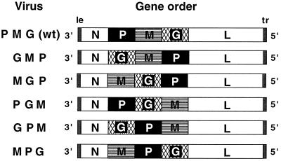
To validate this cloning strategy and to verify that the individual genes encoded functional proteins, we used the same procedure to recreate the wild-type gene order in parallel with the rearranged cDNAs and then tested all six plasmids for their ability to yield infectious viruses, as described before (47). Viable viruses were recovered that had the P, M, and G genes in all six possible orders (Fig. 1). Virus recovered from the wild-type plasmid was designated VSV N1 (PMG) and was used throughout this work as the wild-type virus. Its replication in cell culture was indistinguishable from that of nonrecombinant VSV Indiana (data not shown).
FIG. 1.
Schematic representations (not to scale) of the gene orders of wild-type VSV and the rearranged variant viruses. The wild-type (wt) virus used throughout this work was recovered from a cDNA clone that was reconstructed in parallel with the rearranged viruses by using the same individual gene clones; it was previously designated VSV N1 (PMG) to emphasize this distinction (45). The variant viruses were named according to the order of their central three genes. le, leader; tr, trailer.
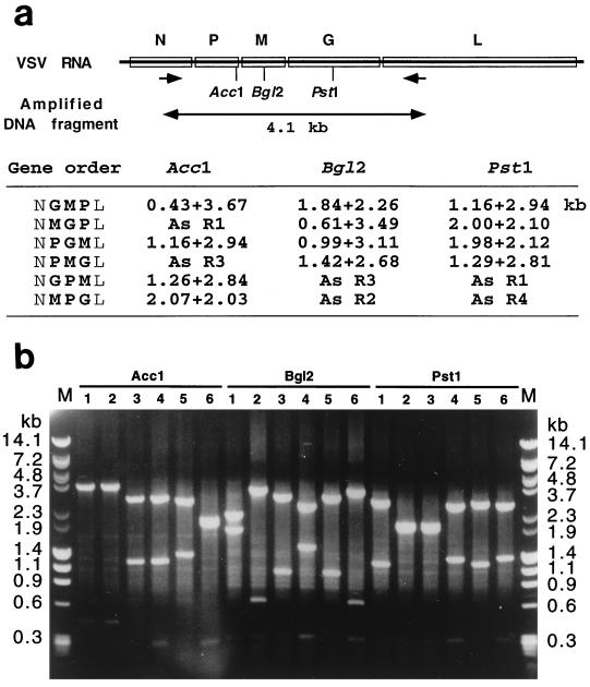
The gene orders of the recovered viruses were determined after three passages in cell culture by amplifying a 4.1-kb fragment encompassing the rearranged portions of the viral genomes by reverse transcription and PCR, followed by restriction enzyme analysis of the PCR products. PCR was carried out with primers located in the N and L genes. After cleavage with restriction endonuclease AccI, BglII, or PstI, which cleave uniquely in the P, M, or G gene, respectively, the observed sizes of the digestion products were found to be exactly as predicted (Fig. 2a and b). The data in Fig. 2 show that the gene orders of the recovered viruses corresponded to the cDNA clones from which they were recovered. There was no evidence for the reappearance of the wild-type gene order among the variants.
FIG. 2.
Determination of gene order in recovered viral genomes. Genomic RNA was isolated from recovered viruses and analyzed by reverse transcription and PCR, followed by restriction enzyme analysis of the PCR products. (a) Schematic diagram of the VSV genome showing positions of PCR primers that annealed to the N or L genes, respectively (shown by the arrows) and restriction enzyme cleavage sites and predicted fragment sizes. (b) Products after digestion with indicated enzymes of the cDNAs of viral RNA from viruses GMP, MGP, PGM, PMG, GPM, and MPG are shown in lanes 1 through 6, respectively. Fragments were analyzed by electrophoresis on a 1% agarose gel in the presence of ethidium bromide. Lanes M = marker DNA fragments with sizes as indicated.
Synthesis of viral RNAs and proteins.
The recovered viruses were next examined for their levels of gene expression. Synthesis of viral RNAs and proteins by the variant viruses was examined by metabolic incorporation of [3H]uridine or [35S]methionine into infected cells, and analysis of the radiolabeled products by gel electrophoresis. The same species of viral RNAs were made in cells infected with the wild-type virus and with each of the variants: the 11.16-kb genomic RNA and mRNAs representing the L, G, N, P, and M genes (Fig. 3a). The latter two mRNAs are similar in size and comigrated during electrophoresis (44). No novel or aberrant RNA species were found in cells infected with the variant viruses, showing that the virus preparations were free of DI particles (Fig. 3a). Moreover, the similarities among the RNA patterns reinforced the idea that the behavior of the viral polymerase during transcription across the intergenic junctions was determined exclusively by local sequence elements at these positions, with no detectable long-range influences (4, 5, 38). In accordance with the RNA patterns, the viral proteins made by the variant viruses also resembled qualitatively those made during wild-type infection (Fig. 3b).
FIG. 3.
(a) Viral RNAs synthesized in BHK-21 cells that were infected with the wild-type and variant viruses. Viral RNAs were labeled with [3H]uridine as described in Materials and Methods, resolved by electrophoresis on an agarose-urea gel, and detected by fluorography. The infecting viruses are shown above the lanes, and the viral RNAs are identified on the left. (b) Viral proteins synthesized in BHK-21 cells that were infected with the wild-type and variant viruses. Viral proteins were labeled with [35S]methionine as described in Materials and Methods, resolved by electrophoresis on an SDS-polyacrylamide gel, and detected by autoradiography. The infecting viruses are shown above the lanes, and the viral proteins are identified on the left. Uninf, uninfected cells.
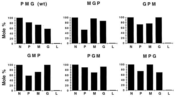
Although the RNA and protein profiles of cells infected with the wild-type and variant viruses were qualitatively similar, measurement of the relative levels of the different RNAs and proteins showed that the variant viruses expressed their genes in molar ratios that differed from both the wild type and one another (Fig. 4). Normalizing the expression of each protein to that of the promoter-proximal N gene for each variant virus showed that the relative expression level of a gene depended primarily on its location in the genome and thus on its distance from the promoter, just as predicted by the model of progressive transcriptional attenuation. This is clearly exemplified by comparison of the molar ratios of proteins expressed by the wild-type (PMG) with variant GMP in which the order of the three internal genes is reversed (Fig. 4). A similar quantitative analysis of the mRNA profiles was complicated by the lack of resolution of the M and P mRNAs (Fig. 3a), but measurement of the L, G, and N mRNA levels reinforced the conclusion that the proximity of a gene to the 3′ end of the viral genome was the major determinant of its level of transcription. For example, the differences in the relative abundance of G mRNA reflected the position of the G gene (Fig. 3a). The RNA profiles also showed that the level of RNA replication, as measured by the abundance of the 11.1-kb genomic and antigenomic RNAs, differed substantially among the variants (Fig. 3a).
FIG. 4.
Molar ratios of viral proteins synthesized in BHK-21 cells that were infected with the wild-type and variant viruses. Proteins were labeled as described in Materials and Methods, resolved on SDS-polyacrylamide gels as shown in Fig. 3b, and quantitated by phosphorimaging. Molar ratios were calculated after normalizing for the methionine contents of the individual proteins: N-14, P-5, M-11, G-10, and L-60 (35).
Replication of viruses with rearranged genomes.
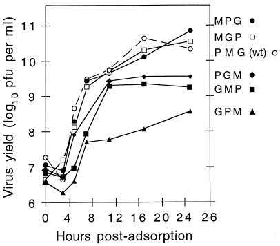
The variant viruses were compared for their ability to replicate under the conditions of plaque formation and single-cycle growth. Although some of the viruses such as MGP and MPG were indistinguishable from the N1 wild-type virus (PMG) in these assays, others such as GMP, GPM, and PGM formed significantly smaller plaques than the wild type on monolayers of BSC-1 cells (Table 1). Moreover, GMP plaques ceased to grow after 24 h when those of the wild-type virus and the other variants were still increasing in size (Table 1). The impaired replication of GMP, GPM, and PGM was also demonstrated during single-cycle growth on BSC-1 cells (Fig. 5). At 17 h postinfection, the incremental yields of the variants averaged over three independent growths and expressed as percentages of the wild-type were as follows: for MGP, 107%; for MPG, 51%; for GMP, 23%; for PGM, 21%; and for GPM, 1.6% (Table 2).
TABLE 1.
Plaque diameter (mean ± standard error)a
| Gene order | 24 h | 30 h |
|---|---|---|
| P M G (wild type) | 4.02 ± 0.12 | 4.81 ± 0.19 |
| G M P | 3.08 ± 0.17 | 3.10 ± 0.18 |
| M G P | 3.96 ± 0.19 | 4.97 ± 0.18 |
| P G M | 3.36 ± 0.12 | 3.86 ± 0.14 |
| G P M | 2.26 ± 0.09 | 3.16 ± 0.13 |
| M P G | 3.85 ± 0.18 | 5.43 ± 0.17 |
Plaque diameters were measured from photographs taken at approximately twofold magnification of groups of 50 (24 h) or 70 (30 h) viral plaques formed at 37°C on monolayers of BSC-1 cells.
FIG. 5.
Single-step growth curves of wild-type VSV and the rearranged variants in BSC-1 cells. Viral titers were measured in duplicate at each time point during three independent single-step growth experiments at 37°C, and the results were averaged.
TABLE 2.
Summary of properties of variant viruses
Virulence in mice.
Intracerebral or intranasal inoculation of wild-type VSV into mice causes fatal encephalitis. Since 1938, when Sabin and Olitsky first described the neuropathology and comparative susceptibility of mice to VSV encephalitis as a function of age and route of inoculation (36), young mice have served as a convenient and sensitive small animal model for comparing the lethality of VSV and its mutants (42, 49). We therefore examined the pathogenesis of the variant viruses in mice.
Intranasal inoculation of wild-type VSV into 3- to 4-week-old mice causes encephalitis, paralysis, and death after 7 to 11 days (42), with the LD50 dose being about 10 PFU. We compared the virulence of the variant viruses by inoculating groups of mice by this route with serial 10-fold dilutions ranging from 0.1 to 1,000 PFU per dose and observing them twice daily. Viral gene orders were verified on viruses recovered shortly after death from the brains of inoculated mice by using the methodology shown in Fig. 2. In each case, the gene order of the recovered virus corresponded to that of the inoculum (data not shown).
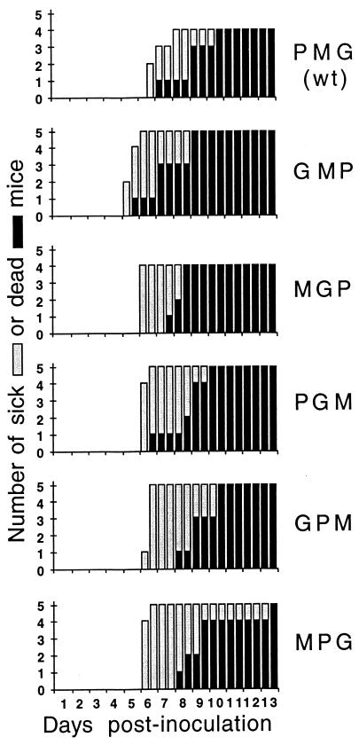
The LD50 doses for the variant viruses were similar to that of the wild type, with viruses GPM, GMP, and MGP requiring slightly higher (1.5- to 2-fold) doses (Table 2). These experiments were repeated three times, and the results of a representative experiment (Fig. 6) show the time of appearance of illness and death at a dose of 100 PFU per mouse. The wild-type-infected animals first appeared sick at 6 days postinoculation, rapidly became paralyzed, and died within 2 weeks. Recombinants GMP and MGP elicited reproducibly faster pathogenesis, with symptoms developing 24 to 36 h earlier than in wild-type-infected animals, whereas the onset of death from infection with MPG and GPM occurred 24 to 36 h later (Fig. 6). In general, the paralysis that is typical of infection with wild-type VSV was less apparent with the variant viruses, but there was no evidence of persistent nervous system disease such as that produced by some M protein mutants (17).
FIG. 6.
Pathogenesis of wild-type (wt) and variant viruses following intranasal inoculation into mice. The time course of morbidity (gray bars) and mortality (black bars) in animals that received intranasal inoculation of 100 PFU of each of the variant viruses is shown. No further changes occurred after the time periods shown.
Virulence in mice could not be predicted from the cell culture phenotypes of the variant viruses (Table 2). Of the three recombinants whose replication in cell culture was most compromised (GMP, PGM, and GPM), one (GPM) required twofold more virus for an LD50 than the wild type and showed slightly delayed killing in mice, whereas GMP induced faster onset of symptoms and death, and PGM was indistinguishable from wild type. This lack of correlation between the behavior of viruses in cell culture and their properties in animals is a familiar observation among different animal viruses but is interesting in this context, in which the only differences between the viruses were the relative levels of wild-type proteins they expressed.
DISCUSSION
The results presented above show that the three central genes in the VSV genome can be rearranged into all six possible orders (one of which is the wild-type order) without eliminating infectivity. This work extends our earlier recovery of viable variants in which the VSV N protein gene was moved from its promoter-proximal position to become the second, third, or fourth gene in the genome (45), and together with the recovery of infectious viruses with the gene orders GNPML and GPMNL (data not shown), it brings to 10 the total number of viable gene order variants of VSV constructed to date.
Evidently, the VSV genome can accommodate a significant degree of reorganization. Our results show that several different gene orders are compatible with virus replication, despite the fact that the natural gene order is strongly conserved among the Mononegavirales (32). Comparisons across the entire order of viruses suggest that little, if any, homologous recombination has occurred during their evolution from a presumed common ancestor, and all experimental attempts to detect homologous recombination have failed (30). Although the total number of viral genes differs among the Mononegavirales, there is little evidence that the conserved intergenic junction sequences have served as recombination sites to mediate gene rearrangement (30, 32). Our results indicate that this is not because rearrangement is necessarily lethal or even detrimental to the virus, because MGP and MPG replicated as well as the wild-type virus. Instead, it seems more likely that conservation results from the ancestral gene order being immobilized by the lack of a mechanism for homologous recombination.
A consequence of this is that natural processes should not be able to reverse experimental gene rearrangements such as those described above. Given the intrinsically high error rates of the viral RNA polymerases, their lack of proofreading mechanisms and the resulting genetic instability of these viruses (11), changing the gene order may be a unique way to create stable variants with desirable phenotypes. Indeed, we have shown that moving the VSV N gene from its promoter-proximal position progressively further down the viral genome systematically attenuates the virus without compromising its immunogenicity, thus creating a promising prototype for a novel live attenuated vaccine (45). The ability of the VSV genome to accommodate significant reorganization enhances the prospects of the use of the virus as a vector.
Since the variants described here were constructed by rearranging the viral genes without introducing other mutations, the phenotypic consequences could be unambiguously attributed to translocation of the genes and the resulting effects on their expression. The levels of viral protein synthesis accurately reflected the positions of the genes with respect to the 3′ end of the genome (Fig. 4), directly demonstrating that gene expression was regulated primarily by transcriptional attenuation. Moreover, the results confirmed that the level of attenuation at the first three intergenic junctions was determined principally by the conserved sequence elements in their immediate vicinity, with no detectable long-range influences (5, 38, 40). However, in the wild-type virus, transcriptional attenuation is greater at the G-L boundary than at the preceding junctions, and the same was true at the P-L and M-L junctions in the pertinent variants (Fig. 3a and 4). Since the same effect was seen at an N-L junction (45), these results suggest that the unusually high transcriptional attenuation at this position may be due to cis-acting signals in the L gene itself.
Our previous experience of the sensitivity of VSV RNA replication to the N:P:L protein ratios led us to anticipate that some of the gene rearrangements would be lethal (28). In particular, we expected severe phenotypic consequences to result from both the underexpression of P protein, which acts as a chaperone to incorporate N into assembling nucleocapsids (10, 16, 19), and the overexpression of M protein, which inhibits viral transcription (8, 9, 21) and condenses the viral nucleocapsids in preparation for particle formation (22, 24). Although more severe perturbations of the levels of viral proteins would doubtless have more profound effects on the viral phenotype, variant MGP, which combined these features, replicated in cell culture about as well as the wild-type virus and had a similar LD50 in mice (Fig. 5 and Table 2). Variant MPG also resembled the wild-type virus both in cell culture and in mice. In contrast, the two variants that underexpressed M protein (PGM and GPM) replicated the least well in cell culture (Fig. 5), although they were not correspondingly attenuated in mice (Fig. 6 and Table 2). Contrary to expectations, therefore, the level of M protein expression correlated most closely with the level of replication in cell culture. The lack of a straightforward concordance between the replication of the variants in cell culture and their virulence in mice illustrates the well-known limitations of cell culture methods for studying the biological properties of animal viruses.
One key to understanding these disparate phenotypes may be provided by the properties of variant GMP, which underexpressed P protein compared with the wild-type virus (Fig. 4), and whose plaques ceased to grow before those of the other viruses (Table 1). Similar plaquing behavior was seen with some conventional mutants of VSV that were defective in shutting off host protein synthesis (14, 39, 46, 49). Unlike the wild-type virus, these mutants induced high levels of interferon, which prematurely arrested plaque development, and it is possible that variant GMP also interacted anomalously with the interferon system, either by inducing unusually high levels of interferon or by being particularly sensitive to its action. Alternatively, variations in the rate of DI particle generation could have contributed to the differences in plaquing behavior, although the input virus stocks were free of DI particles as shown by their intracellular RNA profiles (Fig. 3a). Experiments are in progress to examine these possibilities.
Plaques of variant MGP, unlike those of GMP, did not stop growing prematurely (Table 1). This may be attributable to the overexpression of M protein by the former variant and the documented ability of M to repress the transcription of cellular genes (2, 6, 25), including that for interferon β (13). Similarly, differences in the shut-off of host cell transcription and translation and the consequent effects on interferon induction and action could also explain why variants PGM and GPM replicated so poorly in cell culture. However, direct measurement of the kinetics of host cell shut-off and interferon induction and action will be necessary to substantiate these ideas, and further studies will be required to fully explain the altered phenotypes of the variant viruses in molecular terms.
Although the gene orders of viruses recovered from the brains of dead mice were found invariably to correspond to those of the inoculated viruses, serial passage of the variants either in cell culture or in animals may select for mutations that compensate for the unbalanced patterns of gene expression. Recent work has shown that mutating the conserved nucleotides at the intergenic junction of a model bicistronic VSV replicon can profoundly affect the degree of both transcriptional attenuation (4, 40) and termination (4, 5, 40). In the genome of a variant virus, however, such mutations would confer only a limited benefit, because they would necessarily have pleiotropic effects on the expression of all downstream genes. Nevertheless, if compensating mutations arise during serial passage of the variants either in cell culture or in animals, they should provide valuable insights into the mechanism of VSV gene regulation and the nature of the selective pressures encountered by viruses in these distinct environments.
In summary, these results show that despite the strong conservation of gene order among the Mononegavirales, several variants of VSV with different gene orders are viable, and some of them show altered phenotypes both in cell culture and in mice. This ability to accommodate major genetic changes more readily than might have been expected from phylogenetic data augments VSV’s potential for development as an expression vector and recombinant vaccine. The same method of gene rearrangement could be applied to any of the Mononegavirales for which an infectious cDNA clone is available, and many aspects of virus biology, including replication, pathogenesis, and evolution, should be illuminated by detailed studies of these and similar viruses. In general, the approach of rearranging viral genes to effect changes in gene expression provides an opportunity for the generation of viral phenotypes that may prove valuable for the analysis of virus-host interactions and for optimizing the potential of these viruses as vectors in many different contexts.
ACKNOWLEDGMENTS
We thank Seán Whelan for suggesting the cloning strategy that was used to rearrange the genes and Fenglan Li for excellent technical help.
This work was supported by Public Health Service grants R37 AI 12464 to G.W.W. and R37 AI 18270 to L.A.B.
REFERENCES
- 1.Abraham G, Banerjee A K. Sequential transcription of the genes of vesicular stomatitis virus. Proc Natl Acad Sci USA. 1976;73:1504–1508. doi: 10.1073/pnas.73.5.1504. [DOI] [PMC free article] [PubMed] [Google Scholar]
- 2.Ahmed M, Lyles D S. Effect of vesicular stomatitis virus matrix protein on transcription directed by host polymerases I, II, and III. J Virol. 1998;72:8413–8419. doi: 10.1128/jvi.72.10.8413-8419.1998. [DOI] [PMC free article] [PubMed] [Google Scholar]
- 3.Ball L A, White C N. Order of transcription of genes of vesicular stomatitis virus. Proc Natl Acad Sci USA. 1976;73:442–446. doi: 10.1073/pnas.73.2.442. [DOI] [PMC free article] [PubMed] [Google Scholar]
- 4.Barr J N, Whelan S P, Wertz G W. Role of the intergenic dinucleotide in vesicular stomatitis virus RNA transcription. J Virol. 1997;71:1794–1801. doi: 10.1128/jvi.71.3.1794-1801.1997. [DOI] [PMC free article] [PubMed] [Google Scholar]
- 5.Barr J N, Whelan S P J, Wertz G W. The cis-acting signals involved in termination of VSV mRNA synthesis include the conserved AUAC and the U7 signal for polyadenylation. J Virol. 1997;71:8718–8725. doi: 10.1128/jvi.71.11.8718-8725.1997. [DOI] [PMC free article] [PubMed] [Google Scholar]
- 6.Black B L, Lyles D S. Vesicular stomatitis virus matrix protein inhibits host cell-directed transcription of target genes in vivo. J Virol. 1992;66:4058–4064. doi: 10.1128/jvi.66.7.4058-4064.1992. [DOI] [PMC free article] [PubMed] [Google Scholar]
- 7.Blondel D, Harmison G G, Schubert M. Role of matrix protein in cytopathogenesis of vesicular stomatitis virus. J Virol. 1990;64:1716–1725. doi: 10.1128/jvi.64.4.1716-1725.1990. [DOI] [PMC free article] [PubMed] [Google Scholar]
- 8.Carroll A R, Wagner R R. Role of the membrane (M) protein in endogenous inhibition of in vitro transcription of vesicular stomatitis virus. J Virol. 1979;29:134–142. doi: 10.1128/jvi.29.1.134-142.1979. [DOI] [PMC free article] [PubMed] [Google Scholar]
- 9.Clinton G M, Little S P, Hagen F S, Huang A S. The matrix (M) protein of vesicular stomatitis virus regulates transcription. Cell. 1978;15:1455–1462. doi: 10.1016/0092-8674(78)90069-7. [DOI] [PubMed] [Google Scholar]
- 10.Davis N L, Arnheiter H, Wertz G W. Vesicular stomatitis virus N and NS proteins from multiple complexes. J Virol. 1986;59:751–754. doi: 10.1128/jvi.59.3.751-754.1986. [DOI] [PMC free article] [PubMed] [Google Scholar]
- 11.Domingo E, Escarmis C, Sevilla N, Moya A, Elena S F, Quer J, Novella I S, Holland J J. Basic concepts in RNA virus evolution. FASEB J. 1996;10:859–864. doi: 10.1096/fasebj.10.8.8666162. [DOI] [PubMed] [Google Scholar]
- 12.Emerson S U, Yu Y-H. Both NS and L proteins are required for in vitro RNA synthesis by vesicular stomatitis virus. J Virol. 1975;15:1348–1356. doi: 10.1128/jvi.15.6.1348-1356.1975. [DOI] [PMC free article] [PubMed] [Google Scholar]
- 13.Ferran M, Lucas-Lenard J M. The vesicular stomatitis virus matrix protein inhibits transcription from the human beta interferon promoter. J Virol. 1997;71:371–377. doi: 10.1128/jvi.71.1.371-377.1997. [DOI] [PMC free article] [PubMed] [Google Scholar]
- 14.Francoeur A M, Poliquin L, Stanners C P. The isolation of interferon-inducing mutants of vesicular stomatitis virus with altered viral P function for the inhibition of total protein synthesis. Virology. 1987;160:236–245. doi: 10.1016/0042-6822(87)90065-1. [DOI] [PubMed] [Google Scholar]
- 15.Fuerst T R, Niles E G, Studier F W, Moss B. Eukaryotic transient-expression system based on recombinant vaccinia virus that synthesizes bacteriophage T7 RNA polymerase. Proc Natl Acad Sci USA. 1986;83:8122–8126. doi: 10.1073/pnas.83.21.8122. [DOI] [PMC free article] [PubMed] [Google Scholar]
- 16.Howard M, Wertz G. Vesicular stomatitis virus RNA replication: a role for the NS protein. J Gen Virol. 1989;70:2683–2694. doi: 10.1099/0022-1317-70-10-2683. [DOI] [PubMed] [Google Scholar]
- 17.Hughes J V, Johnson T C, Rabinowitz S G, Dal Canto M C. Growth and maturation of a vesicular stomatitis virus temperature-sensitive mutant and its central nervous system isolate. J Virol. 1979;29:312–321. doi: 10.1128/jvi.29.1.312-321.1979. [DOI] [PMC free article] [PubMed] [Google Scholar]
- 18.Iverson L E, Rose J K. Localized attenuation and discontinuous synthesis during vesicular stomatitis virus transcription. Cell. 1981;23:477–484. doi: 10.1016/0092-8674(81)90143-4. [DOI] [PubMed] [Google Scholar]
- 19.La Ferla F M, Peluso R W. The 1:1 N-NS protein complex of vesicular stomatitis virus is essential for efficient genome replication. J Virol. 1989;63:3852–3857. doi: 10.1128/jvi.63.9.3852-3857.1989. [DOI] [PMC free article] [PubMed] [Google Scholar]
- 20.Lefkowitz E J, Pattnaik A K, Ball L A. Complementation of a vesicular stomatitis virus glycoprotein G mutant with wild-type protein expressed from either a bovine papilloma virus or a vaccinia virus vector system. Virology. 1990;178:373–383. doi: 10.1016/0042-6822(90)90334-n. [DOI] [PubMed] [Google Scholar]
- 21.Li Y, Luo L, Wagner R R. Transcription inhibition site on the M protein of vesicular stomatitis virus located by marker rescue of mutant tsO23(III) with M-gene expression vectors. J Virol. 1989;63:2841–2843. doi: 10.1128/jvi.63.6.2841-2843.1989. [DOI] [PMC free article] [PubMed] [Google Scholar]
- 22.Lyles D S, McKenzie M O, Kaptur P E, Grant K W, Jerome W G. Complementation of M gene mutants of vesicular stomatitis virus by plasmid-derived M protein converts spherical extracellular particles into native bullet shapes. Virology. 1996;217:76–87. doi: 10.1006/viro.1996.0095. [DOI] [PubMed] [Google Scholar]
- 23.Meier E, Harmison G G, Schubert M. Homotypic and heterotypic exclusion of vesicular stomatitis virus replication by high levels of recombinant polymerase protein L. J Virol. 1987;61:3133–3142. doi: 10.1128/jvi.61.10.3133-3142.1987. [DOI] [PMC free article] [PubMed] [Google Scholar]
- 24.Odenwald W F, Arnheiter H, Dubois-Dalcq M, Lazzarini R A. Stereo images of vesicular stomatitis virus assembly. J Virol. 1986;57:922–932. doi: 10.1128/jvi.57.3.922-932.1986. [DOI] [PMC free article] [PubMed] [Google Scholar]
- 25.Paik S-Y, Banerjea A C, Harmison G G, Chen C-J, Schubert M. Inducible and conditional inhibition of human immunodeficiency virus proviral expression by vesicular stomatitis virus matrix protein. J Virol. 1995;69:3529–3537. doi: 10.1128/jvi.69.6.3529-3537.1995. [DOI] [PMC free article] [PubMed] [Google Scholar]
- 26.Pal R, Grinnell R W, Snyder R M, Wagner R R. Regulation of viral transcription by the matrix protein of vesicular stomatitis virus probed by monoclonal antibodies and temperature-sensitive mutants. J Virol. 1985;56:386–394. doi: 10.1128/jvi.56.2.386-394.1985. [DOI] [PMC free article] [PubMed] [Google Scholar]
- 27.Pattnaik A K, Ball L A, LeGrone A W, Wertz G W. Infectious defective interfering particles of VSV from transcripts of a cDNA clone. Cell. 1992;69:1011–1020. doi: 10.1016/0092-8674(92)90619-n. [DOI] [PubMed] [Google Scholar]
- 28.Pattnaik A K, Wertz G W. Cells that express all five proteins of vesicular stomatitis virus from cloned cDNAs support replication, assembly, and budding of defective interfering particles. Proc Natl Acad Sci USA. 1991;88:1379–1383. doi: 10.1073/pnas.88.4.1379. [DOI] [PMC free article] [PubMed] [Google Scholar]
- 29.Pattnaik A K, Wertz G W. Replication and amplification of defective interfering particle RNAs of vesicular stomatitis virus in cells expressing viral proteins from vectors containing cloned cDNAs. J Virol. 1990;64:2948–2957. doi: 10.1128/jvi.64.6.2948-2957.1990. [DOI] [PMC free article] [PubMed] [Google Scholar]
- 30.Pringle C R. The genetics of paramyxoviruses. In: Kingsbury D, editor. The paramyxoviruses. New York, N.Y: Plenum Press; 1991. pp. 1–39. [Google Scholar]
- 31.Pringle C R. The genetics of vesiculoviruses. Arch Virol. 1982;72:1–34. doi: 10.1007/BF01314447. [DOI] [PubMed] [Google Scholar]
- 32.Pringle C R, Easton A J. Monopartite negative strand RNA genomes. Semin Virol. 1997;8:49–57. [Google Scholar]
- 33.Reed E J, Muench H. A simple method of estimating fifty percent endpoints. Am J Hyg. 1938;27:493–497. [Google Scholar]
- 34.Richardson J H, Barkley W E, editors. Biosafety in microbiological and biomedical laboratories. 2nd ed. Atlanta, Ga. and Bethesda, Md.: USDHHS: Centers for Disease Control and Prevention, and National Institutes of Health; 1988. [Google Scholar]
- 35.Rose J, Schubert M. Rhabdovirus genomes and their products. In: Wagner R R, editor. The rhabdoviruses. New York, N.Y: Plenum Press; 1987. pp. 127–166. [Google Scholar]
- 36.Sabin A, Olitsky P. Influence of host factors on neuroinvasiveness of vesicular stomatitis virus. J Exp Med. 1938;67:201–227. doi: 10.1084/jem.67.2.229. [DOI] [PMC free article] [PubMed] [Google Scholar]
- 37.Sambrook J, Fritsch E F, Maniatis T. Molecular cloning: a laboratory manual. Cold Spring Harbor, N.Y: Cold Spring Harbor Laboratory Press; 1989. [Google Scholar]
- 38.Schnell M J, Buonocore L, Whitt M A, Rose J K. The minimal conserved transcription stop-start signal promotes stable expression of a foreign gene in vesicular stomatitis virus. J Virol. 1996;70:2318–2323. doi: 10.1128/jvi.70.4.2318-2323.1996. [DOI] [PMC free article] [PubMed] [Google Scholar]
- 39.Stanners C P, Francoeur A M, Lam T. Analysis of VSV mutant with attenuated cytopathogenicity: mutation in viral function, P, for inhibition of protein synthesis. Cell. 1977;11:273–281. doi: 10.1016/0092-8674(77)90044-7. [DOI] [PubMed] [Google Scholar]
- 40.Stillman E A, Whitt M A. Mutational analysis of the intergenic dinucleotide and the transcriptional start sequence of vesicular stomatitis virus (VSV) define sequences required for efficient termination and initiation of VSV transcripts. J Virol. 1997;71:2127–2137. doi: 10.1128/jvi.71.3.2127-2137.1997. [DOI] [PMC free article] [PubMed] [Google Scholar]
- 41.Villarreal L P, Breindl M, Holland J J. Determination of molar ratios of vesicular stomatitis virus induced RNA species in BHK21 cells. Biochemistry. 1976;15:1663–1667. doi: 10.1021/bi00653a012. [DOI] [PubMed] [Google Scholar]
- 42.Wagner R R. Pathogenicity and immunogenicity for mice of temperature-sensitive mutants of vesicular stomatitis virus. Infect Immun. 1974;10:309–315. doi: 10.1128/iai.10.2.309-315.1974. [DOI] [PMC free article] [PubMed] [Google Scholar]
- 43.Wertz G W, Davis N. Characterization of mapping of RNase III cleavage sites in VSV genome RNA. Nucleic Acids Res. 1981;9:6487–6503. doi: 10.1093/nar/9.23.6487. [DOI] [PMC free article] [PubMed] [Google Scholar]
- 44.Wertz G W, Davis N L. RNase III cleaves vesicular stomatitis virus genome-length RNAs but fails to cleave viral mRNA’s. J Virol. 1979;30:108–115. doi: 10.1128/jvi.30.1.108-115.1979. [DOI] [PMC free article] [PubMed] [Google Scholar]
- 45.Wertz G W, Perepelitsa V P, Ball L A. Gene rearrangement attenuates expression and lethality of a non-segmented negative strand RNA virus. Proc Natl Acad Sci USA. 1998;95:3501–3506. doi: 10.1073/pnas.95.7.3501. [DOI] [PMC free article] [PubMed] [Google Scholar]
- 46.Wertz G W, Youngner J S. Interferon production and inhibition of host synthesis in cells infected with vesicular stomatitis virus. J Virol. 1970;6:476–484. doi: 10.1128/jvi.6.4.476-484.1970. [DOI] [PMC free article] [PubMed] [Google Scholar]
- 47.Whelan S P J, Ball L A, Barr J N, Wertz G T W. Recovery of infectious vesicular stomatitis virus entirely from cDNA clones. Proc Natl Acad Sci USA. 1995;92:8388–8392. doi: 10.1073/pnas.92.18.8388. [DOI] [PMC free article] [PubMed] [Google Scholar]
- 48.Whitt M A, Chong L, Rose J K. Glycoprotein cytoplasmic domain sequences required for rescue of a vesicular stomatitis virus glycoprotein mutant. J Virol. 1989;63:3569–3578. doi: 10.1128/jvi.63.9.3569-3578.1989. [DOI] [PMC free article] [PubMed] [Google Scholar]
- 49.Youngner J S, Wertz G. Interferon production in mice by vesicular stomatitis virus. J Virol. 1968;2:1360–1361. doi: 10.1128/jvi.2.11.1360-1361.1968. [DOI] [PMC free article] [PubMed] [Google Scholar]


