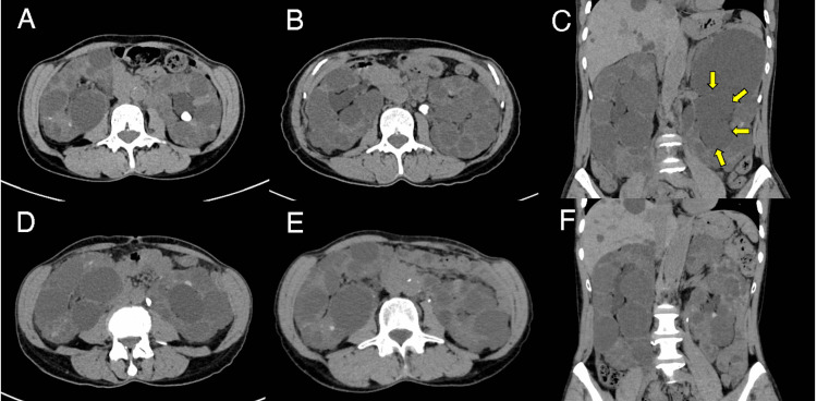
Figure 1. Patient's abdominal CT.
Abdominal CT revealed (A) a 10.5 mm renal pelvic stone and (B) a 10.3 mm stone in the ureteropelvic junction. CT revealed (C) left hydronephrosis (arrows) and (D) a 9.4 mm ureteric stone at the level of L3/L4. (E and F) A second TUL successfully fragmented and extracted almost all the stone fragments and improved left hydronephrosis.
CT: computed tomography, TUL: transurethral lithotripsy

