ABSTRACT
Familial Mediterranean fever (FMF) is a hereditary disorder that presents with recurrent fever, rash, and polyserosal inflammation. The nonspecific symptoms of FMF allow it to mimic a large variety of diseases including metastatic Crohn's disease (MCD). MCD is a rare extraintestinal manifestation of Crohn's disease characterized by the presence of cutaneous noncaseating granulomas that are noncontiguous within the gastrointestinal tract. We describe a patient who had a delay in diagnosis of FMF as her clinical presentation mimicked MCD.
KEYWORDS: familial mediterranean fever, Crohn's disease mimicker, metastatic Crohn's disease
INTRODUCTION
Familial Mediterranean fever (FMF) is an inherited disease due to an autosomal recessive mutation in the Mediterranean Fever (MEFV) gene found most commonly in patients of Mediterranean descent.1 This mutation leads to dysregulation of the innate immune response causing periodic fever, rash, abdominal pain, peritonitis, pleuritis, and monoarthritis.2,3 The nonspecific nature of these symptoms allows FMF to imitate metastatic Crohn's disease (MCD) as both diseases can have integumentary and gastrointestinal system involvement. However, the cutaneous lesions seen in MCD histologically reveal noncaseating granulomas while FMF has erysipelas-like erythema with histology revealing lymphocytic and neutrophilic infiltration.4,5 We report a case of FMF with clinical symptoms and histologic findings that closely resemble MCD.
CASE REPORT
A 28-year-old White woman presented to the dermatology clinic 13 years ago with symptoms of recurrent episodes of facial erythema, labial edema, oral ulcers, abdominal pain, and fevers. She was evaluated by gynecology for labial erythema/edema and associated fevers and symptoms were originally attributed to recurrent soft-tissue skin infections. Oral mucosa biopsy revealed noncaseating granulomatous infiltrates within the dermis composed of lymphocytes, histiocytes, giant cells, plasma cells, and neutrophils. Colonoscopy showed no evidence of gastrointestinal involvement; however, the patient was started on adalimumab for presumed MCD solely based on skin biopsies, elevated C-reactive protein (11.7 mg/L), and her clinical symptoms of oral ulcers and abdominal pain. Following the initiation of adalimumab, she reported mild improvement in her symptoms with initial reduction of vulvar erythema and edema, but continued oral ulcers.
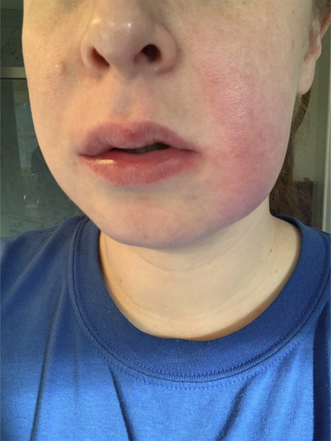
However, after being on adalimumab for 5 years, she was referred to a gastroenterology clinic for worsening flare-ups of her symptoms. Detailed history revealed episodic abdominal pain lasting 1–3 days accompanied by fever up to 104 °F, nausea, vomiting, non-bloody diarrhea, intermittent abdominal distension, labial swelling, facial rash, and oral ulcers. Skin examination showed erythematous nodules in the buccal mucosa and an erythematous indurated plaque along the left cheek (Figure 1). Given she had 5 unremarkable colonoscopies and 2 esophagogastroduodenoscopies, a normal computed tomography enterography, and a negative inflammatory bowel disease gene panel, her diagnosis of MCD was questioned.
Figure 1.

Erythematous indurated plaque along the left cheek.
Retrospective review of symptomatology raised suspicion of FMF. The patient revealed her ancestors originated from the Mediterranean region but denied any family history of FMF. Extensive genetic testing revealed a mutation in the MEFV gene. She was empirically treated with colchicine 1.2 mg daily, which dramatically improved her symptoms after 1 month of therapy. She was ultimately diagnosed with FMF as she satisfied Tel-Hashomer criteria (recurrent febrile episodes, erysipelas-like erythema, and response to colchicine).
Her quality of life improved as she continued to be symptom-free on a maintenance dose of colchicine. She follows with rheumatology, gastroenterology, and dermatology on a regular basis.
DISCUSSION
Diagnosis of FMF is based on symptomatology, a positive response to colchicine treatment, and genetic testing. Diagnostic criteria such as Tel-Hashomer can also aid in making the diagnosis.6 Despite having these criteria, the diagnosis of FMF is sometimes difficult in patients with atypical symptoms. Our patient had episodic fever associated with abdominal pain and skin rash that was originally diagnosed with MCD. However, she was minimally responsive to adalimumab and had numerous negative endoscopic evaluations, causing the diagnosis of MCD to be questioned despite the skin biopsy results being consistent with MCD.
Comprehensive knowledge about FMF and MCD can allow one to differentiate the 2 diseases when one imitates the other. FMF has the highest prevalence among people of Turkish, Armenian, North African, Jewish, and Arabic descent, and 90% of patients typically experience their first attack before age 20.7,8 FMF flares are characterized by episodic fever, abdominal pain, skin involvement, and joint pain and are generally self-limited, lasting 1–3 days, and vary in severity and symptomatology.6 The most common symptoms are fever, abdominal pain (90%), arthralgias (75%), and skin involvement (45%).9–11 MCD is defined as cutaneous lesion in areas noncontinuous with the gastrointestinal tract that can present during, after, and rarely proceeding the Crohn's disease (CD).5 Since MCD is a manifestation of CD, it can still present with abdominal pain (seen in approximately 33% of patients) and arthritis, which are symptoms that are seen in primary CD.5,12
FMF may be easily mistaken for CD, especially in non-Mediterranean individuals who have a low risk of FMF since both have skin and gastrointestinal involvement.13 For instance, Asakura et al reported a case of FMF with gastrointestinal lesions mimicking CD.14 They found multiple linear erosions in the stomach and longitudinal ulcers in the terminal ileum; however, the patient had no cutaneous manifestations. Arasawa et al also reported a FMF case with similar gastrointestinal biopsy findings.13 In contrast to these patients, our patient represents the only case reported where a cutaneous manifestation of FMF mimics MCD.
The typical skin eruptions usually differ between the 2 diseases. FMF can have various cutaneous features which include an erysipelas-like skin eruption that occurs in 12%–40% of patients as well as urticaria, diffuse erythema, maculopapular eruption, and subcutaneous nodules.4,15 Skin biopsy of these lesions may show heavy neutrophil infiltrates.16 By contrast, skin involvement in MCD typically appears as cutaneous ulcerations and plaques, and these skin lesions usually occurs on skin folds and legs.5 Biopsy typically shows inflammatory infiltrate consisting of noncaseating sarcoid-type granulomas.5 In our patient presenting with an erythematous plaque, MCD was suspected initially due to an atypical FMF rash that closely resembled MCD both histologically and clinically. Our patient's skin biopsy revealed noncaseating granulomas consistent with MCD and represents a novel presentation of FMF.
MCD may be considered in the differential diagnosis of a young patient presenting with abdominal pain and skin rash showing noncaseating granulomas. However, FMF should be included on the differential, and therefore, it is important to obtain a detailed history when evaluating such patients. An alternative diagnosis should be considered in patients who are being treated for MCD and are found to be refractory to biological therapy particularly when there is a lack of gastrointestinal pathology typically seen in Crohn's disease on endoscopies and biopsies.
DISCLOSURES
Author contributions: HS Patel consented the patient and performed literature review, initial text draft, and edits. J. Srivastav performed text review and edits. R. Thapa wrote and edited the manuscript and reviewed the literature. HS Patel, J. Srivastav and R. Thapa edited the manuscript and revised the manuscript for intellectual content. HS Patel is the article guarantor.
Financial disclosure: None to report.
Previous Presentation: This case was presented at the American College of Gastroenterology Annual Scientific Meeting; October 23–28, 2020; Virtual.
Informed consent was obtained for this case report.
Contributor Information
Hiral S. Patel, Email: hiralspatelmd@gmail.com.
Jigisha Srivastav, Email: jsrivast@wakehealth.edu.
Rupak Thapa, Email: rthapa@wakehealth.edu.
REFERENCES
- 1.Sönmez HE, Batu ED, Özen S. Familial Mediterranean fever: Current perspectives. J Inflamm Res. 2016;9:13–20. [DOI] [PMC free article] [PubMed] [Google Scholar]
- 2.Tidow N, Chen X, Müller C, et al. Hematopoietic-specific expression of MEFV, the gene mutated in familial Mediterranean fever, and subcellular localization of its corresponding protein, pyrin. Blood. 2000;95(4):1451–5. [PubMed] [Google Scholar]
- 3.Stehlik C, Reed JC. The PYRIN connection: Novel players in innate immunity and inflammation. J Exp Med. 2004;200(5):551–8. [DOI] [PMC free article] [PubMed] [Google Scholar]
- 4.Tsuruma H, Sato H, Hasegawa E, et al. An adult case of atypical familial Mediterranean fever (pyrin-associated autoinflammatory disease) similar to adult-onset Still's disease. Clin Case Rep. 2019;7(4):801–5. [DOI] [PMC free article] [PubMed] [Google Scholar]
- 5.Aberumand B, Howard J, Howard J. Metastatic Crohn's disease: An approach to an uncommon but important cutaneous disorder. Biomed Res Int. 2017;2017:8192150. [DOI] [PMC free article] [PubMed] [Google Scholar]
- 6.Bashardoust B. Familial Mediterranean fever; diagnosis, treatment, and complications. J Nephropharmacol. 2015;4(1):5–8. [PMC free article] [PubMed] [Google Scholar]
- 7.Sarkisian T, Ajrapetian H, Beglarian A, Shahsuvarian G, Egiazarian A. Familial Mediterranean fever in Armenian population. Georgian Med News. 2008;156:105–11. [PubMed] [Google Scholar]
- 8.Ben‐Chetrit E, Touitou I. Familial Mediterranean fever in the world. Arthritis Rheum. 2009;61(10):1447–53. [DOI] [PubMed] [Google Scholar]
- 9.Sohar E, Gafni J, Pras M, Heller H. Familial Mediterranean fever: A survey of 470 cases and review of the literature. Am J Med. 1967;43(2):227–53. [DOI] [PubMed] [Google Scholar]
- 10.Garcia-Gonzalez A, Weisman MH. The arthritis of familial Mediterranean fever. Semin Arthritis Rheum. 1992;22(3):139–50. [DOI] [PubMed] [Google Scholar]
- 11.Maggio MC, Corsello G. FMF is not always “fever”: From clinical presentation to treat to target. Ital J Pediatr. 2020;46(1):7. [DOI] [PMC free article] [PubMed] [Google Scholar]
- 12.Palamaras I, El-Jabbour J, Pietropaolo N, et al. Metastatic Crohn's disease: A review. J Eur Acad Dermatol Venereol. 2008;22(9):1033–43. [DOI] [PubMed] [Google Scholar]
- 13.Arasawa S, Nakase H, Ozaki Y, Uza N, Matsuura M, Chiba T. Mediterranean mimicker. Lancet. 2012;380(9858):2052. [DOI] [PubMed] [Google Scholar]
- 14.Asakura K Yanai S Nakamura S, et al. Familial Mediterranean fever mimicking Crohn disease: A case report. Medicine. 2018;97(1):e9547. [DOI] [PMC free article] [PubMed] [Google Scholar]
- 15.Drenth JP, Van Der Meer JW. Hereditary periodic fever. New Engl J Med. 2001;345(24):1748–57. [DOI] [PubMed] [Google Scholar]
- 16.Kolivras A, Provost P, Thompson CT. Erysipelas-like erythema of familial Mediterranean fever syndrome: A case report with emphasis on histopathologic diagnostic clues. J Cutan Pathol. 2013;40(6):585–90. [DOI] [PubMed] [Google Scholar]


