


Abstract
Introduction and importance
Iatrogenic injury to the cavernous nerve and its branches results in post-operative erectile dysfunction in up to 85 % of men undergoing a radical prostatectomy. Here, we describe using a novel fluorescence-imaging system developed to detect nerve autofluorescence in a 66-year-old gentleman with prostate adenocarcinoma (Gleason Score 8 [4 + 4], prognostic group 4, indicating a highly-aggressive prostate cancer) who underwent laparotomic radical prostatectomy.
Case presentation
Under general anesthesia, a laparotomic radical prostatectomy was performed using standard operative techniques. During surgery, a Dendrite imaging camera (Dendrite® Imaging, Germany) was employed to permit the surgical team to toggle freely between standard operating room (white) light and near-ultraviolet light (NUVL), with the specific purpose of enhancing visualization of the periprostatic nerve plexus, including the cavernous nerve and all its branches. Under white light, neither the cavernous nerve nor any of its branches were clearly visible. However, under NUVL, all fluoresced brightly and were easily avoided during prostate resection. Prostate resection proceeded with no intra-operative or post-operative complications. Moreover, upon one-month follow-up in the surgery clinic, the patient reported no erectile dysfunction, difficulties voiding, or other neurological or non-neurological complaints.
Clinical discussion
In this case, autofluorescence of the cavernous nerve and its branches during radical prostatectomy aided in their visualization and appeared to help prevent post-operative erectile dysfunction and all other potential neurological deficits.
Conclusion
Novel intra-operative technology enabling nerves to auto-fluoresce warrants larger series and comparative trials to assess its effectiveness reducing iatrogenic nerve injury during radical prostatectomies.
Keywords: Case report, Prostatectomy, Fluorescence imaging, Nerve preservation, Erectile dysfunction, Nerve autofluorescence
Highlights
-
•
Erectile dysfunction occurs in up to 80 % of men undergoing radical prostatectomy.
-
•
To prevent nerve injury, a novel camera was used to detect nerve autofluorescence.
-
•
In fluorescent light, visualization of all critical neural structures was enhanced.
-
•
Enhanced nerve visualization helped to prevent iatrogenic nerve injury.
-
•
Our patient had no erectile dysfunction or other nerve deficits post-operatively.
1. Introduction
Radical prostatectomy has been documented to produce excellent long-term survival in select patients with prostate cancer, reducing the risk of pelvic recurrence and metastatic spread [1,2]. However, during prostatectomy procedures, the cavernous nerve, which divides into several smaller branches to provide innervation to the penis, can sustain crush, tension, and resection injury, resulting in erectile dysfunction in up to 85 % of patients [3,4]. This occurs because the nerve and its branches are small, localized between the prostatic capsule and prostatic fascia, and thus difficult to visualize and avoid within a tight anatomical space.
In recent years, a novel fluorescence imaging system has been developed (Dendrite® Imaging, Germany) that makes nerves auto-fluoresce when visualized in near-ultraviolet light (NUVL) [5]. Its use has been documented in vivo in both Wistar rats, where nerves are generally very small [5] and in patients undergoing a broad range of surgical procedures, including parotidectomies, thyroidectomies, spinal, and peripheral nerve surgeries [6]. Most recently, this imaging system was found to be 100 % sensitive and 100 % specific identifying 81 recurrent laryngeal nerves in 65 patients undergoing a thyroidectomy [7].
Here, we describe our use of this innovative imaging system to visualize the cavernous nerves and its branches within the surgical field and prevent their iatrogenic injury, thereby avoiding the severe complication of erectile dysfunction post radical prostatectomy.
2. Case presentation
This case report has been drafted in full compliance with 2023 SCARE guidelines [8].
A 66-year-old gentleman with Grade II obesity underwent a routine medical evaluation. He was asymptomatic, and his only notable history was chronic, unmedicated hypertension. He had no prior surgical history and was taking no medication. Digital rectal examination revealed a diffusely-enlarged prostate. Since subsequent testing revealed an elevated serum level of prostate-specific antigen (PSA), further diagnostic work-up was considered necessary.
Ultrasound imaging revealed bulbous enlargement of the prostate. Histological analysis of biopsy samples revealed acinar adenocarcinoma Grade 2, with a summed Gleason score of 7 (3 + 4) with perineural invasion, indicating an intermediate-grade cancer. Multiparametric magnetic resonance imaging (MRI) again demonstrated prostatic enlargement with a lesion assessed as PIRADS (Prostate Imaging Reporting and Data System) level 5 (high-risk) with potential extracapsular extension. However, bone scintigraphy was not indicative of any metastatic spread. The patient was thus scheduled for a laparoscopic radical prostatectomy.
The operation was performed using the Dendrite® imaging system to permit surgical field visualization both under standard operating room (white) light and NUVL. For surgery, the patient was placed in the dorsal lithotomy position and a lower midline abdominal incision was made. Standard operating procedures followed, except for use of the Dendrite camera, permitting the surgeon to toggle freely between standard operating room light and NUVL, as desired. Under white light, neither the cavernous nerve nor any of its branches were clearly visible (Fig. 1A). However, under NUVL, all fluoresced brightly (Fig. 1B), permitting the surgical team to visualize all critical neural structures, including the periprostatic nerve plexus and multiple branches of the cavernous nerve bilaterally. Delicate manipulation and dissection were, thus, employed to preserve nerve integrity.
Fig. 1.
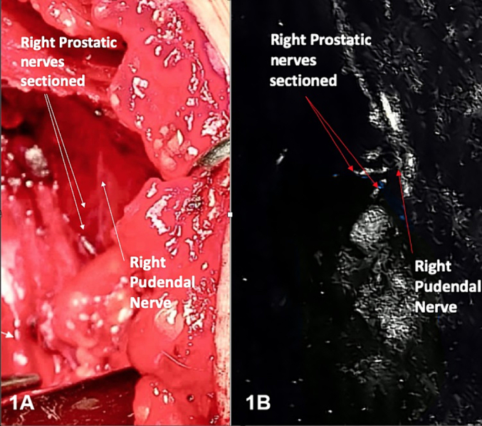
A: Surgical field of the right hemipelvis in standard operating room light after radical prostatectomy.
B: Surgical field in near-ultraviolet light, revealing autofluorescence of the right peri-prosthetic nerve plexus and right pudendal nerve.
The patient's postoperative course was uncomplicated, and he was discharged home following an uneventful recovery. Later follow-up in the surgery clinic within one month of surgery revealed no complaints of erectile or urethral dysfunction, and no other neurological or non-neurological symptoms. Histological evaluation revealed acinar adenocarcinoma (Gleason 8 [4 + 4]), accompanied by intraductal comedo-necrosis and signet-ring cells.
Prior to participating in this study, the patient provided signed informed consent, in compliance with the most recent Declaration of Helsinki.
3. Discussion
Autofluorescence has been documented in a few other critical anatomical structures, most notably the parathyroid glands, which are susceptible to iatrogenic injury during thyroidectomy procedures [9]. In one randomized clinical trial comparing parathyroid visualization during a thyroidectomy using standard operating room (white light) alone versus adding a near-infrared light (NIRL) fluorescence imaging system, toggling from white to NIRL increased the number of glands detected from 2.6 to 3.5 per patient (p < 0.001), revealed at least one gland not seen in white light in 67.1 % of patients, and reduced the rate of post-operative hypocalcaemia requiring treatment ten-fold (p = 0.005).
Nerves, however, do not auto-fluoresce in NIRL. Nor is their visualization enhanced using fluorescent dyes like indocyanine green, the exception being large nerves with highly-vascular perineurium. Initially in vitro [10], then in vivo in Wistar rats [5], and most recently in vivo in human patients intra-operatively [6,7], evidence has mounted that nerves auto-fluoresce when visualized under NUVL using a recently-developed nerve imaging system. In a study involving 65 thyroidectomy patients published in 2024 [7], in which fluorescence intensity was measured using ImageJ software of the recurrent laryngeal nerve (RLN), thyroid gland, and other background tissues, all 81 RLNs sought (16 bilateral resections) were clearly distinguished from both thyroid and all other background tissues, with mean relative fluorescence unit (RFU) scores of 134.3, 33.7, and 14.4 (p < 0.001), respectively. Moreover, under NUVL, 76 % more RLNs and more than twice as many RLN branches were observed with NUVL than with white light, and the average length of nerve visualized was 32 % greater (mean nerve lengths = 2.5 vs. 1.9 cm, respectively), all differences statistically significant at p < 0.001 [7]. Nerves as small as 1 mm in width were visualized under NUVL. And the lowest RFU count recorded for any of the 81 RLNs visualized under NUVL was higher than the highest RFU count recorded for any of the background tissues used for comparison for those 81 RLNs.
Throughout the current patient's radical prostate resection, this same Dendrite camera was pivotal to illuminating the nerves, which fluoresced distinctly against surrounding tissue. This included the many small branches of the cavernous nerve, which is consistent with the 1 mm-width nerves detected during thyroidectomies [7]. Moreover, using this technology permitted the prostatectomy surgical team to perform a meticulous nerve-preserving technique that prevented erectile dysfunction and all other neurological dysfunction in the patient post-operatively. Given that either temporary or permanent erectile dysfunction has been reported in up to 85 % of radical prostatectomy patients, the lack of such a complaint in our patient, though clearly not definitively related to the use of nerve autofluorescence, warrants further examination in additional patients.
4. Conclusion
In our laparotomic radical prostatectomy patient, nerve auto-fluorescence under NUVL clearly enhanced the visualization of nerves typically damaged during this invasive procedure and, as such, might have prevented nerve injury and resultant post-operative erectile dysfunction. Larger case series and comparative clinical trials seem warranted.
Ethical approval
Since this was a single patient receiving standard surgical care, no application was required by the institution's ethics committee.
Funding
This study received no funding from any external source.
Author contribution
RD, KPW, and JH were the main authors of the paper. All authors contributed to study design and edited the manuscript prior to submission.
Guarantor
Dr. Jorge Harraca is the study guarantor.
Research registration number
This is not the first time that Dendrite camera use has been reported for a human patient.
Conflict of interest statement
None of the authors has any conflicts of interest to disclose.
References
- 1.Bill-Axelson A., Holmberg L., Garmo H., Taari K., Busch C., Nordling S., et al. Radical prostatectomy or watchful waiting in prostate cancer - 29-year follow-up. N. Engl. J. Med. 2018;379:2319–2329. doi: 10.1056/NEJMoa1807801. [DOI] [PubMed] [Google Scholar]
- 2.Hamdy F.C., Donovan J.L., Lane J.A., Metcalfe C., Davis M., Turner E.L., et al. Fifteen-year outcomes after monitoring, surgery, or radiotherapy for prostate cancer. N. Engl. J. Med. 2023;388:1547–1558. doi: 10.1056/NEJMoa2214122. [DOI] [PubMed] [Google Scholar]
- 3.Emanu J.C., Avildsen I.K., Nelson C.J. Erectile dysfunction after radical prostatectomy: prevalence, medical treatments, and psychosocial interventions. Curr. Opin. Support. Palliat. Care. 2016;10:102–107. doi: 10.1097/SPC.0000000000000195. [DOI] [PMC free article] [PubMed] [Google Scholar]
- 4.Kendirci M., Hellstrom W.J. Current concepts in the management of erectile dysfunction in men with prostate cancer. Clin. Prostate Cancer. 2004;3:87–92. doi: 10.3816/cgc.2004.n.017. [DOI] [PubMed] [Google Scholar]
- 5.Dip F., Bregoli P., Falco J., White K.P., Rosenthal R.J. Nerve autofluorescence in near-ultraviolet light markedly enhances nerve visualization in vivo. Surg. Endosc. 2022;36:1999–2005. doi: 10.1007/s00464-021-08484-0. [DOI] [PubMed] [Google Scholar]
- 6.Dip F., Rosenthal D., Socolovsky M., Falco J., De la Fuente M., White K.P., et al. Nerve autofluorescence under near-ultraviolet light: cutting-edge technology for intra-operative neural tissue visualization in 17 patients. Surg. Endosc. 2022;36:4079–4089. doi: 10.1007/s00464-021-08729-y. [DOI] [PubMed] [Google Scholar]
- 7.Dip F., Falco J., White K., Rosenthal R. Fluorescence imaging to visualize the recurrent laryngeal nerve during thyroidectomy procedures: analysis of 65 cases and 81 nerves. Surg. Endosc. 2024;38:1406–1413. doi: 10.1007/s00464-023-10627-4. [DOI] [PubMed] [Google Scholar]
- 8.Sohrabi C., Mathew G., Maria N., Kerwan A., Franchi T., Agha R.A. The SCARE 2023 guideline: updating consensus Surgical CAse REport (SCARE) guidelines. Int. J. Surg. 2023;109:1136–1140. doi: 10.1097/JS9.0000000000000373. [DOI] [PMC free article] [PubMed] [Google Scholar]
- 9.Dip F., Falco J., Verna S., Prunello M., Loccisano M., Quadri P., et al. Randomized controlled trial comparing white light with near-infrared autofluorescence for parathyroid gland identification during total thyroidectomy. J. Am. Coll. Surg. 2019;228:744–751. doi: 10.1016/j.jamcollsurg.2018.12.044. [DOI] [PubMed] [Google Scholar]
- 10.Dip F., Aleman R., Socolovsky M., Villalba N., Falco J., Menzo E.L., et al. Nerve spectroscopy: understanding peripheral nerve autofluorescence through photodynamics. Surg. Endosc. 2021;35:7104–7111. doi: 10.1007/s00464-020-08227-7. [DOI] [PubMed] [Google Scholar]