Abstract
Lentiviruses have in their transmembrane glycoprotein (TM) a highly immunogenic structure referred to as the principal immunodominant domain (PID). The PID forms a loop of 5 to 7 amino acids between two conserved cysteines. Previous studies showed that envelope (Env) glycoprotein functions of feline immunodeficiency virus (FIV) could be retained after extensive mutation of the PID loop sequence, in spite of its high conservation. In order to compare Env function in different lentiviruses, either random mutations were introduced in the PID loop sequence of human immunodeficiency virus type 1 (HIV-1) or the entire HIV-1 PID loop was replaced by the corresponding PID loop of FIV or simian immunodeficiency virus (SIV). In the macrophage-tropic HIV-1 ADA Env, mutations impaired the processing of the gp160 Env precursor, thereby abolishing viral infectivity. However, 6 of the 108 random Env mutants that were screened retained the capacity to induce cell membrane fusion. The SIV and FIV sequences and five random mutations were then introduced in the context of T-cell-line-adapted HIV-1 LAI which, although phenotypically distant from HIV-1 ADA, has an identical PID loop sequence. In contrast to the situation for HIV-1 ADA mutants, the cleavage of the Env precursor was unaffected in most HIV-1 LAI mutants. Such mutations, however, resulted in increased shedding of the gp120 surface glycoprotein (SU) from the gp41 TM. The HIV-1 LAI Env mutants showed high fusogenic efficiency. Three Env mutants retained the capacity to mediate virus entry in target cells, although less efficiently than the wild-type Env, and allowed the reconstitution of infectious molecular clones. These results indicated that in HIV-1, like FIV, the conserved PID sequence can be changed without impairing Env function. However, functional constraints on the PID of HIV-1 vary depending on the structural context of Env, presumably in relation to the role of the PID in the interaction of the SU and TM subunits and the stability of the Env complex.
The envelopes (Env) of lentiviruses, despite wide sequence divergence, share certain structural features (14, 15, 30). In particular, the transmembrane glycoprotein (TM) of lentiviruses has a Cys(X)5–7Cys motif which appears to be folded as a loop between two cysteines linked by a disulfide bond (14, 27). Remarkably, this domain elicits a strong antibody response in all lentiviral infections and constitutes the principal immunodominant domain (PID) of the TM of lentiviruses (3, 8, 16, 26, 29, 52). The PID, located in the middle of the TM ectodomain, forms a hinge between two α-helical domains which are thought to be involved in the transition of the Env complex from the native to the fusogenic state (6, 47). The conservation of this structure suggests that it has an essential function in the viral life cycle. On the basis of immunological and computer modeling data, it has been suggested that the PID comes in contact with the C terminus of the surface glycoprotein (SU) and participates in the noncovalent association of the SU and TM subunits of the Env oligomeric complex (22, 39, 40).
In spite of these peculiar structural and immunological characteristics, the functional relevance of the PID remains unclear. The presence of the loop structure appears to be essential, because disruption of the cysteine loop affected Env processing both in human immunodeficiency virus type 1 (HIV-1) and in feline immunodeficiency virus (FIV) (10, 28, 46). Nevertheless, extensive changes, including mutation of four of the six highly conserved residues located between the cysteines, could be introduced in the FIV PID without disrupting Env functions, such as the capacities to induce syncytia and to mediate infection in cell cultures. Conversely, these mutations modified the antigenic properties of the FIV PID (32; unpublished observations).
The functional tolerance of PID changes in FIV was surprising, considering the extreme phylogenetic conservation of this domain. In order to verify whether our observations for FIV held true for other lentiviruses, we wished to analyze PID functions in HIV-1. Indeed, HIV-1 is of special interest in this regard. First, the PID sequence is highly conserved among different HIV-1 isolates despite structural and functional differences in the Env of macrophage-tropic (M-tropic; now called R5) and T-cell-line-tropic (T-tropic; now called X4) isolates (2, 42, 48). Thus, the influence of the PID on Env functions in different contexts may be addressed. Second, antibodies directed against the PID of HIV-1 enhance in vitro infection (5, 13, 34, 35). It may therefore be of interest to modify the immunological properties of the PID to diminish the potential enhancing response for vaccine purposes.
In this study, we performed extensive mutagenesis of the PID of HIV-1 in the context of two different HIV-1 isolates, the M-tropic isolate HIV-1 ADA and the T-cell-line-adapted (TCLA) isolate HIV-1 LAI. The functional consequences of the mutations were analyzed with regard to Env expression and processing, syncytium-forming ability, and infectivity. Although the PID is identical in the isolates studied, we observed that identical mutations have different consequences in the two different Env backgrounds.
MATERIALS AND METHODS
Plasmids.
The env expression vector pSV7d, containing the HIV-1 ADA envelope, and the pNL-Luc-E−R+ vector, containing the env-deleted HIV-1 provirus into which the luc gene has been introduced (9), were kindly provided by T. Dragic and E. Landau (Aaron Diamond AIDS Research Center, New York, N.Y.). The HIV-1 LAI env expression vector (pMA243/105 vector) (41) and the LAI provirus molecular clone (90.1) (1) were gifts from M. Alizon (Institut Cochin de Génétique Moléculaire, Paris, France). The rev expression vector pcREV (23) was provided by B. Shacklett (Aaron Diamond AIDS Research Center).
Cell cultures.
COS, 293T, and HeLa cells were cultured in Dulbecco’s modified Eagle’s medium (DMEM) supplemented with 10% fetal calf serum (FCS). The HeLa P4 cell line, which stably expresses the CD4 receptor and contains an integrated Tat-inducible LacZ gene, and the HeLa P5 cell line, which is derived from HeLa P4 and expresses the chemokine receptor CCR5 (33), were kindly provided by M. Alizon. The MT4 cell line was cultured in RPMI 1640 supplemented with 10% FCS.
Human monocyte-derived macrophages (MDM) were isolated from fresh human peripheral blood mononuclear cells from healthy, HIV-1-seronegative blood donors by the countercurrent centrifugal elutriation technique with a J-6 MC induction-drive centrifuge and a JE-5.0 rotor (Beckman Instruments Inc.). Cell suspensions were more than 97% monocytes, as determined by cell surface staining with anti-CD14 (Medarex Inc., West-Lebanon, N.H.) and anti-CD11b (Immunotech S.A., Marseille, France) monoclonal antibodies. The monocytes were cultured as adherent cell monolayers (106 cells/ml) in the presence of RPMI 1640 containing 10% FCS. The monocytes were allowed to differentiate into macrophages for 7 days (48-well dishes) before being infected.
PID mutagenesis. (i) Random mutagenesis of the PID of ADA Env.
An EagI site was introduced into ADA env (positions 1808 to 1813) contained in plasmid pSV7d-ADA, without changing the encoded amino acids, by use of the ExSite PCR-based site-directed mutagenesis kit (Stratagene) according to the manufacturers’ instructions. The oligonucleotides 5′-GGTGCAGATGAGTTTTCCAGAGCAACCCCAAAT-3′ and 5′-ACGGCCGTGCCTTGGAATGCTAGTTGGAGTAATA-3′ (containing the EagI site and 5′ phosphorylated) were used as mutagenesis primers. The plasmid obtained, pSV7d-ADA-E, was used to clone DNA fragments containing PID mutations (see below) between the AvrII (positions 1767 to 1772) and EagI sites.
We performed random mutagenesis of either the 5 amino acids situated between the two cysteines (mutants designated p, for PID) or the 3 amino acids following the N-terminal cysteine (mutants designated t, for terminal) of the PID loop by use of two oligonucleotides degenerate in either all five codons or the three N-terminal codons of the PID loop (oligonucleotide 1 [positions 1758 to 1821], 5′-TCAACAGCTCCTAGGCATATGGGGTTGCNNNNNNNNNNNNNNNTGCACCACGGCCGTGCCTTGG-3′; oligonucleotide 2 [positions 1758 to 1821], 5′-TCAACAGCTCCTAGGCATATGGGGTTGCNNNNNNNNNCTCATCTGCACCACGGCCGTGCCTTGG-3′).
Both oligonucleotides contained an NdeI site introduced at position 1772 upstream of the PID loop (positions 1773 to 1778), without changing amino acids. Double-stranded DNA inserts were generated from the oligonucleotides by use of an extension primer (5′-CCAAGGCACGGCCGTGGT-3′; positions 1821 to 1804) and Klenow treatment. The inserts were then digested with AvrII and EagI, purified with Gene Clean (Ozyme), and cloned into pSV7d-ADA-E (previously digested with AvrII and EagI and purified to eliminate the AvrII-EagI fragment) [these ADA Env mutants were designated p(n)A or t(n)A].
(ii) Construction of chimeric PIDs of ADA Env.
The AvrII and EagI sites were also used to generate PID mutants in which the sequences between the cysteines were replaced with the corresponding sequences of FIV env and simian immunodeficiency virus (SIV) env (these mutants were designated FivA and SivA). For this purpose, two sets of oligonucleotides (also containing the NdeI site) were hybridized with each other to generate double-stranded DNA fragments that were cloned into pSV7d-ADA-E. The oligonucleotides were 5′-CTAGGCATATGGGGTTGCAATCAAAATCAATTCTTCTGCACCAC-3′ and 5′-GGCCGTGGTGCAGAAGAATTGATTTTGATTGCAACCCCATATGC-3′ for the FIV PID and 5′-CTAGGCATATGGGGTTGCGCGTTTAGACAAGTCTGCACCAC-3′ and 5′-GGCCGTGGTGCAGACTTGTCTAAACGCGCAACCCCATATGC-3′ for the SIV PID. Minipreparations of plasmids (minipreps) were prepared by use of the Wizard Minipreps DNA purification system. Recombinant clones were identified by NdeI digestion and used to transfect COS cells.
(iii) Site-directed mutagenesis of LAI env.
The PID sequences of four fusogenic mutants of ADA Env (p34A, t7A, t24A, and t25A) and of three nonfusogenic mutants (t35A and the chimeric PIDs containing the FIV and SIV PID sequences [see Table 2]) were introduced into LAI env by site-directed mutagenesis by an adaptation of the method described by Kunkel (20). For this purpose, the SmaI-BamHI fragment (positions 6818 to 8068) of pMA243/105 that contains a 1,950-bp fragment of LAI env (positions 6818 to 8068) was introduced into pTZ18 and used as a target for oligonucleotide-directed mutagenesis. The EagI site was introduced into the mutagenesis oligonucleotides (positions 7600 to 7650), without changing the encoded amino acids, in order to enable the selection of mutant clones. The NheI-BamHI fragments (positions 6853 to 8068) containing the mutations were then introduced into expression vector pMA243/105. These LAI env mutants were designated FivL, SivL, t35L, p34L, t7L, t24L, and t25L.
TABLE 2.
Amino acid sequences of mutated PID in ADA Env
| PID source | Sequencea |
|---|---|
| Wild type | CSGKLIC |
| Nonfusogenic mutants | |
| p8A | CGRGQLC |
| p9A | CAYRKGC |
| p36A | CHAQEGC |
| p42A | CN*AGFC |
| p59A | CTYSKLC |
| p81A | CYEAALC |
| t21A | CQDN**C |
| t27A | CPDG**C |
| t35A | CC*I**C |
| FivA | CNQNQFFC |
| SivA | CAFRQVC |
| Fusogenic mutants | |
| p34A | CGDA**C |
| t2A | CHAS**C |
| t7A | CRAM**C |
| t9A | CRKA**C |
| t24A | CRAT**C |
| t25A | C**E**C |
Asterisks represent positions between the cysteines that are identical to those in wild-type PID.
(iv) Sequencing.
The LAI and ADA Env mutants were entirely sequenced to confirm the mutations introduced into the PIDs and to ensure that no other changes were introduced elsewhere during mutagenesis.
Construction of infectious molecular clones.
The LAI Env mutants that were shown to maintain high fusogenicity (p34L, t7L, t24L, and t25L) and the FIV and SIV sequences were introduced into the LAI infectious molecular clone (90.1) by use of SalI and BamHI sites (positions 5367 and 8068, respectively).
Cell transfection.
Transfection was performed by the calcium phosphate precipitation method (36). COS, 293T, or HeLa cells were plated either at 250,000 cells per well in a six-well plate or at 1.2 × 10⁶ cells per 10-cm-diameter petri dish on the day before transfection. Under these conditions, cells were confluent on day 3 after transfection.
Detection and quantitation of Env glycoproteins by an enzyme-linked immunosorbent assay (ELISA).
For the screening procedure and to detect potentially functional ADA Env mutants, samples were prepared as follows. COS cells plated in six-well dishes were transfected with 500 ng of both env expression vectors and the pNL-Luc-E−R+ vector. On the following day, the culture medium was changed; 48 h later, supernatants were harvested, centrifuged for 10 min at 2,000 × g, treated with Empigen BB (0.25% final concentration) (Calbiochem), and centrifuged again for 30 min at 13,000 × g. Cells were washed with cold phosphate-buffered saline (PBS), lysed with 0.5% Empigen BB in 0.5 ml of PBS, and centrifuged for 10 min at 1,500 × g and for 1 h at 13,000 × g. Sample dilutions were made in DMEM–10% FCS, maintaining the final concentration of Empigen BB at 0.25%.
To measure gp120 released in the supernatants of transfected cells, samples were prepared as follows. HeLa P5 cells plated in six-well dishes were transfected in triplicate with 2 μg of env expression vectors (containing the tat and rev genes) per well for the LAI mutants or both env expression vectors and the rev expression vector pcREV (2 μg of each per well) for the ADA mutants. The samples were then prepared as described above.
To measure virion-associated Env glycoproteins, supernatants from 293T cells transfected with wild-type or mutant HIV-1 LAI proviruses were centrifuged for 10 min at 2,000 × g and filtered through 0.45-μm-pore-size membranes. Virions were pelleted by centrifugation through a 20% sucrose cushion for 35 min at 45,000 rpm (Kontron TST60.4 rotor). Pellets were dissolved in DMEM–10% FCS containing 0.5% Empigen BB.
To quantify HIV-1 Env glycoproteins, we used a sandwich ELISA (24). Two affinity-purified sheep antibodies, anti–HIV-1 gp41 (D7323) and anti–HIV-1 gp120 (D7324) (Aalto Bio Reagents Ltd., Dublin, Ireland), were used as capture antibodies. These antibodies are raised against conserved peptides within gp41 and gp120 (peptides IPRRIRQGLERILL and APTKAKRRVVQREKR, respectively). The capture antibodies were used to coat 96-microwell plates (Nunc F-16 Maxisorp) overnight at room temperature at a final concentration of 5 μg/ml in 100 μl of 100 mM NaHCO3 (pH 9.6) per well. The wells were blocked for 4 h with a solution of 2% nonfat milk powder in PBS. Aliquots (100 μl) of diluted samples were added to duplicate wells and incubated overnight at 4°C. Supernatants or lysates from mock-transfected cultures were used as controls. After five washes with PBS containing 0.5% Tween 20 (washing buffer), 100 μl of pooled sera obtained from HIV-1-seropositive patients was added per well at a 1/500 dilution in PBS containing 4% nonfat milk and 0.5% Tween 20 and incubated at room temperature for 1 h. After five washes, peroxidase-labelled goat anti-human immunoglobulin (Calbiochem), diluted in PBS containing 4% nonfat milk and 0.5% Tween 20, was added at 100 μl per well and incubated for 1 h. After five additional washes, the reaction was visualized with 2,2′-azinobis(3-ethylbenzthiazoline)-6-sulfonic acid (ABTS) (at 0.4 mg/ml) and read at 405 nm after 30 min. Quantification of gp120 in samples was performed with, as calibration standards, either recombinant HIV-1 gp120 (HIV-1 strain GB08; kindly provided by M. Girard, Institut Pasteur, Paris, France) or a supernatant that was obtained from HeLa243env cell cultures that stably expressed HIV-1 LAI Env (kindly provided by M. Alizon) (41) and that was previously calibrated. Calibration standards were included in each microplate. The threshold of detection of the ELISA was 6 ng of gp120 per ml. For all cell lysate samples, the protein concentrations were determined with a Micro BCA protein assay reagent kit (Pierce).
Fusion assays.
The fusion assay for the ADA mutants consisted of cotransfecting in triplicate the mutated env expression vectors and pNL-Luc-E−R+, which expresses in trans the Rev and Tat proteins, into the 293T cell line (2 μg of each plasmid per well) and coculturing the transfected cells with an equal number (250,000 cells per well) of HeLa P5 cells. Env-induced syncytia were detected by an in situ β-galactosidase assay as described previously (11). For the LAI mutants, 293T cells were transfected with 3 μg of DNA from env expression vectors derived from pMA243/105, which contains the tat and rev genes, per well. Cells were then cocultured with the HeLa P4 or P5 cell line, and syncytia were visualized as described above.
RIPA.
Either 293T cells or HeLa P4 or P5 cells plated on 10-cm petri dishes were transfected with 10 μg of env expression vector. In the case of the ADA constructions, 5 μg of pcREV was cotransfected. At 36 h after transfection, cells were harvested in methionine-cysteine-deprived medium (ICN) for 1 h and then labelled overnight with 100 μCi of Pro-mix l-35S-labelling mix (Amersham) per ml. The supernatants (4 ml/dish) were centrifuged for 10 min at 1,500 × g, mixed with 400 μl of 10× radioimmunoprecipitation assay (RIPA) buffer (RB) (50 mM Tris-HCl [pH 7.5], 150 mM NaCl, 5 mM KCl, 1% Triton X-100, 0.5% sodium deoxycholate, 1 mM Pefabloc [Boehringer Mannheim Biochemicals], 5 μg of aprotinin [Boehringer] per ml, 5 μg of leupeptin [Boehringer] per ml), and centrifuged again at 3,000 × g for 1 h. Cells were lysed in RB, and nuclei and cell fragments were eliminated by centrifugation (10 min at 1,200 × g and 1 h at 17,000 × g). Cell lysates and supernatants were then incubated overnight with 50 μl of protein G-agarose suspension in RB. After centrifugation, all samples were incubated for 3 h with D7323 and D7324 antibodies, both at a final concentration of 10 μg/ml, and overnight with 50 μl of protein G-agarose. After centrifugation, pellets were washed twice with RB, twice with a high-salt buffer (50 mM Tris-HCl [pH 7.5], 500 mM NaCl, 0.1% Triton X-100, 0.05% sodium deoxycholate), and once with a low-salt buffer (50 mM Tris-HCl [pH 7.5], 0.1% Triton X-100, 0.05% sodium deoxycholate). Immunoprecipitates were eluted by boiling protein G beads in 50 μl of Laemmli buffer (18). All steps before elution were performed at 4°C. Immunoprecipitates were analyzed by sodium dodecyl sulfate-polyacrylamide gel electrophoresis on a 10% gel. For quantification of Env glycoproteins, gels were dried and exposed on a Molecular Dynamics Storage Fluor screen. Band radioactivity was measured with a PhosphorImager SI and ImageQuant software.
Virus entry complementation assay.
293T cells were plated at 1.2 × 10⁶ cells per 10-cm-diameter petri dish on the day before transfection. Wild-type and mutant env expression vectors were cotransfected with pNL-Luc-E−R+ (7.5 μg of each) to generate Env-pseudotyped virions. Supernatants were collected after 48 h, and p24gag levels were measured with a commercial ELISA kit (Innogenetics, Zwijndrecht, Belgium). Supernatants of cells transfected only with pNL-Luc-E−R+ were used as controls. Target cells plated in either 24-well dishes for HeLa P4 or HeLa P5 cells (105 cells per well) and MT4 cells (106 cells per well) or 48-well dishes for MDM were incubated overnight in triplicate with 1 ml of viral supernatant containing 100 ng of p24. Cells were lysed for either 2 days (HeLa P4, HeLa P5, and MT4) or 3 days (MDM) after infection with 100 μl of luciferase lysis buffer (Promega Corp., Madison, Wis.). The amount of luciferase activity in 20 μl of lysate was measured by use of commercially available reagents (Promega) in a luminometer.
Infectivity assay.
Viral supernatants were obtained by transfecting 293T cells (plated as indicated above) with 15 μg of DNA from LAI wild-type and mutant proviruses. Medium was replaced at 6 and 24 h after transfection. Supernatants were collected 24 h later, and the concentration of p24 was measured by an ELISA. Cells (MT4 cells plated at 2 × 10⁵ cells per well in 96-well dishes or HeLa P4 cells plated at 2 × 10⁵ cells per well in 6-well dishes) were infected for 3 h with viral supernatant containing 100 ng of p24 per ml and then washed with PBS to eliminate the input p24. HeLa P4 cells were split every 3 days. Kinetics of infection were monitored in triplicate by measurement of p24 contained in the culture supernatants.
RESULTS
Selection of random mutants of the ADA Env PID.
In order to select recombinant clones producing Env and thus to discard nonfunctional constructions (i.e., containing stop codons or frameshifts), clones derived from random mutagenesis were screened by detection of Env products in cell lysates and supernatants of COS cells after transfection with the DNA recovered from minipreps corresponding to single colonies. To achieve Env expression, Rev was produced in trans by cotransfecting pNL-Luc-E−R+. A total of 108 clones derived from mutagenesis of the env expression vector pSV7d-ADA were analyzed. Seventy-two clones were obtained by random mutagenesis of all five codons of the cysteine loop of the PID, and 36 clones were obtained by random mutagenesis of the three N-terminal codons of the cysteine loop. At 72 h after transfection, the presence of Env glycoproteins in cell extracts and supernatants was tested by an ELISA with both anti-SU (D7324) and anti-TM (D7323) antibodies. Env products were detected in cell lysates from most transfections (86 of 108). For 24 clones, Env reactivity was also found in supernatants, suggesting Env processing and gp120 production. The 32 clones (19 p clones and 13 t clones) showing the highest Env production in cell lysates were selected for further analysis.
Capacity of HIV-1 ADA Env mutants for fusion and complementation of virus entry.
The two chimeric ADA Env mutants in which the sequence of the HIV-1 PID loop was replaced by the corresponding sequences of FIV and SIV (FivA and SivA) and the 32 clones selected following random mutagenesis were analyzed by a syncytial assay for their ability to induce cell membrane fusion. Env mutants were cotransfected with the pNL-Luc-E−R+ vector into 293T cells. HeLa P5 target cells were then added, permitting the detection of fusion events by in situ β-galactosidase staining. Six mutant clones were capable of inducing cell fusion, although with less efficiency than the wild-type envelope. Table 1 shows the results of a representative syncytium-forming assay. The 28 other mutant clones, including FivA and SivA, did not induce cell fusion. The sequences of the fusogenic mutants are shown in Table 2; for comparison, the sequences of the PIDs of some nonfusogenic clones are also presented. All of the fusogenic clones contained mutations in the three N-terminal residues, while none bore mutations in the two C-terminal residues, although mutations in these positions were present in several clones tested (Table 2). No other mutation outside the PID was found by sequencing of the Env mutants.
TABLE 1.
Syncytium formation induced by ADA Env mutantsa
| Clone | No. of syncytia | No. of nuclei/syncytium (range) |
|---|---|---|
| Wild type | 5,833 ± 465 | 2–30 |
| FivA | 0 | |
| SivA | 0 | |
| t35A | 0 | |
| p34A | 809 ± 322 | 2–5 |
| t7A | 912 ± 173 | 2–5 |
| t24A | 1,859 ± 553 | 2–5 |
| t25A | 2,207 ± 323 | 2–5 |
293T cells plated in six-well plates were transfected in triplicate with ADA wild-type Env or Env mutants. After coculturing with HeLa P5 cells (CD4+ CCR5+), fusion events were detected by β-galactosidase staining. Syncytia were counted independently by two investigators using a grid (486 squares/well). Mean ± standard deviation numbers of syncytia in three wells are shown. In three wells with mock-transfected cells, there were fewer than 10 syncytia. The syncytium-forming capacities of the t2A and t9A mutants were analyzed in separate experiments. The percentages of syncytia were approximately 10 and 3% (for t2A and t9A, respectively) that found with wild-type Env.
To determine whether the ADA Env mutants were capable of mediating cell entry, HIV-1 particles containing a luciferase reporter gene were pseudotyped with the FivA and SivA Env mutants and the 32 Env mutants selected after random mutagenesis. Pseudotyped virions were tested in single-cycle infectivity assays. HeLa P5 target cells and MDM (owing to the tropism of the ADA envelope for macrophages) were used as target cells in these assays. Wild-type Env permitted the reconstitution of infectious virus, as revealed by luciferase activity measured in infected cells. In a representative assay, 177,250 ± 48,000 relative light units (RLU)/min versus 517 RLU/min of background and 185,000 ± 50,000 RLU/min versus 520 RLU/min of background were obtained in HeLa P5 and MDM target cells, respectively; in contrast, none of the viruses pseudotyped with the Env mutants produced luciferase activity over the background level in either HeLa P5 or MDM cells.
Capacity of HIV-1 LAI Env mutants for fusion and complementation of virus entry.
Some of the mutations analyzed in the context of HIV-1 ADA Env were introduced into Env of the TCLA isolate HIV-1 LAI in order to determine whether the effect of PID changes was the same in a different Env background. The selected PID mutations represented four (of six) fusogenic (p34A, t7A, t24A, and t25A) and three nonfusogenic (t35A, FivA, and SivA) ADA Env mutants (Table 2).
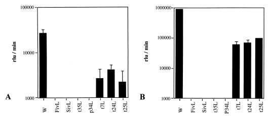
The fusogenic properties of the HIV-1 LAI Env mutants were analyzed after transfection of 293T cells and coculturing with the HeLa P5 cell line. The number of syncytia and the number of nuclei in the syncytia are presented in Table 3. In contrast to their ADA counterparts, four LAI Env mutants (p34L, t7L, t24L, and t25L) showed a syncytium-forming capacity similar to that of the wild-type LAI Env. Three other LAI Env mutants (t35L, FivL, and SivL), containing mutations which abolished fusogenic capacity in the context of ADA Env, were able to form syncytia. Similar results were obtained when HeLa P4 cells were used as target cells (data not shown). The capacity of the HIV-1 LAI Env mutants to mediate the transduction of pseudotyped viral particles was assessed with either HeLa P4 or MT4 cell lines. As Fig. 1 shows, luciferase activity was detected in both target cell lines with mutants t7L, t24L, and t25L but not with mutants p34L, t35L, FivL, and SivL. However, the signal obtained with viral particles pseudotyped with the Env mutants was reduced by approximately 1 log unit in both MT4 (Fig. 1A) and HeLa P4 (Fig. 1B) cells compared to the signal obtained with wild-type Env.
TABLE 3.
Syncytium formation induced by LAI Env mutantsa
| Clone | No. of syncytia | No. of nuclei/syncytium (range) |
|---|---|---|
| Wild type | 20,648 ± 1,227 | 2–>30 |
| FivL | 4,949 ± 937 | 2–5 |
| SivL | 11,677 ± 3,490 | 2–5 |
| t35L | 8,429 ± 1,071 | 2–5 |
| p34L | 18,405 ± 2,555 | 2–>30 |
| t7L | 20,648 ± 2,237 | 2–>30 |
| t24L | 22,349 ± 2,806 | 2–>30 |
| t25L | 18,560 ± 1,448 | 2–>30 |
293T cells plated in six-well plates were transfected in triplicate with LAI wild-type Env or Env mutants. After coculturing with HeLa P5 cells (CD4+ CCR5+), fusion events were detected by β-galactosidase staining. Syncytia were counted independently by two investigators using a grid (486 squares/well). Mean ± standard deviation numbers of syncytia in three wells are shown. In three wells with mock-transfected cells, there were fewer than 10 syncytia.
FIG. 1.
Ability of HIV-1 LAI Env mutants to complement virus entry. Virus entry in MT4 cells (A) or HeLa P4 (CD4+) cells (B) was measured by quantification of luciferase activity. Cells were inoculated in triplicate with luciferase reporter viruses pseudotyped with HIV-1 LAI wild-type (W) or mutant Env. The inoculum consisted of supernatants obtained after cotransfection of 293T cells with wild-type and mutant env expression vectors and the env-defective pNL-Luc-E−R+ vector (100 ng of p24 per infection). The background was measured by inoculating cells with supernatants obtained after transfection of 293T cells with the pNL-Luc-E−R+ vector alone. The values indicated were those obtained after background subtraction. rlu, relative light units.
Infectivity of LAI mutant viruses.
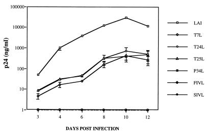
To analyze the infectious properties of the LAI Env mutants in greater detail, the p34L, t7L, t24L, t25L, FivL, and SivL mutations were introduced into the HIV-1 LAI infectious molecular clone, yielding the P34L, T7L, T24L, T25L, FIVL, and SIVL mutant viruses. The kinetics of replication of the mutant viruses were monitored with the MT4 cell line. Supernatants containing equal amounts of p24 were used for infections. As shown in Fig. 2, the T7L, T24L, and T25L mutant viruses were able to establish a productive infection. The level of replication of the infectious mutant viruses, as evaluated by measurement of p24 in supernatants, appeared to be inferior by approximately 1 log unit to that of the wild-type virus. This difference was similar to that observed in entry assays. This decrease in the infectivity of the mutant viruses in relation to the wild-type virus was confirmed when the experiment was performed with HeLa P4 cells (data not shown).
FIG. 2.
Infectivity of LAI mutant viruses. MT4 cells were infected in triplicate with viral stocks obtained by transfection of 293T cells with wild-type or mutant HIV-1 LAI molecular clones and normalized for p24 content (100 ng of p24 per infection). The kinetics of infection were measured by p24 monitoring in infected MT4 supernatants on the indicated days.
Influence of PID mutations on Env processing and stability.
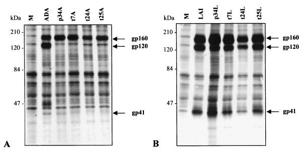
To analyze the expression and processing of the mutant Env glycoproteins, 293T cells were transfected either with HIV-1 ADA wild-type Env and four mutants (p34A, t7A, t24A, and t25A) or with HIV-1 LAI wild-type Env and the four corresponding mutants (p34L, t7L, t24L, and t25L). After 48 h, Env products were analyzed by a RIPA with two sheep antibodies, anti–HIV-1 gp120 (D7324) and anti–HIV-1 gp41 (D7323). Figure 3A shows the results of a RIPA performed on lysates of cells transfected with ADA Env mutants. Although the mutant Env precursors (Pr) were produced at levels comparable to those of wild-type Env, SU gp120 was not detectable for any of the mutants. The impaired processing of the ADA mutants contrasted with the efficient cleavage of the Pr into SU and TM subunits that was observed for the four LAI mutants (Fig. 3B).
FIG. 3.
RIPA analysis of Env glycoproteins expressed upon transfection of 293T cells with env expression vectors containing mutated or wild-type env genes. Cells were 35S labelled overnight, and Env glycoproteins were immunoprecipitated with a mixture of anti-gp120 and anti-gp41 sheep antibodies. The names of the Env mutants are indicated above the lanes. M, mock transfection. Molecular mass standards are indicated on the left. (A) HIV-1 ADA Env mutants. The weak gp41 band detected for wild-type ADA Env and the p34A mutant was also visible for the t7A, t24A, and t25A mutants after overexposure of the film (data not shown). (B) HIV-1 LAI Env mutants.
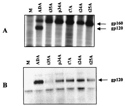
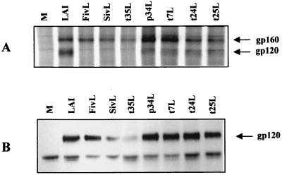
Not only gp120 but also Env Pr were precipitated from the supernatants of 293T cells transfected with either ADA or LAI wild-type or Env mutants (data not shown), suggesting that these Env products were partly released from dead cells, thus perturbing measurement of the shedding of gp120 in supernatants. Indeed, some mortality was noted in 293T cell cultures after transfection with HIV-1 env and was possibly related to toxic effects due to a very high level of expression of Env. To analyze this point and to verify that the defect in the processing of ADA Env mutants was not dependent upon a particular cell line, the HeLa P5 cell line was used in a new set of RIPA experiments. Transfection of these cells with HIV-1 ADA or LAI wild-type or Env mutants, in spite of syncytium formation, did not cause the toxicity observed for 293T cells and the consequent release of intracellular Env products. Indeed, an ELISA performed with the anti-gp41 antibody (D7323) as a capture antibody did not reveal any trace of Pr or TM gp41 products in the supernatants of HeLa P5 cells after transfection (Table 4). Under these conditions, while gp120 was not visible in cell lysates from cells transfected with ADA Env mutants (Fig. 4A), gp120 was detected in supernatants for four of five mutants, although the signals were weaker than that of ADA wild-type Env (Fig. 4B). An ELISA performed with the anti-gp120 antibody (D7324) confirmed the RIPA results (data not shown). These findings confirmed that the cleavage of ADA Env mutant Pr occurs to a lower extent than that of wild-type Pr and indicated that the gp120 produced is released into supernatants. However, the defect of Pr cleavage in ADA mutants obscured evaluation of the shedding of gp120 in relation to the total amount of gp120 produced. Thus, the analysis of the effects of the PID mutations on SU-TM association and gp120 shedding was continued with LAI Env mutants.
TABLE 4.
Env processing and stability in LAI Env mutants
| Clone | Processing indexa | Association indexa | Env glycoprotein concn (ng/ml) inb:
|
|
|---|---|---|---|---|
| Lysates | Supernatants | |||
| Wild type | 1.00 | 1.00 | 953 ± 92 | 61 ± 5 |
| FivL | 0.44 | 0.01 | ND | ND |
| SivL | 0.26 | 0.16 | ND | ND |
| t35L | 0.66 | 0.05 | ND | ND |
| p34L | 0.75 | 0.08 | 1,220 ± 34 | 280 ± 52 |
| t7L | 0.88 | 0.07 | 1,220 ± 51 | 186 ± 46 |
| t24L | 1.35 | 0.13 | 1,000 ± 69 | 188 ± 2 |
| t25L | 1.06 | 0.19 | 903 ± 106 | 336 ± 5 |
The amounts of Env glycoproteins immunoprecipitated after transfection of HeLa P5 cells were evaluated by PhosphorImager analysis of sodium dodecyl sulfate-polyacrylamide gels. Background radioactivity was subtracted from specific band radioactivity. The processing index (4) was calculated as follows: [(total gp120)mutant × (gp160)wild type]/[(gp160)mutant × (total gp120)wild type]. The association index (4), calculated as [(mutant gp120)cell × (wild-type gp120)supernatant]/[(mutant gp120)supernatant × (wild-type gp120)cell], reflects the stability of the SU-TM association relative to that of wild-type glycoproteins.
To measure the release of SU gp120 from HIV-1 LAI Env mutants, HeLa P5 cells plated in six-well plates were transfected in triplicate with wild-type or mutant env expression vectors, and Env glycoproteins were quantified in cell lysates and in supernatants by an ELISA with anti–HIV-1 gp120 D7324 as the capture antibody. ELISA values were normalized in relation to the protein content of the cell lysates. The anti-gp120 reactivity in cell lysates reflects both gp160 and gp120 reactivities; in culture supernatants, it reflects only the presence of gp120. This finding was confirmed by an ELISA with anti–HIV-1 gp41 D7323: no reactivity was detected in the supernatants, indicating that no detectable gp41 or gp160 was present. ND, not done.
FIG. 4.
RIPA analysis of wild-type or mutant HIV-1 ADA Env glycoproteins expressed upon transfection of HeLa P5 cells. M, mock transfection. RIPA was performed on cell lysates (A) or supernatants (B). Note the presence of a band unrelated to the HIV-1 Env glycoproteins in the supernatants of all transfections, including the mock transfection.
A RIPA analysis after transfection of HeLa P5 cells with LAI Env mutants (Fig. 5) revealed that, in relation to the respective Env Pr, the gp120 signals in cell lysates of all the Env mutants were weaker than that of wild-type Env (Fig. 5A). In particular, cell-associated gp120 of the FivL, SivL, and t35L mutants was not detected in this experiment. This reduction of cell-associated gp120 for LAI Env mutants was confirmed by a RIPA analysis performed after transfection of HeLa P4 cells (data not shown) but was not evident in transfections of 293T cells (Fig. 3B), presumably owing to the very high level of production of Env glycoproteins in 293T cells. The level of gp120 shed into supernatants also appeared to be higher for LAI Env mutants than for wild-type Env (Fig. 5B).
FIG. 5.
RIPA analysis of Env glycoproteins from cell lysates (A) or supernatants (B) of HeLa P5 cells transfected with env expression vectors containing mutant or wild-type HIV-1 LAI env genes. M, mock transfection. The lower band present in the supernatants of all transfections, including the mock transfection, is unrelated to the HIV-1 Env glycoproteins.
The gp160 Pr processing and the SU-TM association of the Env mutants were evaluated by PhosphorImager quantification of the Env glycoproteins immunoprecipitated from cell lysates and supernatants. Table 4 shows the processing and association indices for LAI Env mutants in comparison with those of wild-type Env. While gp160 Pr processing was not affected for the p34L, t7L, t24L, and t25L mutants and was only partially reduced for the FivL, SivL, and t35L mutants, the cell-associated gp120 levels were strongly reduced for all the Env mutants, as indicated by low association indices (Table 4). The increased shedding of gp120 by the four LAI Env mutants which exhibited normal gp160 Pr processing was also confirmed by a quantitative ELISA. After transfection of HeLa P5 cells, the amount of gp120 in supernatants was determined. The quantity of gp120 relative to cell-associated Env glycoproteins in supernatants was larger in Env mutant transfections (Table 4).
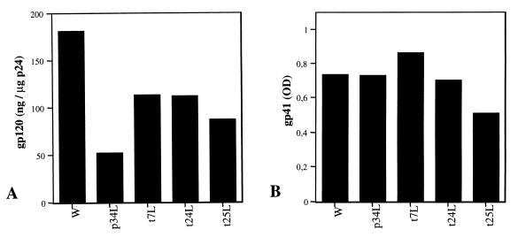
Levels of virion-associated Env glycoproteins in viral pellets of HIV-1 LAI or mutant proviruses were evaluated by an ELISA. gp120 levels were reduced in mutant viruses in relation to the wild-type virus (Fig. 6A). In particular, a strong reduction was observed for the p34L mutant. Conversely, such differences were not observed for the levels of virion-associated gp41, as evaluated by reactivity with the anti-gp41 antibody (Fig. 6B). These results suggest that HIV-1 LAI and mutant viruses produced by 293T cells incorporate similar levels of Env glycoproteins but that gp120 shedding from the virion surface is increased in mutant viruses in relation to the wild-type virus.
FIG. 6.
ELISA analysis of Env glycoproteins in viral pellets of HIV-1 LAI wild-type (W) or mutant viruses from supernatants of transfected 293T cells. Either anti-gp120 D7324 (A) or anti-gp41 D7323 (B) was used as the capture antibody. Tests were run in duplicate; the mean values are shown. p24 in viral pellets was measured with a commercial ELISA kit. Three independent experiments were performed and yielded similar results; a representative assay is shown. (A) The amount of gp120 in viral pellets was quantified with recombinant gp120 as a calibration standard. Results are expressed as nanograms of gp120 per microgram of p24. (B) Anti-gp41 reactivity was measured in a sample dilution (0.7 μg of p24 per ml) which produced optical density (OD) values in the dynamic range of the curve.
DISCUSSION
We have previously shown that the PID loop of FIV, despite its conservation in natural isolates, can be modified without impairing Env function and viral infectivity (32). In this study, we have examined the effect of mutations in the PID loop on the functional properties of HIV-1 Env in an attempt to define the structural constraints that could account for the high level of conservation of this domain in lentiviruses. PID mutations were introduced in the Env from an M-tropic HIV-1 isolate and a TCLA isolate of HIV-1 to analyze the role of the PID in different structural backgrounds. Our results revealed that, although HIV-1 Env can tolerate extensive changes in the PID, functional permissivity varies between different isolates.
Changes in the PID in the context of ADA Env resulted in a loss of the capacity to mediate virus entry into target cells. Some mutants, however, partially retained fusogenic ability. When introduced into HIV-1 LAI, identical mutations neither impaired syncytium-forming ability nor abolished the capacity of the virus to infect cells. Indeed, infectious LAI mutant viruses containing up to three nonconservative changes in the sequence contained between the cysteines were obtained. Moreover, Env chimeras with unrelated sequences from the PIDs of other lentiviruses (FIV and SIV) exhibited fusogenic capacity.
Biochemical analysis of the expression and maturation of the Env mutants revealed that PID mutations affected the intracellular processing of HIV-1 ADA Env by reducing the cleavage of gp160 Env Pr, while the same mutations had no appreciable effect on HIV-1 LAI Env Pr cleavage. These data strongly suggest that the defects in syncytium-forming ability and infectivity observed in ADA mutants are a direct consequence of the decreased efficiency of Env Pr processing.
Measurements of the release of SU gp120 in cell supernatants and of the association index for the LAI Env mutants revealed that PID mutations caused destabilization of the Env complex and a consequent increase in SU shedding. An increase in gp120 shedding was also indicated by the reduction of gp120 associated with mutant virions. This finding may be the major cause of the diminished infectivity of the LAI mutants. Accordingly, the p34L mutant, exhibiting the lowest level of virion-associated gp120 (Fig. 6A), was not infectious.
These results support the hypothesis that the PID loop is involved in the association between the SU and TM subunits (40). Even a single change in the PID, from a basic to an acidic residue (K to E in the t25L mutant), was sufficient to cause an augmentation of SU shedding. Other mutations in TM outside the PID loop have also been shown to affect the SU-TM association (4, 7), suggesting either that several points of contact exist between the two glycoproteins or that these mutations cause structural modifications which reduce the fitness of the subunit interaction.
If the reduction in the SU-TM association upon mutation of the PID is expected considering the putative position of the PID at the interface of the two subunits, it is surprising that the same mutations have such different effects on the processing of gp160 of ADA and LAI. Compared to that of M-tropic isolates, including HIV-1 ADA, the SU of TCLA isolates is more prone to be shed from the TM (38, 39, 43, 44, 50). The intracellular conformation of Pr may reflect properties of the spatial structure assumed later by the SU and TM subunits at the cell and virion surfaces. Like the CD4 binding properties of the SU, which are already present in Env Pr (12), the instability of the SU-TM interaction in the mature Env complex of TCLA isolates might already be determined in the Pr structure. For LAI Env, the early steps of folding could leave a loose contact between the N- and C-terminal portions of Env Pr, destined to become the SU and TM subunits, thus permitting modification of the PID without alteration of its transport and cleavage. In contrast, in the ADA context, the PID would be involved in a closer interaction with the N-terminal half of the Pr such that mutation of the PID would diminish the probability of correct folding of the Pr. Increased retention of misfolded Pr molecules in the rough endoplasmic reticulum would follow, thus preventing the endoproteolytic cleavage in SU and TM which occurs in the Golgi complex (45, 49).
Adaptation of HIV-1 to replicate in T-cell lines, as well as changes in tropism, has been related to amino acid substitutions in the SU (17, 19, 42, 48, 51). Such mutations mainly affect gp120 binding and interaction with cell membrane receptors. Our results suggest that adaptation to T-cell cultures is also concurrent with a decrease in structural constraints on the PID. SU-TM interactions are critical in the postbinding rearrangements of the Env that initiate the fusion process. Indeed, an early step in the conformational changes triggered by Env binding to soluble CD4 in TCLA isolates involves the dissociation of the SU from the TM, with exposure of cryptic epitopes of the TM, including epitopes located in the PID (25, 37). Weaker structural constraints on the PID of HIV-1 LAI than on the PID of HIV-1 ADA could be related to diversity in the nature of the conformational changes occurring during entry in different cell types. While further studies with additional isolates of HIV-1 are needed to address this hypothesis, the HIV-1 LAI PID mutants that showed a reduced SU-TM association without a disruption of fusion capacity and infectivity will be useful for analyzing the relationship between Env stability and conformational changes in greater detail.
We have previously proposed that, given the observed tolerance for mutations in the PID, the conservation of its sequence might be due in part to selective pressure for the conservation of epitopes eliciting enhancing antibodies (32). Several anti-PID antibodies obtained from HIV-1-infected patients enhance cell culture infection by either T- or M-tropic viruses (5, 13, 34, 35), and anti-PID reactivity has been correlated with the probability of mother-to-child transmission of HIV-1 infection (21, 31). In this study, we did not analyze the immunological properties of the mutated PID. Should mutations modify the immunogenicity of the PID of HIV-1, as was the case for FIV mutants of PID (32), the use as immunogens of HIV-1 Env mutants with mutations in the PID, as presented here, to avoid the induction of potentially enhancing anti-PID antibodies while maintaining a functional conformation able to induce neutralizing antibodies could be advantageous for AIDS vaccine development.
ACKNOWLEDGMENTS
We thank E. Gomas and F. Letourneur for performing DNA sequencing. We are grateful to T. Dragic, E. Landau, M. Alizon, and B. Shacklett for providing HIV-1 vectors and cells and to M. Girard and the Agence Nationale de Recherche sur le SIDA for rgp120. We are indebted to M. Alizon and J. Richardson for helpful discussion and critical reading of the manuscript and to D. Strosberg for continuous support.
This work was supported by the Agence Nationale de Recherche sur le SIDA and a Biomed 2 grant from the European Economic Community. R.M. was supported by grants from la Fondation pour la Recherche Médicale/SIDAction-Ensemble Contre le SIDA, Paris, France.
REFERENCES
- 1.Adachi A, Gendelman H E, Koenig S, Folks T, Willey R, Rabson A, Martin M A. Production of acquired immunodeficiency syndrome-associated retrovirus in human and nonhuman cells transfected with an infectious molecular clone. J Virol. 1986;59:284–291. doi: 10.1128/jvi.59.2.284-291.1986. [DOI] [PMC free article] [PubMed] [Google Scholar]
- 2.Berger E A, Doms R W, Fenyö E-M, Korber B T M, Littman D R, Moore J P, Sattentau Q J, Schuitemaker H, Sodroski J, Weiss R A. A new classification for HIV-1. Nature. 1998;391:240. doi: 10.1038/34571. [DOI] [PubMed] [Google Scholar]
- 3.Bertoni G, Zahno M-L, Zanoni R, Vogt H-R, Peterhans E, Ruff G, Cheevers W P, Sonigo P, Pancino G. Antibody reactivity to the immunodominant epitopes of the caprine arthritis-encephalitis virus gp38 transmembrane glycoprotein associates with the development of arthritis. J Virol. 1994;68:7139–7147. doi: 10.1128/jvi.68.11.7139-7147.1994. [DOI] [PMC free article] [PubMed] [Google Scholar]
- 4.Cao J, Bergeron L, Helseth E, Thali M, Repke H, Sodroski J. Effects of amino acid changes in the extracellular domain of the human immunodeficiency virus type 1 gp41 envelope glycoprotein. J Virol. 1993;67:2747–2755. doi: 10.1128/jvi.67.5.2747-2755.1993. [DOI] [PMC free article] [PubMed] [Google Scholar]
- 5.Cavacini L A, Emes C L, Wisnewski A V, Power J, Lewis G, Montefiori D, Posner M R. Functional and molecular characterization of human monoclonal antibody reactive with the immunodominant region of HIV type 1 glycoprotein 41. AIDS Res Hum Retroviruses. 1998;14:1271–1280. doi: 10.1089/aid.1998.14.1271. [DOI] [PubMed] [Google Scholar]
- 6.Chan D C, Fass D, Berger J M, Kim P S. Core structure of gp41 from the HIV envelope glycoprotein. Cell. 1997;89:263–273. doi: 10.1016/s0092-8674(00)80205-6. [DOI] [PubMed] [Google Scholar]
- 7.Chen S S-L. Functional role of the zipper motif region of human immunodeficiency virus type 1 transmembrane protein gp41. J Virol. 1994;68:2002–2010. doi: 10.1128/jvi.68.3.2002-2010.1994. [DOI] [PMC free article] [PubMed] [Google Scholar]
- 8.Chong Y-H, Ball J M, Issel C J, Montelaro R C, Rushlow K E. Analysis of equine humoral responses to the transmembrane envelope glycoprotein (gp45) of equine infectious anemia virus. J Virol. 1991;65:1013–1018. doi: 10.1128/jvi.65.2.1013-1018.1991. [DOI] [PMC free article] [PubMed] [Google Scholar]
- 9.Connor R I, Benjamin K C, Choe S, Landau N R. Vpr is required for efficient replication of human immunodeficiency virus type-1 in mononuclear phagocytes. Virology. 1995;206:935–944. doi: 10.1006/viro.1995.1016. [DOI] [PubMed] [Google Scholar]
- 10.Dedera D, Gu R, Ratner L. Conserved cysteine residues in the human immunodeficiency virus type 1 transmembrane envelope protein are essential for precursor envelope cleavage. J Virol. 1992;66:1207–1209. doi: 10.1128/jvi.66.2.1207-1209.1992. [DOI] [PMC free article] [PubMed] [Google Scholar]
- 11.Dragic T, Alizon M. Different requirements for membrane fusion mediated by the envelopes of human immunodeficiency virus types 1 and 2. J Virol. 1993;67:2355–2359. doi: 10.1128/jvi.67.4.2355-2359.1993. [DOI] [PMC free article] [PubMed] [Google Scholar]
- 12.Earl P L, Moss B, Doms R W. Folding, interaction with GRP78-BiP, assembly, and transport of the human immunodeficiency virus type 1 envelope proteins. J Virol. 1991;65:2047–2055. doi: 10.1128/jvi.65.4.2047-2055.1991. [DOI] [PMC free article] [PubMed] [Google Scholar]
- 13.Eaton A M, Ugen K E, Weiner D B, Wildes T, Levy J A. An anti-gp41 human monoclonal antibody that enhances HIV-1 infection in the absence of complement. AIDS Res Hum Retroviruses. 1994;10:13–18. doi: 10.1089/aid.1994.10.13. [DOI] [PubMed] [Google Scholar]
- 14.Gallaher W R, Ball J M, Barry R F, Griffin M C, Montelaro R C. A general model for the transmembrane proteins of HIV and other retroviruses. AIDS Res Hum Retroviruses. 1989;5:431–440. doi: 10.1089/aid.1989.5.431. [DOI] [PubMed] [Google Scholar]
- 15.Gallaher W R, Ball J M, Garry R F, Martin-Amedee A M, Montelaro R C. A general model for the surface glycoproteins of HIV and other retroviruses. AIDS Res Hum Retroviruses. 1995;11:191–202. doi: 10.1089/aid.1995.11.191. [DOI] [PubMed] [Google Scholar]
- 16.Gnann J W, Jr, Nelson J A, Oldstone M B A. Fine mapping of an immunodominant domain in the transmembrane glycoprotein of human immunodeficiency virus. J Virol. 1987;61:2639–2641. doi: 10.1128/jvi.61.8.2639-2641.1987. [DOI] [PMC free article] [PubMed] [Google Scholar]
- 17.Groenink M, Fouchier R, Broersen S, Baker C, Koot M, van’t Wout A, Huisman J G, Miedema F, Tersmette M, Schuitemaker H. Relation of phenotype evolution of HIV-1 to envelope V2 configuration. Science. 1993;260:1513–1516. doi: 10.1126/science.8502996. [DOI] [PubMed] [Google Scholar]
- 18.Harlow E, Lane D. Antibodies, a laboratory manual. Cold Spring Harbor, N.Y: Cold Spring Harbor Laboratory Press; 1988. [Google Scholar]
- 19.Hwang S S, Boyle T J, Lyerly H K, Cullen B R. Identification of the envelope V3 loop as the primary determinant of cell tropism in HIV-1. Science. 1991;253:71–73. doi: 10.1126/science.1905842. [DOI] [PubMed] [Google Scholar]
- 20.Kunkel T A. Rapid and efficient site-specific mutagenesis without phenotypic selection. Proc Natl Acad Sci USA. 1985;82:488–492. doi: 10.1073/pnas.82.2.488. [DOI] [PMC free article] [PubMed] [Google Scholar]
- 21.Lallemant M, Baillou A, Lallemant-Le Coeur S, Nzingoula S, Mampaka M, M’Pelé P, Barin F, Essex M. Maternal antibody response at delivery and perinatal transmission of human immunodeficiency virus type 1 in African women. Lancet. 1994;343:1001–1005. doi: 10.1016/s0140-6736(94)90126-0. [DOI] [PubMed] [Google Scholar]
- 22.Lopalco L, Longhi R, Ciccomascolo F, De Rossi A, Pelagi M, Andronico F, Moore J P, Schultz T, Beretta A, Siccardi A G. Identification of human immunodeficiency virus type 1 glycoprotein gp120/gp41 interacting sites by the idiotypic mimicry of two monoclonal antibodies. AIDS Res Hum Retroviruses. 1993;9:33–39. doi: 10.1089/aid.1993.9.33. [DOI] [PubMed] [Google Scholar]
- 23.Malim M H, Hauber J, Fenrick R, Cullen B R. Immunodeficiency virus rev trans-activator modulates the expression of the viral regulatory genes. Nature. 1988;335:181–183. doi: 10.1038/335181a0. [DOI] [PubMed] [Google Scholar]
- 24.Moore J M, Jarret R F. Sensitive ELISA for the gp120 and gp160 surface glycoproteins of HIV-1. AIDS Res Hum Retroviruses. 1988;4:369–379. doi: 10.1089/aid.1988.4.369. [DOI] [PubMed] [Google Scholar]
- 25.Moore J P, McKeating J A, Weiss R A, Sattentau Q J. Dissociation of gp120 from HIV-1 virions induced by soluble CD4. Science. 1990;250:1139–1142. doi: 10.1126/science.2251501. [DOI] [PubMed] [Google Scholar]
- 26.Norrby E, Biberfeld G, Chiodi F, von Gegerfeldt A, Nauclér A, Parks E, Lerner R. Discrimination between antibodies to HIV and to related retroviruses using site-directed serology. Nature. 1987;329:248–250. doi: 10.1038/329248a0. [DOI] [PubMed] [Google Scholar]
- 27.Oldstone M B A, Tishon A, Lewicki H, Dyson H J, Feher V A, Assa-Munt N, Wright P E. Mapping the anatomy of the immunodominant domain of the human immunodeficiency virus gp41 transmembrane protein: peptide conformation analysis using monoclonal antibodies and proton nuclear magnetic resonance spectroscopy. J Virol. 1991;65:1727–1734. doi: 10.1128/jvi.65.4.1727-1734.1991. [DOI] [PMC free article] [PubMed] [Google Scholar]
- 28.Pancino G, Camoin L, Sonigo P. Structural analysis of the principal immunodominant domain of the feline immunodeficiency virus transmembrane glycoprotein. J Virol. 1995;69:2110–2118. doi: 10.1128/jvi.69.4.2110-2118.1995. [DOI] [PMC free article] [PubMed] [Google Scholar]
- 29.Pancino G, Chappey C, Saurin W, Sonigo P. B epitopes and selection pressures in feline immunodeficiency virus envelope glycoproteins. J Virol. 1993;67:664–672. doi: 10.1128/jvi.67.2.664-672.1993. [DOI] [PMC free article] [PubMed] [Google Scholar]
- 30.Pancino G, Ellerbrok H, Sitbon M, Sonigo P. Conserved framework of envelope glycoproteins among lentiviruses. Curr Top Microbiol Immunol. 1994;188:77–105. doi: 10.1007/978-3-642-78536-8_5. [DOI] [PubMed] [Google Scholar]
- 31.Pancino G, Leste-Lasserre T, Burgard M, Costagliola D, Ivanoff S, Blanche S, Rouzioux C, Sonigo P. Apparent enhancement of perinatal transmission of human immunodeficiency virus type 1 by high maternal anti-gp160 antibody titer. J Infect Dis. 1998;177:1737–1741. doi: 10.1086/517435. [DOI] [PubMed] [Google Scholar]
- 32.Pancino G, Sonigo P. Retention of viral infectivity after extensive mutation of the highly conserved immunodominant domain of the feline immunodeficiency virus envelope. J Virol. 1997;71:4339–4346. doi: 10.1128/jvi.71.6.4339-4346.1997. [DOI] [PMC free article] [PubMed] [Google Scholar]
- 33.Pleskoff O, Sol N, Labrosse B, Alizon M. Human immunodeficiency virus strains differ in their ability to infect CD4+ cells expressing the rat homolog of CXCR-4 (fusin) J Virol. 1997;71:3259–3262. doi: 10.1128/jvi.71.4.3259-3262.1997. [DOI] [PMC free article] [PubMed] [Google Scholar]
- 34.Robinson W E, Jr, Gorny M K, Xu J-Y, Mitchell W M, Zolla-Pazner S. Two immunodominant domains of gp41 bind antibodies which enhance human immunodeficiency virus type 1 infection in vitro. J Virol. 1991;65:4169–4176. doi: 10.1128/jvi.65.8.4169-4176.1991. [DOI] [PMC free article] [PubMed] [Google Scholar]
- 35.Robinson W E, Jr, Kawamura T, Lake D, Masuho Y, Mitchell W M, Hersh E M. Antibodies to the primary immunodominant domain of human immunodeficiency virus type 1 (HIV-1) glycoprotein 41 enhance HIV-1 infection in vitro. J Virol. 1990;64:5301–5305. doi: 10.1128/jvi.64.11.5301-5305.1990. [DOI] [PMC free article] [PubMed] [Google Scholar]
- 36.Sambrook J, Fritsch E F, Maniatis T. Molecular cloning: a laboratory manual. 2nd ed. Cold Spring Harbor, N.Y: Cold Spring Harbor Laboratory Press; 1989. [Google Scholar]
- 37.Sattentau Q J, Moore J P. Conformational changes induced in the human immunodeficiency virus envelope glycoprotein by soluble CD4 binding. J Exp Med. 1991;174:407–415. doi: 10.1084/jem.174.2.407. [DOI] [PMC free article] [PubMed] [Google Scholar]
- 38.Sattentau Q J, Moore J P. Human immunodeficiency virus type 1 neutralization is determined by epitope exposure on the gp120 oligomer. J Exp Med. 1995;182:185–196. doi: 10.1084/jem.182.1.185. [DOI] [PMC free article] [PubMed] [Google Scholar]
- 39.Sattentau Q J, Zolla-Pazner S, Poignard P. Epitope exposure on functional, oligomeric HIV-1 gp41 molecules. Virology. 1995;206:713–717. doi: 10.1016/s0042-6822(95)80094-8. [DOI] [PubMed] [Google Scholar]
- 40.Schulz T F, Jameson B A, Lopalco L, Siccardi A G, Weiss R A, Moore J P. Conserved structural features in the interaction between retroviral surface and transmembrane glycoproteins? AIDS Res Hum Retroviruses. 1992;8:1571–1580. doi: 10.1089/aid.1992.8.1571. [DOI] [PubMed] [Google Scholar]
- 41.Schwartz O, Alizon M, Heard J-M, Danos O. Impairment of T cell receptor-dependent stimulation in CD4+ lymphocytes after contact with membrane-bound HIV-1 envelope glycoprotein. Virology. 1994;198:360–365. doi: 10.1006/viro.1994.1042. [DOI] [PubMed] [Google Scholar]
- 42.Shioda T, Levy J A, Cheng-Mayer C. Macrophage and T cell-line tropisms of HIV-1 are determined by specific regions of the envelope gp 120 gene. Nature. 1991;349:167–169. doi: 10.1038/349167a0. [DOI] [PubMed] [Google Scholar]
- 43.Stamatatos L, Cheng-Mayer C. Evidence that the structural conformation of envelope gp120 affects human immunodeficiency virus type 1 infectivity, host range, and syncytium-forming ability. J Virol. 1993;67:5635–5639. doi: 10.1128/jvi.67.9.5635-5639.1993. [DOI] [PMC free article] [PubMed] [Google Scholar]
- 44.Stamatatos L, Cheng-Mayer C. Structural modulations of the envelope gp120 glycoprotein of human immunodeficiency virus type 1 upon oligomerization and differential V3 loop epitope exposure of isolates displaying distinct tropism upon virion-soluble receptor binding. J Virol. 1995;69:6191–6198. doi: 10.1128/jvi.69.10.6191-6198.1995. [DOI] [PMC free article] [PubMed] [Google Scholar]
- 45.Stein B, Englemann E. Intracellular processing of the gp160 HIV-1 envelope precursor. J Biol Chem. 1990;265:2640–2649. [PubMed] [Google Scholar]
- 46.Syu W-J, Lee W-R, Du B, Yu Q-C, Essex M, Lee T-H. Role of conserved gp41 cysteine residues in the processing of human immunodeficiency virus envelope precursor and viral infectivity. J Virol. 1991;65:6349–6352. doi: 10.1128/jvi.65.11.6349-6352.1991. [DOI] [PMC free article] [PubMed] [Google Scholar]
- 47.Weissenhorn W, Dessen A, Harrison S C, Skehel J J, Wiley D C. Atomic structure of the ectodomain from HIV-1 gp41. Nature. 1997;387:426–430. doi: 10.1038/387426a0. [DOI] [PubMed] [Google Scholar]
- 48.Westervelt P, Gendelman H E, Ratner L. Identification of a determinant within the human immunodeficiency virus 1 surface envelope glycoprotein critical for productive infection of primary monocytes. Proc Natl Acad Sci USA. 1991;88:3097–3101. doi: 10.1073/pnas.88.8.3097. [DOI] [PMC free article] [PubMed] [Google Scholar]
- 49.Willey R L, Bonifacino J S, Potts B J, Martin M A, Klausner R D. Biosynthesis, cleavage, and degradation of the human immunodeficiency virus type 1 envelope glycoprotein gp160. Proc Natl Acad Sci USA. 1988;85:9580–9584. doi: 10.1073/pnas.85.24.9580. [DOI] [PMC free article] [PubMed] [Google Scholar]
- 50.Willey R L, Theodore T S, Martin M A. Amino acid substitutions in the human immunodeficiency virus type 1 gp120 V3 loop that change viral tropism also alter physical and functional properties of the virion envelope. J Virol. 1994;68:4409–4419. doi: 10.1128/jvi.68.7.4409-4419.1994. [DOI] [PMC free article] [PubMed] [Google Scholar]
- 51.Wrin T, Loh T P, Vennari J C, Schuitemaker H, Nunberg J H. Adaptation to persistent growth in the H9 cell line renders a primary isolate of human immunodeficiency virus type 1 sensitive to neutralization by vaccine sera. J Virol. 1995;69:39–48. doi: 10.1128/jvi.69.1.39-48.1995. [DOI] [PMC free article] [PubMed] [Google Scholar]
- 52.Xu J-Y, Gorny M K, Palker T, Karwowska S, Zolla-Pazner S. Epitope mapping of two immunodominant domains of gp41 transmembrane protein of human immunodeficiency virus type 1, using ten human monoclonal antibodies. J Virol. 1991;65:4832–4838. doi: 10.1128/jvi.65.9.4832-4838.1991. [DOI] [PMC free article] [PubMed] [Google Scholar]


