Abstract
Neutralization resistance of human immunodeficiency virus type 1 (HIV-1) is a major impediment to vaccine development. We have found that residues of HIV-1 MN strain in the C terminus of gp120 and the leucine zipper (LZ) region of gp41 viral envelope proteins interact cooperatively to determine neutralization resistance and modulate infectivity. Further, results demonstrate that this interaction, by which regions of gp120 are assembled onto the LZ, involves amino acid residues intimately related to those which participate in the binding of the envelope to its receptor and coreceptor. Variations in this critical assembly structure determine the concordant, interdependent evolution of increased infectivity efficiency and neutralization resistance phenotypes of the envelopes. The results elucidate important structure-function relationships among epitopes that are important targets of vaccine development.
The induction of a broadly protective neutralizing antibody response is a major goal of efforts to develop a vaccine against human immunodeficiency virus type 1 (HIV-1). However, variation in neutralization epitopes, and neutralization resistance in general, are serious impediments to this goal (10, 22). There are multiple neutralization epitopes on the HIV-1 envelope complex, including the third variable region (V3), and epitopes which overlap the binding site for the receptor for the virus, CD4, or are exposed upon CD4 binding (2, 8, 9, 16, 19, 27, 30). Mutations in these epitopes or at other residues in the envelope proteins may alter the sensitivity of the virus to neutralizing antibodies (1, 14, 15, 17, 18, 23, 29). The mutations may render the virus either resistant to neutralization by epitope-specific antibodies or more globally resistant to antibodies directed at all neutralization epitopes.
In a previous study, we described HIV-1 neutralization escape mutants which were globally resistant to neutralization by all of a large number of HIV-1 antibody positive human sera tested with varied neutralizing antibody profiles against V3 and non-V3 epitopes (21). The envelope gene regions coding for this resistance phenotype were determined by constructing and studying chimeric envelope genes consisting of differing regions of neutralization-sensitive and -resistant parent clones. The regions responsible for the neutralization resistance phenotype were demonstrated to be the C terminus of the gp120 and the leucine zipper (LZ) domain in the N terminus of the gp41 envelope glycoproteins (3, 6, 12, 13, 28, 32, 33). The two regions contained two and four mutations, respectively. An interaction between the two regions affecting neutralization resistance was also demonstrated to affect viral infectivity. We hypothesized that the gp120 and gp41 mutations in these regions were complementary and that studies of clones containing various combinations of these mutations would reveal interactions between these two proteins which were responsible for the phenotypic effects. A number of such mutants were prepared and characterized. The findings presented here demonstrate critical structure-function relationships within the envelope which (i) determine neutralization resistance and high infectivity phenotypes, (ii) lead us to attribute a previously unrecognized role to the LZ motif in the organization of structure and function within the oligomeric complex, and (iii) illustrate the potential power of the covariant evolution of distinct fitness phenotypes.
MATERIALS AND METHODS
Plasmid constructs and chimeric env plasmid construction.
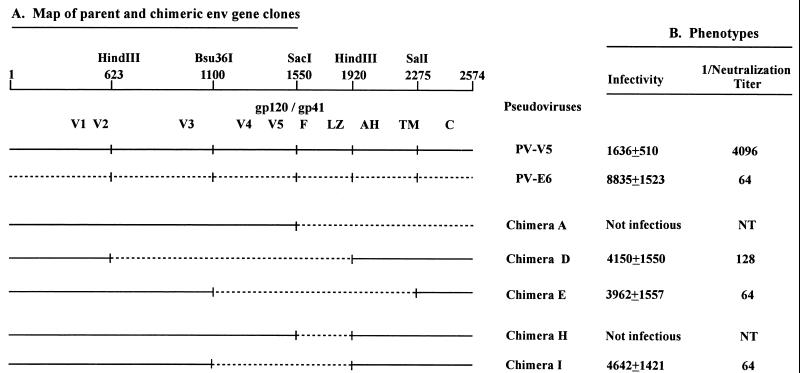
Plasmids pSV-V5 and pSV-E6, which contain env gene derived from neutralization-sensitive and -resistant variants of the HIV-1 MN strain, respectively, have been described previously (21). Chimeric envelope plasmids (chimeras A through G) constructed with these plasmids were also described previously (21). In this study, two additional chimeric env clones were constructed. Chimera H was constructed such that the SacI-HindIII fragment of pSV-V5 was effectively replaced with the corresponding sequence from pSV-E6, as shown schematically in Fig. 1. Similarly, chimera I was constructed such that the Bsu36I-HindIII fragment in pSV-V5 was effectively replaced with that of pSV-E6.
FIG. 1.
(A) Schematic diagram depicting a series of chimeras constructed from V5 and E6 parents. The construction and characterization of clones V5, E6, A, D, and E have been described previously (21). The approximate locations of the gp120 variable regions (V) and the gp41 fusion (F), LZ, membrane proximal alpha-helix (AH), transmembrane (TM), and cytoplasmic (C) domains are indicated. (B) Infectivity and neutralization resistance phenotypes of the clones. Infectivity is expressed as the mean ± the standard deviation of luciferase assay results for lysates of cells inoculated with 1:4 pseudovirus dilutions. The infectivity of the V5 clone was significantly less (P < 0.02 by two-tailed Student t test, assuming equal variances) than all other infectious clones shown, while E6 was significantly more infectious than the chimeras D, E, and I (P < 0.02 for each). The statistical analyses were done in Microsoft Excel. The neutralization titer is the titer of the reference serum (HIV-1 Neutralizing Serum [2]) against pseudovirus expressing each clone (31). NT, not tested.
Site-directed mutagenesis.
Mutagenesis procedures were carried out by using Pfu polymerase (Quick Change Mutagenesis Kit; Stratagene) by following the instructions of the manufacturer. The reactions were performed in an automated thermal cycler (Perkin-Elmer model 2400). Each mutagenized plasmid was then digested with restriction endonucleases, and the fragments containing the introduced mutations were cloned into pSV-V5m (21). For cloning, Bsu36I and SacI were used for mutations in the C terminus of gp120, SacI and SalI were used for mutations present in the N terminus of gp41, and Bsu36I and SalI were used for mutations present in both gp120 and gp41. Nucleotide sequences of entire transferred fragments had been confirmed previously by sequencing by using the ABI PRISM dye-terminator method (Perkin-Elmer).
Pseudovirus construction and infectivity titration.
Pseudoviruses expressing envelope glycoproteins derived from various env plasmids were constructed by using pSV7d-env and pNL-Luc-E−R− plasmids, as described previously (21). Infectivity assays were conducted in triplicate in PM1 cells. The luciferase activity of infected cells was measured in a luminometer.
Neutralization assays.
Neutralization assays were performed in 96-well plates as described previously (21). Positive control human reference sera, the HIV-1 neutralizing serum (1) and serum (2), were serially diluted and incubated with pseudovirus suspensions in triplicate wells at 37°C for 1 h (31). The samples were then used to infect PM1 cells, and the luciferase activity of each well was measured 72 h after infection. The neutralizing endpoint was determined to be the serum dilution which inhibited 90% of viral infectivity compared to the non-neutralized control.
Enzyme immunoassay for envelope glycoprotein.
Medium from cell cultures transfected for pseudovirus production was harvested, filtered through a 45-μm-pore-size sterile filter (Millipore Corp.), and centrifuged at 15,000 rpm for 2 h (Tomy Tech refrigerated centrifuge) to sediment pseudoviruses. Each sample of supernatant and resuspended pellet was tested for viral infectivity. The full infectivity was recovered from the pellet sample, while no infectivity was detected from the supernatant (data not shown). For the antigen assay, the pellets were washed twice with phosphate-buffered saline (PBS) and resuspended in lysis buffer (35). For cell lysate samples, transfected cells were harvested and washed with PBS, and one million cells were counted and resuspended in lysis buffer.
Pseudoviruses purified through sucrose step-gradient centrifugation were also prepared and compared for their antigen expression. Transfected cell culture medium was harvested, filtered as described above, and centrifuged on 20 and 55% sucrose at 30,000 rpm (Beckman L8-70M ultracentrifuge) for 3 h at 4°C. After medium was removed from the top and 55% sucrose was removed from the bottom of the tube, samples were collected and pseudoviruses were sedimented twice after the addition of equal volumes of cold PBS, as described above. Samples were diluted as needed before being dispensed into the wells of microtiter plates.
For antigen assays, each well of microtiter plate (Immulon 2; Dynex) was coated with a human anti-HIV-1 immunoglobulin G (36). After each well was blocked with BLOTTO, each serially diluted preparation of cell lysate or pseudovirus was then added. gp120 antigen was detected with sheep anti-gp120 antibody (7, 21). p24 antigen was detected with rabbit anti-p24 antibody (24). Bound detection antibodies were assayed with biotinylated anti-sheep or anti-rabbit antibody mixed with avidin D-horseradish peroxidase. Orthophenylenediamine was used as a substrate (Abbott Laboratory), the absorbance at 492 nm was measured in a microtiter plate reader, and the data were analyzed by using I-Smart (Packard Corp). Antigens used for HIV MN gp120 and p24 primary reference standards were obtained from the AIDS Research and Reference Reagent Program (contributed by MicroGeneSys, Inc. [catalog number 2967] and K. Steimer [catalog number 382 [26]). In some experiments, HIV gp160 (MicroGeneSys, Inc.) was used as a working standard, with calibration to the gp120 reference.
Western blotting was performed as previously described with the globulin fraction of an HIV-1 immune human serum for antigen detection (7a).
RESULTS
Phenotypes of chimeric envelope clones.
The effects of introducing all six mutations, or the four gp41 mutations, into the neutralization-sensitive clone V5, were tested first by constructing and testing two chimeric envelope genes, H and I, as shown in Fig. 1. The encoded proteins of these and other envelope genes described here were phenotyped by using pseudotyped, replication-incompetent HIV-1 virions carrying a luciferase reporter gene (5). Chimera H, which contains only the N terminus of gp41 derived from the neutralization-resistant parent, clone E6, and the rest of the envelope sequence from the neutralization-sensitive parent, clone V5, was not infectious. However, chimera I, which has both the gp120 C terminus and the gp41 N terminus from clone E6, was infectious and as neutralization resistant as the E6 clone (titer of the reference serum was 1:64 against both but ≥1:4,096 against the V5 clone). The properties of these mutant genes were, therefore, consistent with those predicted based on our earlier study (21).
Mutagenic analysis of infectivity phenotype.
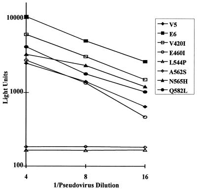
The effects of introduction of single point mutations into the V5 clone on infectivity were then evaluated. Clone V5 mutants which carried each single point mutation were prepared. Pseudoviruses expressing each were repeatedly tested for comparative infectivity. The results of testing of these clones, compared to clones V5 and E6 in a representative experiment, are shown in Fig. 2. Numerous parallel transfections of the V5 and E6 clones have always yielded pseudovirus preparations of E6 with an infectivity generally ca. fivefold higher than that of V5 (range, 2- to 10-fold), as exemplified by the results shown. The V420I mutant had significantly increased infectivity compared to V5, while E460I had an infectivity equivalent to that of V5. The N565H and Q582L mutations increased infectivity moderately, but significantly, while the L544P and A562S single mutations completely eliminated infectivity. In view of the parallelism of the infectivity titrations of the infectious clones, exemplified by the data presented in Fig. 2, relative infectivity values, compared to V5, were assigned to each clone, as shown in Table 1.
FIG. 2.
Quantitation of luciferase activity in cells infected with various clones of the HIV-1 MN strain. Results shown are averages of quadruplicate determinations made at each dilution. The infectivity of each of the V420I, N565H, and Q582L mutants was significantly increased (analysis of variance − repeat major (SAS), P = 0.002, 0.06, and 0.01, respectively; t test (Excel), P < 0.05 for each) compared to V5. Amino acids are identified by using standard single-letter designations.
TABLE 1.
Neutralization and infectivity phenotypes of mutagenized envelopes
| Parent and V5 mutant env clones
|
Relative infectivitya | Relative neutralization resistanceb | |
|---|---|---|---|
| Clone no. or name | E6 mutation(s) introduced into V5 | ||
| V5 | 1 | 1 | |
| E6 | 4.10 | 128 | |
| 1 | V420I | 2.45 | 8 |
| 2 | E460I | 1.06 | 1 |
| 3 | L544P | NI | NT |
| 4 | A562S | NI | NT |
| 5 | N565H | 1.41 | 2 |
| 6 | Q582L | 1.57 | 2 |
| 7 | V420I+E460I | 1.63 | 8 |
| 8 | V420I+L544P | 0.15 | NT |
| 9 | V420I+A562S | NI | NT |
| 10 | V420I+N565H | 1.92 | 8 |
| 11 | V420I+Q582L | 2.54 | 8 |
| 12 | E460I+L544P | NI | NT |
| 13 | E460I+A562S | NI | NT |
| 14 | E460I+Q582L | 1.75 | 4 |
| 15 | A562S+N565H | 0.26 | NT |
| 16 | N565H+Q582L | 1.40 | 4 |
| 17 | V420I+E460I+L544P | 0.15 | NT |
| 18 | V420I+E460I+A562S | NI | NT |
| 19 | V420I+A562S+N565H | 0.28 | NT |
| 20 | V420I+N565H+Q582L | 2.37 | 8 |
| 21 | E460I+A562S+N565H | 0.23 | NT |
| 22 | A562S+N565H+Q582L | 1.27 | 4 |
| 23 | V420I+E460I+A562S+N565H | 1.34 | NT |
| 24 | V420I+L544P+N565H+Q582L | 0.21 | NT |
| 25 | V420I+A562S+N565H+Q582L | 1.83 | 8 |
| 26 | L544P+A562S+N565H+Q582L | NI | NT |
| 27 | V420I+E460I+A562S+N565H+Q582L | 3.88 | 16 |
| 28 | V420I+L544P+A562S+N565H+Q582L | 1.92 | 64 |
| 29 | V420I+E460I+L544P+A562S+N565H+Q582L | 2.12 | 128 |
Relative infectivity was determined as XM/XV5 for a given y, calculated by using the best-fit lines determined by regression analysis of the log-transformed luciferase activity determinations (light units) as a function of the inoculum dilution. XM is the value obtained for the particular mutant, and XV5 is the value obtained for V5. y values used for comparisons were intermediate between the curves being compared. Calculations were performed by using Microsoft Excel. NI, not infectious.
Relative neutralization resistance was determined as NV5/NM, where N is the neutralization titer of the reference serum (31). Minor variation was observed in specific neutralization titers observed in different experiments, but the relative effects of the various mutations on neutralization resistance were highly consistent. NT, not tested.
Mutagenic analysis of the neutralization resistance phenotype.
The effects of the mutations on neutralization resistance are also shown in Table 1. Relative neutralization resistance is expressed in comparison to that of V5. A mutant with neutralization sensitivity equivalent to that of V5 was assigned a relative value of 1, while a mutant equivalent to E6 was 128-fold more resistant than V5. Of the six point mutations tested, the V420I mutation had the largest individual effect, consistently eightfold, on neutralization sensitivity. The N565H and Q582L mutations each had small, but consistent (usually twofold) effects. The E460I mutation had no direct effect.
Interaction between amino acid residues and correlation between high-infectivity and neutralization resistance phenotypes.
The mechanism by which the A562S mutation caused loss of infectivity was probed by constructing and testing clones which contained various combinations of the six E6 mutations. Neither the V420I nor the E460I mutation, either singly or together, complemented the A562S effect which eliminated infectivity (Table 1, mutants 9, 13, and 18). However, the effect was partially abrogated by the introduction of the N565H mutation and fully abrogated by the introduction of both the N565H and Q582L mutations (Table 1, mutants 15 and 22). In the alpha-helical configuration of the LZ, residues 562 and 565 are adjacent to each other but distant from, and not capable of direct interaction with, residue 582 (3, 32). Additional mutants were compared to determine whether the effects of the mutations at residues 562, 565, and 582 might be reflecting interactions between gp120 and gp41. Specifically, we observed that the V420I and E460I mutations together were able to replace functionally the mitigating effects of the Q582L mutation on the lethality of the A562S mutation (Table 1, mutant 23). Moreover, addition of the Q582L mutation to this parent caused a substantial additional increase in infectivity (Table 1, mutant 23 versus mutant 27). These results indicated that the ability of the mutations at residues 565 and 582 to mitigate the lethal effect of the mutation at residue 562 and the lethal effect of the mutation at residue 562 itself were due to interactions of gp41 with gp120.
The mechanism of the lethal effect of the L544P mutation was also probed by constructing and testing additional mutants. The L544P mutation was not compensated for by the E460I mutation (Table 1, mutant 12). However, the introduction of V420I into mutants containing the L544P mutation always enhanced infectivity (Table 1, mutants 8 versus 3, 17 versus 12, and 28 versus 26). Mutants which included V420I with L544P were always at least partially infectious (Table 1, mutants 8, 17, 24, and 28), while those with the L544P mutation which lacked the V420I mutation never had detectable infectivity. The infectivity defect caused by the L544P mutation was only fully compensated for by the addition of the E6 mutations at residues 562, 565, 582, and 420 (Table 1, mutant 28). This quintuple mutant was also eightfold more resistant to neutralization than the quadruple mutant lacking the L544P mutation, an effect equivalent to that of the V420I mutation (Table 1, mutant 25). It is notable that the L544P mutation caused a reduction in infectivity, but an increase in neutralization resistance, when introduced into the respective quintuple mutant (Table 1, mutant 29 versus mutant 27). This is the only case we encountered in which an increase in neutralization resistance was accompanied by a decrease in infectivity.
Possible interactions of the E460I mutation with other mutations were also tested. When it was introduced in combination with the Q582L mutation, it enhanced neutralization resistance two- to fourfold in each case in repeated experiments (Table 1, mutants 14 versus 6, 27 versus 25, and 29 versus 28). The E460I mutation increased infectivity significantly when introduced into the V420I+A562S+N565H+Q582L mutant (Table 1, mutant 25 versus mutant 27; ANOVA, P = 0.005; t test, P <0.05). The E460I mutation had no effect when introduced in the absence of the Q582L mutation (Table 1, mutants 7, 12, 13, 17, 18, and 21 versus mutants 1, 3, 4, 8, 9, and 15, respectively). Reciprocal evidence of a functional interaction between the mutations E460I and Q582L is indicated by the substantially greater infectivity of mutant 27 compared to mutant 23. As indicated by results shown in Fig. 1 and Table 1, and by other comparative results not shown, it is also notable that the sextuple mutant (Table 1, mutant 29) and chimera I were equivalent in neutralization resistance but had consistently lower infectivity than the E6 clone. Since chimera I and the sextuple mutant should be molecularly identical, but lack the mutations which distinguish the E6 and V5 clones from each other in regions other than those studied here, the results suggest an effect on infectivity of mutations in E6 outside of the two regions studied (21).
Since changes in the neutralization and infectivity phenotypes often occurred concordantly, we examined the relationship among these two parameters by comparing all mutated clones which were tested for neutralization sensitivity. Regression analysis indicated a significant correlation between infectivity and neutralization resistance (R2 = 0.432, P = 0.006; analysis was performed using Microsoft Excel). Considering the involvement of similar mutations in the two phenotypes and the close correlation between the phenotypes, it is likely that the neutralization sensitivity and infectivity are based on the same properties of the envelopes.
Envelope glycoprotein expression.
Envelope protein expression was evaluated to determine whether the infectivity and neutralization sensitivity phenotypes could simply reflect envelope processing or density on virion surfaces. V5 and E6 pseudoviruses concentrated by sedimentation and by sucrose gradient centrifugation were analyzed by Western blot and were found to contain equivalent amounts of gp41, indicating gp160 cleavage (data not shown). HIV-1 gp120 and p24 antigen concentrations were quantified by enzyme-linked immunosorbent assay of pseudovirus preparations and transfected cell lysates (Table 2). The amount of p24 expressed on pseudoviruses or in cell lysates did not vary as a function of the envelope with which it was coexpressed. The levels of gp120 from the samples derived from different clones were also similar regardless of their neutralization or infectivity phenotypes. The amount of gp120 detected on both pseudoviruses and cell lysates from V5 was either comparable or greater than E6 in multiple experiments. Envelope clones with no infectivity were also either comparable or greater with respect to gp120 production and incorporation than other mutant clones with various levels of infectivity. This pattern was observed among both sedimented and sucrose gradient-purified pseudovirus preparations. The relative gp120 expression compared with the amount of p24 in the latter preparations was lower than the sedimented samples, possibly due to gp120 dissociation during the process of sample preparation. Thus, neither the infectivity nor the neutralization phenotypes appeared to be determined by the level of envelope expression or incorporation into virus particles.
TABLE 2.
Comparison of HIV-1 gp120 and p24 expression on pseudoviruses and in transfected cells
| Clonesa | Antigen expression in pseudovirus prepn
|
|||||
|---|---|---|---|---|---|---|
| Sedimented (ng/ml)b
|
Sucrose gradient purified (μg/ml)c
|
Antigen expression in cell lysates (ng/106 cells)d
|
||||
| gp120 | p24 | gp120 | p24 | gp120 | p24 | |
| Parents | ||||||
| V5 | 10.2 | 64 | 0.29 | 11.7 | 4.3 | 24 |
| E6 | 7.6 | 76 | 0.29 | 11.4 | 4.3 | 21 |
| E6 mutations in V5 | ||||||
| V420I | 7.6 | 80 | 0.25 | 11.9 | 3.7 | 21 |
| L544P | 8.2 | 72 | 0.24 | 11.8 | 4.3 | 17 |
| A562S | 8.2 | 88 | 0.25 | 11.7 | 5.1 | 20 |
| A562S + N565H | 7.6 | 76 | NTe | NT | 4.7 | 23 |
| V420I + E460I + A562S + N565H + Q582L | NT | NT | 0.28 | 12.1 | NT | NT |
| V420I + L544P + A562S + N565H + Q582L | 8.2 | 80 | NT | NT | 3.8 | 17 |
| V420I + E460I + L544P + A562S + N565H + Q582L | 7.6 | 76 | NT | NT | 3.9 | 17 |
The L544P and A562S mutants were not infectious, the A562S/N565H mutant was minimally infectious. The other clones shown were all significantly more infectious than V5.
The amount of antigen for sedimented pseudovirus preparation is expressed as nanograms per milliliter in each pseudovirus harvest.
For sucrose gradient-purified samples, the amount of antigen is expressed as micrograms per milliliter of each preparation.
The amount of each antigen in cell lysates is expressed as nanograms per 106 cells.
NT, not tested.
DISCUSSION
The potential role of the six individual mutations present in the regions responsible for the neutralization resistance phenotype was studied. The significance of each mutation, with respect to impact on viral fitness phenotype, is most apparent upon consideration of the combined effects of the mutations. Both the N565H and Q582L mutations had small but consistent positive effects on neutralization resistance and infectivity phenotypes when introduced individually into V5, yet both were required before the A562S mutation could be introduced without a negative effect on infectivity. This triple LZ mutation had minor positive effects on the neutralization resistance and infectivity phenotypes compared to V5. The greater importance of this three-mutation complex was that, when it was introduced in the presence of the V420I mutation, it accommodated the introduction of the L544P mutation. In this context the L544P mutation caused an eightfold increase in neutralization resistance, a value equivalent to the effect of V420I. The E460I mutation had no independent effect on either phenotype, but when combined with the Q582L mutation it consistently enhanced neutralization resistance ca. twofold. Thus, all six mutations contributed in concert to the fitness phenotypes.
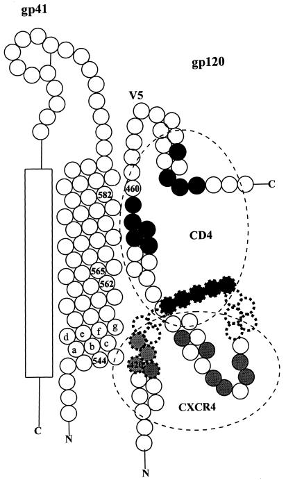
The highly interactive nature of the functional effects of these mutations suggests the strong possibility of direct structural interactions between the regions of gp120 and gp41 involved. There was extensive evidence of functional interaction between residues 420 and 544 and between residues 460 and 582, as described above. Thus, we propose that structural interaction between gp120 and the LZ maintains residue 420 near residue 544 and residue 460 near residue 582, as illustrated in Fig. 3. In the model shown, the amino acid residues are displayed in linear arrays, not in three dimensions. The model is not intended to challenge any known features of the atomic structures of gp120 or gp41 (3, 11, 32, 37).
FIG. 3.
Proposed model for assembly of the receptor-coreceptor binding complex of gp120 on the LZ of gp41. The two-dimensional model is based on the results presented here and on published data regarding the secondary and atomic structures of the HIV-1 envelope (3, 11–13, 21, 25, 28, 32, 33, 37, 38). The amino acid residues are generally represented by circles. The proximal and distal extensions of the amino (N) and carboxyl (C) residues of each polypeptide are indicated. The alpha-helix of gp41 which constitutes the LZ is shown as diagonally stacked rows, alternating three and four residues each, representing the seven residues of each helical turn. One such sequence of residues is lettered a through g to indicate conventional residue designations. Beta sheet regions of gp120 are indicated by diagonally stacked rows of two residues each. Residues which form part of the bridging sheet in the fusion-competent conformation of gp120 are indicated by circles with dashed outlines. Residues which are known to form hydrogen bands or van der Waals interactions with CD4 are shown as black circles. Residues which are thought to be important for coreceptor binding to gp120, based on mutagenesis studies (25), are shown as gray circles. The four residues in the LZ and the two residues in gp120 which were mutagenized in this study are shown as numbered circles. The location of variable region 5 (V5) is shown. The membrane proximal region of the gp41 ectodomain which forms an alpha-helix in the fusion-competent state is shown as a rectangle. Ovals schematically represent the CD4 and CXCR4 binding domains.
The HIV-1 LZ forms an oligomeric, probably trimeric, coiled coil which maintains the complete envelope complex in an oligomeric state. LZs are amphipathic alpha-helical structures in which leucine, isoleucine, or valine residues tend to be present in a pattern which repeats alternately, approximately every three and four residues, respectively. These residues are conventionally considered to occupy the a and d (i.e., first and fourth) positions of each seven-residue wheel of the helix. The hydrophobic face formed by these residues typically interacts with hydrophobic domains on other macromolecules. The faces formed by the a and d residues of the LZs of three gp41 molecules interact with each other to mediate oligomerization, and the three interacting alpha-helices coil around each other to form a coiled coil (3, 28, 32). A change in the conformation of the envelope complex is thought to follow engagement of the receptor binding site with CD4, and this change is thought to result in the alignment of the gp41 membrane proximal alpha-helix in the external groove of the coiled coil (4, 34). The residues of the membrane proximal alpha-helix bond noncovalently, usually to the g and e residues of adjacent LZ motifs in the coiled coil. The residues 544, 562, 565, and 582 are all b, c, and f residues in the helical wheel, appearing on the unoccupied hydrophilic face of the LZ (3, 32).
The localizations of residues 420 and 460 in proximity to the functionally interacting residues, 544 and 582, would localize the CD4 binding site to a position adjacent to the LZ (Fig. 3). Residue 460, which is located in the CD4 binding pocket, is shown associated with the LZ by virtue of the interactions between the residue 460 and 582 regions of the complex (11, 25, 37, 38). Residue 420 is surrounded by residues which are important for envelope-coreceptor binding and is located within the region which forms part of a bridging sheet between inner and outer gp120 domains in the receptor-engaged state of the envelope (25, 37). It is shown associated with the LZ by virtue of the interactions between the residue 420 and 544 regions of the complex (11, 25, 37, 38). The model shown in Fig. 3 is consistent with the interpretation of Rizzuto et al. (25) that the coreceptor binding domain of gp120 is located near the trifold axis of symmetry of the oligomer. They identified residues important for coreceptor binding based on the effects of mutations of specific residues, some of which are indicated in Fig. 3. It is not yet known which of these residues actually bind to the coreceptor. Rizzuto et al. describe the region of gp120 which binds the coreceptor as highly basic in the periphery and hydrophobic in the center. Residue 420 is located at the very center of this region. Our results raise the possibility that this residue may be important for the function of this region by virtue of its putative role in anchoring the region to the amino terminus of the LZ. The use of distinct residues of the LZ for oligomerization, assembly of the gp120 receptor and coreceptor binding domains, and interaction with the gp41 membrane proximal alpha-helical domain may permit variation of phenotypes related to one interdomain interaction without affecting the others. Wyatt et al. (37) have proposed that residues in the CD4 binding pocket, which are not involved in receptor binding, may vary to cause escape mutation. Indeed, this adaptive process probably involves the LZ. Mutations in the LZ may be followed by compensatory mutations in residues associated with the receptor and coreceptor binding domains of gp120.
The model shown in Fig. 3 is consistent with the published atomic structure of gp120 (11, 25, 37, 38). Our data do not address whether the bridging sheet structure, which is a feature of the fusion-competent conformation of the oligomer, is also present in the resting state. However, in describing the atomic structure of gp120, Kwong et al. (11) proposed a dominant role of the inner domain of gp120 in maintaining contacts with gp41. Our data indicate a probable critical role for residues in outer domain and bridging sheet regions which form parts of the receptor and coreceptor binding domains in assembly of gp120 onto gp41.
Structures of macromolecules cannot be proven by phenotypic analyses, and features of the model proposed in Fig. 3 may be incorrect. However, certain principles which are illustrated by the model are clearly supported. Specifically, (i) there is strong evidence of an intimate structural relationship between the LZ and the receptor-coreceptor binding complex of gp120; (ii) mutations in the LZ can modulate functions of the receptor-coreceptor binding complex; and (iii) the LZ serves multiple functions, in addition to its established role in oligomerization. These functions include the maintenance of the highly ordered organization of gp120 and a role in the coordination of the concerted sequence of events after receptor engagement leading to fusion of the viral and cell membranes.
The absence of correlation between envelope glycoprotein content and either infectivity or neutralization resistance phenotypes is revealing with respect to potential mechanisms for the phenotypes. These results indicate that the greater infectivity of the neutralization resistance mutants is apparently the result of enhancement of some aspect of envelope function rather than envelope density. The specific nature of such a functional effect remains to be defined, and diverse possibilities exist. An intimate relationship between the structural elements involved in these phenotypes and the receptor-coreceptor binding and fusion domains provides ample opportunities for effects of these mutations on infectivity functions.
The evolution of new fitness phenotypes under the influence of environmental pressures often results from mutations which may be associated with negative fitness phenotypes in less-adverse circumstances. As a result, the mutations may be dead-end mutations, coding for phenotypes which are unable to evolve further. The convergent evolution of related but distinct fitness phenotypes described here constitutes a mechanism by which a species may continue to evolve along a general evolutionary pathway in spite of dynamics which limit evolution in response to a single environmental factor. It is reasonable to expect that such a powerful evolutionary capability could contribute significantly to the complex pathogenesis of HIV infection. Elucidation of structural variations which contribute to the neutralization resistance of HIV-1 should contribute to our understanding of the epitopes which are critical targets for vaccination strategies.
ACKNOWLEDGMENTS
This work was supported by NIH grant RO1-AI37438 and USUHS grant RO87EZ.
The assistance of Paul Hsieh with statistical evaluation is appreciated.
REFERENCES
- 1.Back N K T, Smit L, Schutten M, Nara P L, Tersmette M, Goudsmit J. Mutations in human immunodeficiency virus type 1 gp41 affect sensitivity to neutralization by gp120 antibodies. J Virol. 1993;67:6897–6902. doi: 10.1128/jvi.67.11.6897-6902.1993. [DOI] [PMC free article] [PubMed] [Google Scholar]
- 2.Chamat S, Nara P, Berquist L, Whalley A, Morrow W J M, Kohler H, Kang C-Y. Two major groups of neutralizing anti-gp120 antibodies exist in HIV-infected individuals. J Immunol. 1992;149:645–654. [PubMed] [Google Scholar]
- 3.Chan D C, Fass D, Berger J M, Kim P S. Core structure of gp41 from the HIV envelope glycoprotein. Cell. 1997;89:263–273. doi: 10.1016/s0092-8674(00)80205-6. [DOI] [PubMed] [Google Scholar]
- 4.Chen C-H, Matthews T J, McDanal C B, Bolognesi D P, Greenberg M L. A molecular clasp in the human immunodeficiency virus (HIV) type 1 TM protein determines the anti-HIV fusion activity of gp41 derivatives: implications for viral fusion. J Virol. 1995;69:3771–3777. doi: 10.1128/jvi.69.6.3771-3777.1995. [DOI] [PMC free article] [PubMed] [Google Scholar]
- 5.Conner R I, Sheridan K E, Lai C, Zhang L, Ho D D. Characterization of the functional properties of env genes from long-term survivors of human immunodeficiency virus type 1 infection. J Virol. 1996;70:5306–5311. doi: 10.1128/jvi.70.8.5306-5311.1996. [DOI] [PMC free article] [PubMed] [Google Scholar]
- 6.Delwart E L, Mosialos G, Gilmore T. Retroviral envelope glycoproteins contain a leucine zipper-like repeat. AIDS Res Hum Retroviruses. 1990;6:703–706. doi: 10.1089/aid.1990.6.703. [DOI] [PubMed] [Google Scholar]
- 7.Hatch W C, Tanaka K E, Cavelli T, Rashbaum W K, Kress Y, Lyman W D. Persistent productive HIV-1 infection of a CD4- human fetal thymocyte line. J Immunol. 1992;148:3055–3061. [PubMed] [Google Scholar]
- 7a.Hendry R M, Parks D E, Campos Mello D L A, Quinnan G V, Galvao-Castro B. Lack of evidence for HIV-2 infection among at-risk individualsin Brazil. J Acquired Immune Defic Syndr. 1991;4:623–627. [PubMed] [Google Scholar]
- 8.Ho D D, Fung M S C, Cao Y, Li X L, Sun C, Chang T W. Another discontinuous epitope on glycoprotein gp120 that is important in human immunodeficiency virus type 1 neutralization is identified by a monoclonal antibody. Proc Natl Acad Sci USA. 1991;88:8949–8952. doi: 10.1073/pnas.88.20.8949. [DOI] [PMC free article] [PubMed] [Google Scholar]
- 9.Ho D, McKeating J, Li X, Moudgil T, Daar E, Sun N-C, Robinson J. Conformational epitope on gp120 important in CD4 binding and human immunodeficiency virus type 1 neutralization identified by a human monoclonal antibody. J Virol. 1991;65:489–493. doi: 10.1128/jvi.65.1.489-493.1991. [DOI] [PMC free article] [PubMed] [Google Scholar]
- 10.Korber B, Wolinsky S, Haynes B, Kunstman K, Levy R, Furtado M, Otto P, Myers G. HIV-1 intrapatient sequence diversity in the immunogenic V3 region. AIDS Res Hum Retroviruses. 1992;8:1461–1465. doi: 10.1089/aid.1992.8.1461. [DOI] [PubMed] [Google Scholar]
- 11.Kwong P D, Wyatt R, Robinson J, Sweet R W, Sodroski J, Hendrickson W A. Structure of an HIV gp120 envelope glycoprotein in complex with the CD4 receptor and a neutralizing human antibody. Nature. 1998;393:648–659. doi: 10.1038/31405. [DOI] [PMC free article] [PubMed] [Google Scholar]
- 12.Lu M, Blacklow S C, Kim P S. A trimeric structural domain of the HIV-1 transmembrane glycoprotein. Nat Struct Biol. 1995;2:1075–1082. doi: 10.1038/nsb1295-1075. [DOI] [PubMed] [Google Scholar]
- 13.Lu M, Kim P S. A trimeric structural subdomain of the HIV-1 transmembrane glycoprotein. J Biomol Struct Dyn. 1997;15:465–471. doi: 10.1080/07391102.1997.10508958. [DOI] [PubMed] [Google Scholar]
- 14.McKeating J A, Gow J, Goudsmit J, Pearl L H, Mulder C, Weiss R A. Characterization of HIV-1 neutralization escape mutants. AIDS. 1989;3:777–784. doi: 10.1097/00002030-198912000-00001. [DOI] [PubMed] [Google Scholar]
- 15.McKeating J A, Schotton C, Cordell J, Graham S, Balfe P, Sullivan N, Charles M, Page M, Bolmstedt A, Olofsson S, Kayman S C, Wu Z, Pinter A, Dean C, Sodroski J, Weiss R A. Characterization of neutralizing monoclonal antibodies to linear and conformation-dependent epitopes within the first and second variable domains of human immunodeficiency virus type 1 gp120. J Virol. 1993;67:4932–4944. doi: 10.1128/jvi.67.8.4932-4944.1993. [DOI] [PMC free article] [PubMed] [Google Scholar]
- 16.Moore J P, Ho D D. Antibodies to discontinuous or conformationally sensitive epitopes on the gp120 glycoprotein of human immunodeficiency virus type 1 are highly prevalent in sera of infected humans. J Virol. 1993;67:863–875. doi: 10.1128/jvi.67.2.863-875.1993. [DOI] [PMC free article] [PubMed] [Google Scholar]
- 17.Nara P L, Smit L, Dunlop N, Hatch W, Merges M, Waters D, Kelliher J, Gallo R C, Fischinger P J, Goudsmit J. Emergence of viruses resistant to neutralization by V3 specific antibodies in experimental human immunodeficiency virus type 1 IIIB infection of chimpanzees. J Virol. 1990;64:3779–3791. doi: 10.1128/jvi.64.8.3779-3791.1990. [DOI] [PMC free article] [PubMed] [Google Scholar]
- 18.O’Brien W A, Chen I S Y, Ho D D, Daar E S. Mapping genetic determinants for human immunodeficiency virus type 1 resistance to soluble CD4. J Virol. 1992;66:3125–3130. doi: 10.1128/jvi.66.5.3125-3130.1992. [DOI] [PMC free article] [PubMed] [Google Scholar]
- 19.Olshevsky U, Helseth E, Furman C, Li J, Haseltine W, Sodrowski J. Identification of individual human immunodeficiency virus type 1 gp120 amino acids important for CD4 binding. J Virol. 1990;64:5701–5707. doi: 10.1128/jvi.64.12.5701-5707.1990. [DOI] [PMC free article] [PubMed] [Google Scholar]
- 20.Page K A, Stearns S M, Littman D R. Analysis of mutations in the V3 domain of gp160 that affect fusion and infectivity. J Virol. 1992;66:524–533. doi: 10.1128/jvi.66.1.524-533.1992. [DOI] [PMC free article] [PubMed] [Google Scholar]
- 21.Park E J, Vujcic L K, Anand R, Theodore T S, Quinnan G V. Mutations in both gp120 and gp41 are responsible for the broad neutralization resistance of variant human immunodeficiency virus type 1 MN to antibodies directed at V3 and non-V3 epitopes. J Virol. 1998;72:7099–7107. doi: 10.1128/jvi.72.9.7099-7107.1998. [DOI] [PMC free article] [PubMed] [Google Scholar]
- 22.Quinnan G V, Zhang P F, Fu D W, Dong M, Margolick J B. Evolution of neutralizing antibody response against HIV type 1 virions and pseudovirions in multicenter AIDS cohort study participants. AIDS Res Hum Retroviruses. 1998;14:939–949. doi: 10.1089/aid.1998.14.939. [DOI] [PubMed] [Google Scholar]
- 23.Reitz M S, Wilson C, Naugle C, Gallo R C, Robert-Guroff M. Generation of a neutralization resistant variant of HIV-1 is due to selection for a point mutation in the envelope gene. Cell. 1988;54:57–63. doi: 10.1016/0092-8674(88)90179-1. [DOI] [PubMed] [Google Scholar]
- 24.Rencher S D, Slobod K S, Dawson D H, Lockey T D, Hurwitz J L. Does the key to a successful HIV type 1 vaccine lie among the envelope sequences of infected individuals? AIDS Res Hum Retroviruses. 1995;11:1131–1133. doi: 10.1089/aid.1995.11.1131. [DOI] [PubMed] [Google Scholar]
- 25.Rizzuto C D, Wyatt R, Hernandez-Ramos N, Sun Y, Kwong P D, Hendrickson W A, Sodroski J. A conserved HIV gp120 glycoprotein structure involved in chemokine receptor binding. Science. 1998;280:1949–1953. doi: 10.1126/science.280.5371.1949. [DOI] [PubMed] [Google Scholar]
- 26.Steimer K S, Puma J P, Power M D, Powers M A, George-Nascimento C, Stephans J C, Levy J A, Sanchez-Pescador R, Luciw P A, Barr P J, et al. Differential antibody responses of individuals infected with AIDS-associated retroviruses surveyed using the viral core antigen p25gag expressed in bacteria. Virology. 1986;150:283–290. doi: 10.1016/0042-6822(86)90289-8. [DOI] [PubMed] [Google Scholar]
- 27.Steimer K S, Scandella J, Skiles P V, Haigwood N L. Neutralization of divergent HIV-1 isolates by conformation-dependent human antibodies to Gp120. Science. 1991;254:105–108. doi: 10.1126/science.1718036. [DOI] [PubMed] [Google Scholar]
- 28.Tan K, Liu J, Wang J, Shen S, Lu M. Atomic structure of a thermostable subdomain of HIV-1 gp41. Proc Natl Acad Sci USA. 1997;94:12303–12308. doi: 10.1073/pnas.94.23.12303. [DOI] [PMC free article] [PubMed] [Google Scholar]
- 29.Thali M, Charles M, Furman C, Cavacini L, Posner M, Robinson J, Sodroski J. Resistance to neutralization by broadly reactive antibodies to the human immunodeficiency virus type 1 gp120 glycoprotein conferred by a gp41 amino acid change. J Virol. 1994;68:674–680. doi: 10.1128/jvi.68.2.674-680.1994. [DOI] [PMC free article] [PubMed] [Google Scholar]
- 30.Thali M, Moore J P, Furman C, Charlis M, Ho D D, Robinson J, Sodroski J. Characterization of conserved human immunodeficiency virus type 1 gp120 neutralization epitopes exposed upon gp120-CD4 binding. J Virol. 1993;67:3978–3988. doi: 10.1128/jvi.67.7.3978-3988.1993. [DOI] [PMC free article] [PubMed] [Google Scholar]
- 31.Vujcic L K, Quinnan G V., Jr Preparation and characterization of human HIV type 1 neutralizing reference sera. AIDS Res Hum Retroviruses. 1995;11:783–787. doi: 10.1089/aid.1995.11.783. [DOI] [PubMed] [Google Scholar]
- 32.Weissenhorn W, Dessen A, Harrison S C, Skehel J J, Wiley D C. Atomic structure of the ectodomain from HIV-1 gp41. Nature. 1997;387:426–430. doi: 10.1038/387426a0. [DOI] [PubMed] [Google Scholar]
- 33.Weissenhorn W, Wharton S A, Calder L J, Earl P L, Moss B, Aliprandis E, Skehel J J, Wiley D C. The ectodomain of HIV-1 env subunit gp41 forms a soluble, alpha-helical, rod-like oligomer in the absence of gp120 and the N-terminal fusion peptide. EMBO J. 1996;15:1507–1514. [PMC free article] [PubMed] [Google Scholar]
- 34.Wild C T, Shugars D C, Greenwell T K, McDanal C B, Matthews T J. Peptides corresponding to a predictive α-helical domain of human immunodeficiency virus type 1 are potent inhibitors of virus infection. Proc Natl Acad Sci USA. 1994;91:9770–9774. doi: 10.1073/pnas.91.21.9770. [DOI] [PMC free article] [PubMed] [Google Scholar]
- 35.Willey R L, Ross E K, Buckler-White A J, Theodore T S, Martin M A. Functional interaction of constant and variable domains of human immunodeficiency virus type 1 gp120. J Virol. 1989;63:3595–3600. doi: 10.1128/jvi.63.9.3595-3600.1989. [DOI] [PMC free article] [PubMed] [Google Scholar]
- 36.Wittek A E, Phelan M A, Wells M A, Vujcic L K, Epstein J S, Lane H C, Quinnan G V., Jr Detection of human immunodeficiency virus core protein in plasma by enzyme immunoassay. Association of antigenemia with symptomatic disease and T-helper cell depletion. Ann Intern Med. 1987;107:286–292. doi: 10.7326/0003-4819-107-2-286. [DOI] [PubMed] [Google Scholar]
- 37.Wyatt R, Kwong P D, Desjardins E, Sweet R W, Robinson J, Hendrickson W A, Sodroski J G. The antigenic structure of the HIV gp120 envelope glycoprotein. Nature. 1998;393:705–711. doi: 10.1038/31514. [DOI] [PubMed] [Google Scholar]
- 38.Wyatt R, Sodroski J. The HIV-1 envelope glycoproteins: fusogens, antigens, and immunogens. Science. 1998;280:1884–1888. doi: 10.1126/science.280.5371.1884. [DOI] [PubMed] [Google Scholar]


