


Heart valve diseases affect more than one hundred million people around the world, which are a serious cause of illness and mortality.[1] Among the valve diseases, mitral valve insufficiency ranks second in the list of valve diseases requiring surgical repair in Europe.[2] Prosthetic valve replacement is the standard treatment in cases where repair is not sufficient for valve diseases.[3] Mechanical and bioprosthetic valves can be preferred for replacement, mechanical valves are more durable and require lifelong use of anticoagulants. The disadvantage of bioprosthetic valves is early degeneration, and infective reoperation is required due to the formation of endocarditis.[4-5] The risk of thromboembolism, lifetime anticoagulant use and complications related to anticoagulation remain disadvantages of valve surgery.[3] The most important complication requiring urgent intervention is valve dysfunction due to thrombosis. It usually occurs due to inappropriate use of the anticoagulants.[6] Surgical treatment is used for severe obstructions, such as thrombi larger than 10 mm. Thrombolytic therapy can be applied in small thrombi and in cases where surgical intervention is risky.[7] Patients who have undergone prosthetic valve surgery may also present to the emergency department with the presence of congestive heart failure; in these cases, classical diagnostic tools may not always provide useful information. A fluoroscopy device can be used to view the status of the valve in cases where the diagnosis cannot be made by transthoracic echocardiography (TTE).[8] This case report aimed to present the diagnosis and treatment process of a patient who had a history of mitral valve replacement and who was brought to the emergency room with symptoms of hypertensive pulmonary edema.
A 40-year-old female patient who complainted of shortness of breath was brought to the emergency room by an ambulance. A history of hypertension, type 2 diabetes mellitus, coronary artery disease and a history of mitral valve replacement were recorded. Her vital signs included blood pressure of 180/100 mmHg (1 mmHg=0.133 kPa), respiratory rate of 28 breaths/min, heart rate of 115 beats/min, temperature of 36.7 °C, and peripheral oxygen saturation (SpO2) of 74%.
According to the physical examination, her general condition was moderate-poor, and she was conscious, agitated, dyspneic, and tachypneic, with coarsening of breath sounds and bibasilar crepitant rales. No immediate pathological findings were observed in other system examinations. There was no finding other than sinus tachycardia in her electrocardiography (ECG). Hypertensive pulmonary edema was initially considered and treatment was started. Oxygen and 40 mg furosemide was administered via intravenous (IV) push and infusion at 80 mg/h. Nitroglycerin infusion was started, and 3 mg of morphine (IV) slow push was administered. A few minutes after morphine administration, the patient who experienced cardiac arrest was intubated in the emergency department. After five minutes of chest compression, the heartbeat was recorded and the patient was connected to a mechanical ventilator. Her arterial blood gas showed pH of 7.24, arterial partial pressure of carbon dioxide (PaCO2) of 61 mmHg, arterial partial pressure of oxygen (PaO2) of 52 mmHg, oxygen saturation (SO2) of 74 mmHg, HCO3- of 23 mmol/L. Blood tests revealed the following parameters: leukocytes 6,150/mm3, Hemoglobin 10.1 g/dL, platelets 476,000/mm3, glucose 172 mg/ dL, troponin 0.03 μg/L (normal ranges 0–0.1 μg/L), international normalized ratio (INR) 1.8. Chest X-rays taken before intubation revealed effacement in the costophrenic sinuses (Supplementary Figure 1). Despite furosemide treatment, the patient whose urine output was not sufficient consulted with cardiologists for echocardiographic evaluation. The valve structure, functions and gradients cannot be evaluated clearly by the TTE due to subcutaneous edema of the average mitral prosthesis resulting from the resuscitation. The gradient was above 20 mmHg. Since TTE was not performed, the patient was transferred to the angiography unit, and the condition of the valve was evaluated with fluoroscopy. Under the scopy, since the prosthetic mitral valve posterior to the leaflet did not move, it was thought that the prosthetic mitral valve was thrombosed (Figure 1A, thrombosed valve; Figure 1B, normally functioning valve). The patient was taken to the emergency room again and since emergency surgery could not be performed under hospital conditions, a total of 100 mg of tissue plasminogen activator (tPA, Actylise, Boehringer Ingelheim, Germany) was given. The drug was administered as a 10 mg bolus, and 90 mg was infused within 90 min. Along with thrombolytic therapy, the patient received heparin at a rate of 1,000 U/h. Vital signs of the patient who was intubated for a few hours after tPA treatment and not sedated stabilized. The patient woke up spontaneously and followed the orders in the examination, had no secretions, and was extubated in the emergency room. A residue was detected on prosthetic valve at control TTE (Figure 2), and the maximum mitral gradient decreased from 21 mmHg to an average of 9.9 mmHg (Supplemantary Figure 2). The patient, who was transferred to the coronary intensive care unit for follow-up, was discharged with oral anticoagulant and dose adjustments so that the INR was 3.0–3.5.
Figure 1.

The results of the fluoroscopy. A: thrombosed valve (black arrow); B: normally functioning valve (black arrow).
Figure 2.
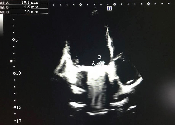
The transthoracic echocardiography (TTE) findings. Control TTE showing residual thrombus on the prosthetic valve.
Prosthetic valve dysfunction is a rare clinical condition that is life-threatening if it develops acutely and is difficult to diagnose in emergency services with classical diagnostic methods.[9] This dysfunction may be caused by pannus tissue or thrombus formation on the lid. If the cause of the pathology is pannus formation, the clinical situation progresses more slowly. However, if the cause is thrombus formation due to insufficent anticoagulation, clinical deterioration occurs suddenly.[10-11] Other rare causes of prosthetic valve dysfunction are endocarditis and valve degeneration. The etiology of the pathology should be identified in order to decide the appropriate therapeutic approach for these patients.[1] In this case, the patient presented with sudden clinical worsening due to valve dysfunction caused by a thrombus. The most important reason for thrombus formation is insufficent anticoagulation.
The patient’s clinical condition was complicated in terms of a clear diagnosis. Patients who present to emergency departments with acute heart failure should be evaluated promptly by experienced clinicians. Patients should receive aggressive treatment together with routine evaluations such as history, physical examination, ECG, chest X-ray, and bedside echocardiography.[12] Care should be taken to intubate and connect patients who develop severe respiratory failure at the time of diagnosis or during follow-up.[13] Non-invasive mechanical ventilation methods are the first choice for airway support in conscious and cooperative patients.[14] Despite the fact that the number of patients admitted to emergency services with acute heart failure clinics is quite high, 14%–29% of patients are misdiagnosed.[15] Although TTE is generally effective in diagnosing such patients, it may not always be sufficient for evaluating patients who have undergone valve replacement.[8] In such cases, valve evaluation with fluoroscopy is an alternative diagnostic method.[2] In this case, the patient’s medical treatment started promptly, and the tests that could be done in the emergency room were performed in order to determine the etiology. Due to the worsening of the clinical condition of the patient, who was not responding to medical intervention, the patient was intubated and connected to a mechanical ventilator. As a result of the inadequacy of the basic diagnostic methods at the stage of etiology determination, fluoroscopy was evaluated, and a definitive diagnosis was made without losing time in the emergency room.
Thrombus-related mechanical valve dysfunctions can be treated with surgical intervention or thrombolytic therapy. In this case, due to the lack of surgical intervention in the hospital and within the province borders, thrombolytic therapy was administered to the patient in this case even though the thrombus was near the surgical margins. Within a few hours, the patient’s clinical condition showed a significant improvement. Following clinical stabilization, the patient was extubated in an emergency care setting, and an echocardiogram revealed significant thrombus regression and valve remobilization.
In conclusion, fluoroscopy may be used when classical diagnostic techniques are ineffective or diagnosis cannot be verified in patients with a history of mechanical prosthetic valve and acute heart failure. This is a quick and non-invasive method that works well without using radiopaque materials. It should be kept in mind as a diagnostic tool, particularly in patients whose valve status cannot be evaluated by TTE.
Footnotes
Funding: There was no financial support or sponsorship.
Ethical approval: Informed consent was obtained from the patient enrolled in this study.
Conflicts of interest: There are no conflicts of interest.
Author contributions: All authors contributed significantly to the writing and revision of this manuscript and approved the final version.
All the supplementary files in this paper are available at http://wjem.com.cn.
REFERENCES
- 1.Dangas GD, Weitz JI, Giustino G, Makkar R, Mehran R. Prosthetic heart valve thrombosis. J Am Coll Cardiol. 2016;68(24):2670–89. doi: 10.1016/j.jacc.2016.09.958. [DOI] [PubMed] [Google Scholar]
- 2.Joint Task Force on the Management of Valvular Heart Disease of the European Society of Cardiology (ESC);European Association for Cardio-Thoracic Surgery (EACTS) Vahanian A, Alfieri O, Andreotti F, Antunes MJ, et al. Guidelines on the management of valvular heart disease (version 2012) Eur Heart J. 2012;33(19):2451–96. doi: 10.1093/eurheartj/ehs109. [DOI] [PubMed] [Google Scholar]
- 3.Zuckerman BD, Sapirstein W. Prosthetic cardiac valve replacement. J Thorac Cardiovasc Surg. 2003;125(6):1563. doi: 10.1016/s0022-5223(03)00120-x. [DOI] [PubMed] [Google Scholar]
- 4.Emrecan B, Taştan H, Şimşek Ş, Çekirdekoğlu K. Prosthetic valve endocarditis:A challenging complication of prosthetic valves. Turk Gogus Kalp Damar Cerrahisi Derg. 2019;27(2):159–164. doi: 10.5606/tgkdc.dergisi.2019.16796. [DOI] [PMC free article] [PubMed] [Google Scholar]
- 5.Eikelboom JW, Connolly SJ, Brueckmann M, Granger CB, Kappetein AP, Mack MJ, et al. Dabigatran versus warfarin in patients with mechanical heart valves. N Engl J Med. 2013;369(13):1206–14. doi: 10.1056/NEJMoa1300615. [DOI] [PubMed] [Google Scholar]
- 6.Laplace G, Lafitte S, Labèque JN, Perron JM, Baudet E, Deville C, et al. Clinical significance of early thrombosis after prosthetic mitral valve replacement:a postoperative monocentric study of 680 patients. J Am Coll Cardiol. 2004;43(7):1283–90. doi: 10.1016/j.jacc.2003.09.064. [DOI] [PubMed] [Google Scholar]
- 7.Zencir C, İnanç MT, Kaya MG, Şarlı B. Recurrent thrombosis due to insufficient anticoagulation in the prosthetic mitral valve and thrombolytic therapy:a case report. Pam Med J. 2009;2:101–3. [Google Scholar]
- 8.Cincin A, Ozben B, Atas H, Tigen K. A case of decompensated heart failure due to prosthetic valve dysfunction:quick diagnosis with fluoroscopy. J Acad Emerg Med Case Reports. 2015;6(3):63–5. [Google Scholar]
- 9.Roudaut R, Serri K, Lafitte S. Thrombosis of prosthetic heart valves:diagnosis and therapeutic considerations. Heart. 2007;93(1):137–42. doi: 10.1136/hrt.2005.071183. [DOI] [PMC free article] [PubMed] [Google Scholar]
- 10.Imasaka KI, Oe M, Baba H, Sumida H. Obstruction of the left ventricular outflow tract due to pannus formation after the implantation of a carbomedics aortic valve. Jpn J Thorac Cardiovasc Surg. 2006;54(7):304–7. doi: 10.1007/pl00022259. [DOI] [PubMed] [Google Scholar]
- 11.Ellensen VS, Andersen KS, Vitale N, Davidsen ES, Segadal L, Haaverstad R. Acute obstruction by pannus in patients with aortic medtronic-hall valves:30 years of experience. Ann Thorac Surg. 2013;96(6):2123–8. doi: 10.1016/j.athoracsur.2013.07.019. [DOI] [PubMed] [Google Scholar]
- 12.Hunter BR, Martindale J, Abdel-Hafez O, Pang PS. Approach to acute heart failure in the emergency department. Prog Cardiovasc Dis. 2017;60(2):178–86. doi: 10.1016/j.pcad.2017.08.008. [DOI] [PubMed] [Google Scholar]
- 13.Pang PS, Zaman M. Airway management and assessment of dyspnea in emergency department patients with acute heart failure. Curr Emerg Hosp Med Rep. 2013;1(2):122–5. doi: 10.1007/s40138-013-0011-9. [DOI] [PMC free article] [PubMed] [Google Scholar]
- 14.Vital FMR, Ladeira MT, Atallah AN. Non-invasive positive pressure ventilation (CPAP or bilevel NPPV) for cardiogenic pulmonary oedema. Cochrane Database Syst Rev. 2013;5:CD005351. doi: 10.1002/14651858.CD005351.pub3. [DOI] [PubMed] [Google Scholar]
- 15.Collins SP, Peacock WF, Lindsell CJ, Clopton P, Diercks DB, Hiestand B, et al. S3 detection as a diagnostic and prognostic aid in emergency department patients with acute dyspnea. Ann Emerg Med. 2009;53(6):748–57. doi: 10.1016/j.annemergmed.2008.12.029. [DOI] [PubMed] [Google Scholar]