



Abstract
BACKGROUND:
Femoral and tibial fractures may result in delayed union and nonunion, posing significant challenges in orthopedic practice. The Ilizarov technique has emerged as a promising solution for managing these complex cases.
OBJECTIVES:
Evaluate the radiographic and functional results of Ilizarov fixation in the treatment of nonunion of tibia and femur fractures.
DESIGN:
Retrospective
SETTINGS:
Hospitals affiliated with a university hospital
PATIENTS AND METHODS:
Patient demographics, fracture characteristics, and treatment details were analyzed for the period from October 2015 to September 2022 in patients who were treated for nonunion of the tibia and femur using the Ilizarov fixator. Clinical and radiological assessments were performed using the Association for the Study and Application of Methods of Ilizarov (ASAMI) criteria. The study focused on assessing the average duration for union and frame removal, bone results, successful union rates, and functional results using the ASAMI criteria, obtaining data from the existing medical records, spanning various medical facilities treating nonunion fractures.
SAMPLE SIZE:
126 patients
RESULTS:
The average duration for union and frame removal was 8 months, with excellent bone results observed in 60.32% of cases. Out of 126 patients, 118 achieved successful union, while there were 2 failure cases necessitating amputation (1.52%). Functional results revealed excellent outcomes in 39.68% of cases. Complications included pin tract infections, ankle and knee stiffness, and limb shortening. External fixation duration and infection eradication were consistent with previous research, emphasizing the technique's effectiveness.
CONCLUSIONS:
The Ilizarov technique proved highly effective in managing nonunion tibia and femur fractures, offering favorable outcomes in terms of union, infection control, pain relief, and functional recovery. While excellent bone outcomes do not guarantee optimal function, this method remains a reliable approach for complex cases.
LIMITATIONS:
Potential biases inherent in retrospective analyses and the need for further randomized controlled trials to comprehensively compare treatment modalities.
INTRODUCTION
Femoral and tibial fractures, often resulting from high-energy trauma are significant limb injuries, and occasionally give rise to delayed union and nonunion as potential complications.1
There are significant differences in interpretation when it comes to the definition of fracture nonunion, which is still up for debate. Our preferred definition identifies a fracture as nonunion when it fails to heal and is unlikely to do so without external intervention.2 Managing nonunion in the context of femoral fractures can be particularly challenging, with an incidence rate of less than 1% among patients who undergo surgical treatment.3
In contrast, tibial nonunion after a fracture can vary considerably, ranging between 2.5% and 17.5%, owing to its anatomical location and the presence of soft-tissue coverage. However, this figure is subject to significant variation based on numerous factors.4 Nonunion stands as one of the most common clinical challenges faced by orthopedic surgeons. It consumes significant time and resources, extends the period of morbidity, delays the return to functional status, and impacts the psychological response to the injury. Moreover, nonunion gives rise to a host of associated problems, including insufficient soft-tissue coverage, bone loss, infection, and residual deformity. Advances in antimicrobial therapy and microvascular surgery, coupled with the application of Ilizarov's principles of distraction osteogenesis, have preserved the functionality of many limbs that would otherwise have faced amputation.5 Nonunion can be categorized based on various factors, including its hypertrophic or atrophic nature, stiffness or mobility, the presence or absence of bone defects, and the involvement of infection.6
Treatment options for nonunion vary widely and encompass a range of approaches. These include nail dynamization,7 exchange nailing,8 plate osteosynthesis,9 the induced membrane technique also known as the Masquelet technique,10 external fixation, and adjunctive therapies like electrical stimulation11 or ultrasound stimulation.12 Additionally, treatment may involve bone grafting using either autogenous or allogenic bone grafts13 and the use of bone morphogenetic proteins (BMPs).14 The use of external fixation as the definitive method of stabilization in cases of fracture nonunion has been documented at a rate of 50%. This percentage notably escalates to 90% when addressing the management of infected fracture nonunions.15
This retrospective case series evaluates the radiographic outcomes and functional results associated with Ilizarov fixation in the treatment of tibia and femur fracture nonunions. Through a meticulous analysis of these results, we aimed to contribute to the ongoing discourse surrounding optimal strategies for managing these challenging orthopedic conditions.
PATIENTS AND METHODS
The study obtained approval from the Institutional Review Board under registration number Pat._3Med.Research.0000003, ensuring ethical compliance and participant protection. It adhered to the Helsinki Declaration ethical guidelines for medical research involving human subjects.
This study represents a retrospective assessment spanning seven years, from October 2015 to September 2022, involving patients treated for nonunion of the tibia and femur at hospitals affiliated with Al Azhar University in Cairo, Egypt (Al Hussien and Bab Al Shariah). The inclusion criteria encompassed cases of nonunion of the femur or tibia (either proximal, shaft, or distal) treated with an Ilizarov fixator at least 1 year after the end of treatment.
Patients underwent clinical evaluation every two weeks for pin site infection and frame stability. Radiological assessments were conducted every four weeks to monitor the nonunion site and consolidation regeneration. The fixator was removed once the regenerate had consolidated, and corticotomy was performed on three of four sides to address the nonunion, as confirmed by anteroposterior and lateral X-rays. Functional and radiographic results were assessed using the ASAMI (Association for the Study and Application of the Method of Ilizarov) scoring system16 a widely accepted method for evaluating limb reconstruction results (Tables 1 and 2).
Table 1.
Bone results using the Association for the Study and Application of Methods of Ilizarov (ASAMI) scoring system.
| Bone result | Description |
|---|---|
| Excellent | Union, no infection, deformity <7°, limb length discrepancy <2.5 cm |
| Good | Union + any two of the following: Absence of infection <7° deformity and limb length inequality <2.5 cm |
| Fair | Union + only one of the following: Absence of infection, deformity <7° and limb length inequality <2.5 cm |
| Poor | Non-union/refracture/union + infection + deformity >7° + limb length inequality >2.5 cm |
Table 2.
Functional results using the Asociation for the Study and Application of Methods of Ilizarov (ASAMI) scoring system.
| Functional result | Description |
|---|---|
| Excellent | Active, no limp, minimum stiffness (loss of <15° knee extension/<15° dorsiflexion of ankle), no RSD, insignificant pain |
| Good | Active, with one or two of the following: Limp, stiffness, RSD, significant pain |
| Fair | Active, with three or all of the following: Limp, stiffness, RSD , significant pain |
| Poor | Poor inactive (unemployment or inability to perform daily activities because of injury) |
| Failure | Amputation |
RSD: Radiographic Structural Deviation
RESULTS
A total of 211 patients met the predefined inclusion criteria for the study. Unfortunately, 85 patients were lost to follow-up, and efforts to track their whereabouts and treatment progress proved infeasible. The exact status of these individuals, whether they continued their treatment or sought care elsewhere, remains unknown. Of the remaining participants, 17 were females (13.49%), and 109 were males (86.51%) (Table 3). The average age at the time of surgery was 38 years (range, 11 to 65 years). The mean follow-up time was 19.77 (2.98) months (range 12 to 24 months). The femur was involved in 22 cases (17.46%), while the tibia was involved in 104 cases (82.54%). Nonunion type was atrophic in 102 cases (80.95%) and hypertrophic in 24 cases (19.05%). Initial fractures were closed in 74 cases (58.73%) and open in 52 cases (41.27%). According to Gustilo-Anderson's classification for open injuries,17 2 patients had Grade II, and 50 had Grade III. Infected nonunion was present in 93 cases (73.81%). Nonunion with additional deformity (varus, valgus, rotation, shortening) was observed in 48 cases (38.06%). The average duration before the operation was 9.77 months.
Table 3.
Patient demographics.
| Characteristic | Variant |
|---|---|
| Total patients | 126 |
| Gender (M:F) | 109 (86.51):17 (13.49) |
| Age (years) | 38 (11 to 65) |
| Fracture location | |
| Femur | 22 (17.46) |
| Tibia | 104 (82.54) |
| Nonunion type | |
| Atrophic | 102 (80.95) |
| Hypertrophic | 24 (19.05) |
| Fracture type | |
| Closed | 74 (58.73) |
| Open | 52 (41.27) (Ga 2 patients, Gb 50 patients) |
| Infected Nonunion | 93 (73.81) |
| Nonunion with deformity | 48 (38.06) |
| Average duration pre-op (months) | 9.8 |
Data are median (range) or numbers (percentage). G: Gustilo-Anderson Grading (
Gustillo grade II and
Gustillo grade III).
Twenty-nine patients underwent plastic intervention for soft tissue procedures. Distraction was employed in 22 cases, compression in 68 cases, bone transport in 31 cases, and the induced membrane technique in 5 cases. The surgical technique depended on the initial clinical assessment and radiographs. If initial metalwork was present, it was removed, and in patients with previous intramedullary nails, reaming was performed. If soft tissue coverage was required, plastic surgeons performed the procedure during the same sitting. Regarding additional techniques, fibular osteotomy was performed in 66 cases. Bone grafting was carried out in 9 cases, and combined fibular osteotomy and bone grafting were performed in 29 cases. In most instances, immediate weight-bearing was encouraged to stimulate early bone healing. Physiotherapy was initiated to maintain or regain a normal range of motion in the knee and ankle joints.
The average duration for union and frame removal was 8 months, ranging from 4 to 14 months. Bone results revealing excellent outcomes in 76 cases (60.32%), good outcomes in 20 cases (15.87%), fair outcomes in 23 cases (18.25%), and poor outcomes in 7 cases (5.56%) (Table 4). Remarkably, 118 patients achieved successful union after Ilizarov removal. There were excellent functional results in 50 cases (39.68%), good functional results in 46 cases (36.51%), fair functional results in 23 cases (18.25%), and poor functional results in 5 cases (3.97%), and 2 cases (1.58%) resulted in failure necessitating amputations.
Table 4.
Results and complications.
| Aspect | Result | Number of cases |
|---|---|---|
| Union and frame removal duration (months) | Average: 8 (4-14) | |
| Bone results (ASAMI scoring) | Excellent | 76 (60.32) |
| Good | 20 (15.87) | |
| Fair | 23 (18.25) | |
| Poor: | 7 (5.56) | |
| Functional results (ASAMI scoring) | Excellent | 50 (39.68) |
| Good | 46 (36.51) | |
| Fair | 23 (18.25) | |
| Poor | 5 (3.97) | |
| Failure | 2 (1.58) | |
| Complications | Pin tract infections: Successfully treated | 60 (47.62) |
| Ankle stiffness: improved by physiotherapy | 82 (65.08) | |
| Knee stiffness: improved by physiotherapy | 26 (20.63) | |
| Limb shortening | 22 (17.46) | |
| Nonunion | 6 (4.76) | |
| Equinus deformity | 1 (0.79) | |
| Recurvatum deformity | 3 (2.38) | |
| Procurvatum deformity | 1 (0.79) | |
| External Rotation deformity | 1 (0.79) |
Data are numbers (percentage).
The most common complication was pin tract infection. We successfully managed this with local dressing and oral antibiotics. Additionally, we noted varying degrees of ankle stiffness in 82 cases and knee stiffness in 26 cases. Fortunately, these stiffness issues showed improvement with physiotherapy. Insignificant limb shortening occurred in 22 cases. Other deformities included one patient with an equinus deformity, three patients with recurvatum deformities, and one patient with a procurvatum deformity. One patient also presented with an external rotation deformity (Table 4).
Case presentation
A 28-year-old male presented in February 2016 with a Gustilo-Anderson type 2 open fracture of the left distal tibia. The initial management involved meticulous debridement, a course of antibiotics, and the application of an external fixator (Figure 1). After one month and consolidation of the soft tissue envelope, the external fixator was removed, and intramedullary nailing was performed (Figure 2). However, upon regular follow-up after 7 months, the patient's fracture exhibited signs of nonunion, indicating a lack of bone healing. A revision surgery was then undertaken, during which the intramedullary nail was replaced with a plate and an autogenous bone graft. However, union remained elusive, and the patient developed varus angulation, recurvatum deformity (Figure 3), and a troubling infection at the surgical site. The persistence of these complications necessitated the removal of the plate. Subsequently, the patient underwent treatment with the Ilizarov technique (Figure 4). The Ilizarov frame was meticulously adjusted to gradually correct the varus angulation and recurvatum deformity, while also applying controlled compression to stimulate the healing process. After 5 months the patient's fracture finally achieved union, and the infection that had plagued his recovery was successfully eradicated, and the deformity was significantly corrected (Figure 5).
Figure 1.

Radiograph demonstrating the initial management of a Gustilo-Anderson type 2 open fracture of the left distal tibia, utilizing an external fixator for the tibia fracture and a plate for the distal fibula.
Figure 2.
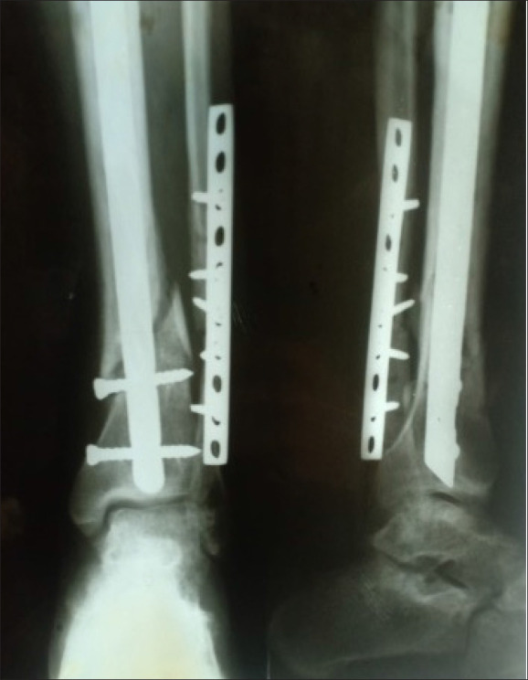
Radiograph showing the management after one month, with removal of the external fixator and subsequent intramedullary nailing.
Figure 3.

Clinical image depicting varus angulation and recurvatum deformity following revision surgery with placement of a plate and autogenous bone graft.
Figure 4.
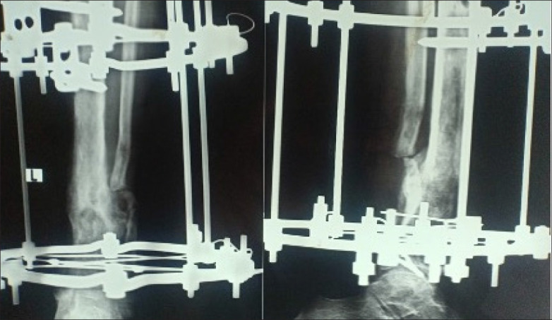
Radiograph illustrating the application of the Ilizarov technique following removal of the plate due to persistent complications.
Figure 5.

Radiograph demonstrating union of the fracture, successful eradication of infection, and significant correction of deformity after 5 months.
DISCUSSION
The Ilizarov technique, initially developed in Kurgan, Russia, in 1950 by Gavril Ilizarov, has emerged as a highly effective and reliable treatment for challenging orthopedic conditions, including tibial and femoral nonunion. These complex cases often necessitate intricate and time-consuming surgical interventions.18
General indications for the use of external fixation in cases of fracture nonunion encompass specific clinical scenarios. These include percutaneous correction of alignment, particularly when addressing deformities at the nonunion site or those proximate to a joint, or when mechanical stimulation of the nonunion site through compression or distraction is required. External fixation is also used for fixation of juxta-articular or ‘Emmental' bone fragments and in anticipation of staged procedures for bone defect or soft tissue reconstruction. It is particularly useful in cases characterized by a narrow medullary canal and instances involving infection at the fracture site. Furthermore, specific anatomical indications exist where external fixation functions as an offloading frame or augmentation of fixation. This comprises situations such as subtrochanteric femur fractures and scenarios involving juxta-articular fractures associated with stiffness or deformity near a joint. In light of a growing comprehension of the outcomes associated with both external and internal fixation methods, there is an ongoing refinement of the relative indications for the use of external fixation in managing fracture nonunion.15
In our study, we employed ASAMI criteria, widely recognized for their comprehensive assessment of orthopedic procedures. Our findings align with prior research, indicating that the Ilizarov technique consistently produces favorable results in terms of union, infection control, pain relief, and the patients' ability to resume daily activities.
Sahu et al conducted a study involving 60 patients with complex tibial nonunion treated using the Ilizarov technique. Their research demonstrated an average time to successful union of 7 months, with bone results categorized as excellent in 83.33% of cases, good in 11.67%, fair in 3.33%, and poor in 1.67%. Functional results were equally promising, with excellence noted in 75% of cases, good in 16.67%, fair in 5%, and poor in 3.33%.19
Similarly, Zaidi et al treated 48 patients with nonunion of the femur using the Ilizarov technique. The average external fixator application time was 8 months, and bone results were excellent in 31.25% of cases, good in 62.5%, and poor in 4.17%. Functional results showed excellence in 20.83% of cases, good in 52.08%, fair in 14.85%, and poor in 10.42%.20
Chattopadhyay et al addressed difficult non-union cases of long bones with the Ilizarov technique, achieving a mean union time of 34 weeks. Bone results in their study were excellent in 50% of cases, good in 33.33%, and fair in 16.67%. Functional results included excellence in 33.33%, good in 33.33%, and fair in 33.33%.21
Furthermore, Szelerski et al conducted a retrospective study involving 102 cases treated with the Ilizarov technique for tibial nonunion. The mean duration of Ilizarov stabilization was 7.9 months. Their ASAMI bone scores were excellent in 86.27% of cases, good in 8.82%, and poor in 4.9%. ASAMI functional scores were excellent in 65.69%, good in 29.41%, and poor in 4.9%.22
In our study, the average external fixation duration was 8 months, consistent with previous research. We also achieved complete eradication of infection, mirroring findings from other studies. Our bone results were excellent in 60.32%, good in 15.87%, fair in 18.25%, and poor in 5.56% of cases, similar to previous reports. Likewise, our functional results showed excellence in 39.68%, good in 36.51%, fair in 18.25%, and poor in 5.56% of cases, in line with previous research.
We observed that excellent bone outcomes do not necessarily guarantee good functional results. This underscores the influence of soft tissue and neurovascular structures on functional results. However, our study does not directly compare with other treatment options, emphasizing the need for further randomized controlled trials to draw more valuable conclusions.
This study has a few limitations. Firstly, it employed a retrospective design, relying on existing medical records, which may introduce biases and missing data. Additionally, as a chart review, surgeries done in another institution without patient follow-up may not have been documented. Furthermore, the relatively small sample size of 126 patients may impact the robustness of the findings, particularly when examining results across different variables. The study's use of various techniques within the Ilizarov method introduces variability in the treatment approach. It lacked a control group for comparison with other treatments. Moreover, demographic information on comorbidities, such as the number of smokers and patients with diabetes, was not collected or included in the analysis. These variables might have played a role in understanding their effect on the radiographic and functional. Lastly, there was limited discussion of long-term complications and secondary procedures beyond the immediate post-operative period. These limitations should be considered when interpreting the study's results and their broader implications.
This retrospective analysis underscores the effectiveness of the Ilizarov technique in managing nonunion tibia and femur fractures, offering a promising solution to a challenging orthopedic problem. Our findings align with existing research, indicating consistently favorable outcomes in terms of union, infection control, pain relief, and the restoration of daily activities. This reaffirms the Ilizarov technique as a reliable approach, particularly in complex cases requiring intricate interventions.
While our study presents positive results, it is essential to acknowledge that achieving excellent bone outcomes does not always translate into optimal functional results. The interplay of soft tissue and neurovascular structures plays a crucial role in functional recovery. However, our study lacks a direct comparative analysis with other treatment modalities, emphasizing the need for further randomized controlled trials to provide more conclusive evidence.
Nonetheless, our study contributes insights into the potential clinical advantages of the Ilizarov technique for managing nonunion tibia and femur fractures. It offers hope for improved patient outcomes and the preservation of limb functionality. Ongoing research and refinement of treatment approaches will continue to deepen our understanding of this orthopedic method.
Funding Statement
None.
Footnotes
CONFLICT OF INTEREST: None.
REFERENCES
- 1.Cleveland KB.. Delayed union and nonunion of fractures. In: Canale & Beaty: Campbell's operative orthopaedics. 12th ed. Ch. 59: Elsevier/Mosby; 2013. p. 4253. [Google Scholar]
- 2.Wittauer M, Burch MA, McNally M, Vandendriessche T, Clauss M, Della Rocca GJ, et al.. Definition of long-bone nonunion: A scoping review of prospective clinical trials to evaluate current practice. Injury. 2021. Nov;52(11):3200–5. [DOI] [PubMed] [Google Scholar]
- 3.Basile G, Fozzato S, Petrucci QA, Gallina M, Bianco Prevot L, Accetta R, et al.. Treatment of Femoral Shaft Pseudarthrosis, Case Series and Medico-Legal Implications. J Clin Med. 2022. Dec 14;11(24):7407. [DOI] [PMC free article] [PubMed] [Google Scholar]
- 4.Tzioupis C, Giannoudis PV.. Prevalence of long-bone non-unions. Injury. 2007. May;38 Suppl 2: S3–9. [DOI] [PubMed] [Google Scholar]
- 5.Bose D, Kugan R, Stubbs D, McNally M.. Management of infected nonunion of the long bones by a multidisciplinary team. Bone Joint J. 2015. Jun;97-B(6):814–7. [DOI] [PubMed] [Google Scholar]
- 6.Wu CC, Chen WJ.. A revised protocol for more clearly classifying a nonunion. J Orthop Surg (Hong Kong). 2000. Jun;8(1):45–52. [DOI] [PubMed] [Google Scholar]
- 7.Wu CC. The effect of dynamization on slowing the healing of femur shaft fractures after interlocking nailing. J Trauma. 1997. Aug;43(2):263–7. [DOI] [PubMed] [Google Scholar]
- 8.Furlong AJ, Giannoudis PV, DeBoer P, Matthews SJ, MacDonald DA, Smith RM.. Exchange nailing for femoral shaft aseptic non-union. Injury. 1999. May;30(4):245–9. [DOI] [PubMed] [Google Scholar]
- 9.Choi YS, Kim KS.. Plate augmentation leaving the nail in situ and bone grafting for non-union of femoral shaft fractures. Int Orthop. 2005. Oct;29(5):287–90. [DOI] [PMC free article] [PubMed] [Google Scholar]
- 10.Soliman ME, Elzohairy MM, AbdelWahab AM, Khaira YM, Elbatrawy Y, Abdalla UG, et al.. Masquelet's Induced Membrane Technique for Reconstruction of Large Extra-Articular Intercalary Bone Defect. Journal of Limb Lengthening & Reconstruction [Internet]. 2021. Dec [cited 2023 Oct 9];7(2):125. Available from: https://journals.lww.com/jllr/Fulltext/2021/07020/Masquelet_s_Induced_Membrane_Technique_for.7.aspx [Google Scholar]
- 11.Griffin XL, Warner F, Costa M.. The role of electromagnetic stimulation in the management of established non-union of long bone fractures: what is the evidence? Injury. 2008. Apr;39(4):419–29. [DOI] [PubMed] [Google Scholar]
- 12.Jingushi S, Mizuno K, Matsushita T, Itoman M.. Low-intensity pulsed ultrasound treatment for postoperative delayed union or nonunion of long bone fractures. J Orthop Sci. 2007. Jan;12(1):35–41. [DOI] [PubMed] [Google Scholar]
- 13.Wang JW, Weng LH.. Treatment of distal femoral nonunion with internal fixation, cortical allograft struts, and autogenous bone-grafting. J Bone Joint Surg Am. 2003. Mar;85(3):436–40. [DOI] [PubMed] [Google Scholar]
- 14.Kanakaris NK, Lasanianos N, Calori GM, Verdonk R, Blokhuis TJ, Cherubino P, et al.. Application of bone morphogenetic proteins to femoral non-unions: a 4-year multicentre experience. Injury. 2009. Dec;40 Suppl 3: S54–61. [DOI] [PubMed] [Google Scholar]
- 15.Simpson AHRW, Robiati L, Jalal MMK, Tsang STJ.. Non-union: Indications for external fixation. Injury. 2019. Jun;50 Suppl 1:S73–8. [DOI] [PubMed] [Google Scholar]
- 16.Paley D, Catagni MA, Argnani F, Villa A, Benedetti GB, Cattaneo R.. Ilizarov treatment of tibial nonunions with bone loss. Clin Orthop Relat Res. 1989. Apr;(241):146–65. [PubMed] [Google Scholar]
- 17.Gustilo RB, Anderson JT.. Prevention of infection in the treatment of one thousand and twenty-five open fractures of long bones: retrospective and prospective analyses. J Bone Joint Surg Am. 1976. Jun;58(4):453–8. [PubMed] [Google Scholar]
- 18.Govindasamy R, Gnanasundaram R, Kasirajan S, Sengani P, Meleppuram JJ.. A retrospective study of ilizarov fixation in infected non-union tibia: 36 cases. Int J Res Orthop [Internet]. 2016. Sep 3 [cited 2023 Oct 9];2(3):70. Available from: http://www.ijoro.org/index.php/ijoro/article/view/57 [Google Scholar]
- 19.Sahu RL, Ranjan R.. Treatment of complex nonunion of the shaft of the tibia using Ilizarov technique and its functional outcome. Niger Med J [Internet]. 2016. [cited 2023 Oct 9];57(2):129–33. Available from: https://www.ncbi.nlm.nih.gov/pmc/articles/PMC4872491/ [DOI] [PMC free article] [PubMed] [Google Scholar]
- 20.Zaidi IH, Sahito B, Tariq SM, Kumar D, Ali M.. Outcome of Nonunion Femur with Ilizarov Technique. Journal of Surgery Pakistan. 2015; [Google Scholar]
- 21.Chattopadhyay P, Mandal P, Sabui KK, Banka PK.. Treatment of Difficult Non-union of Long Bones using the Ilizarov Technique. 2017;4(12). [Google Scholar]
- 22.Szelerski £, Pajchert Koz³owska A, ¯arek S, Górski R, Mochocki K, Dejnek M, et al.. A new criteri111on for assessing Ilizarov treatment outcomes in nonunion of the tibia. Arch Orthop Trauma Surg. 2021. May;141(5):879–89. [DOI] [PMC free article] [PubMed] [Google Scholar]