
Fig. 2. In situ cryoET of tau deposits in vitrified postmortem AD brain.
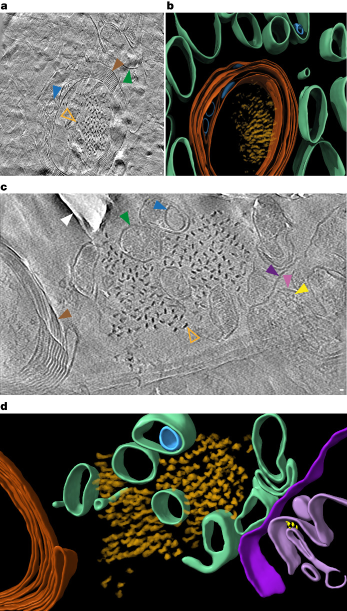
a, Tomographic slice of intracellular tau pathology in postmortem AD brain cryo-section (Supplementary Video 4). Open orange arrowhead, filament oriented axially (z axis) within the tomogram; brown arrowhead, myelinated axon; green arrowhead, subcellular compartment; blue arrowhead, intracellular membrane-bound organelle. Scale bar, 10 nm. b, Segmentation of tomogram coloured as in a. c, Tomographic slice of extracellular tau pathology in AD postmortem brain cryo-section (Supplementary Video 5). Open orange arrowhead, tau filament oriented axially (z axis) within the tomogram; brown arrowhead, myelinated axon; dark and light purple arrowheads, outer and inner membranes of damaged mitochondrion, respectively; yellow arrowhead, putative Fo-F1 ATPase; dark green arrowhead, subcellular compartment; blue arrowhead, intracellular membrane-bound organelle; white arrowhead, knife damage. Scale bar, 10 nm. d, Segmentation of tomogram coloured as in c.