Abstract
Objective
Cerebral artery dissection (CeAD) is a rare but serious disease. Genetic risk assessment for CeAD is lacking in Chinese population. We performed genome-wide association study (GWAS) and computed polygenic risk score (PRS) to explore genetic susceptibility factors and prediction model of CeAD based on patients in Huashan Hospital.
Methods
A total of 210 CeAD patients and 280 controls were enrolled from June 2017 to September 2022 in Department of Neurology, Huashan Hospital, Fudan University. We performed GWAS to identify genetic variants associated with CeAD in 140 CeAD patients and 210 control individuals according to a case and control 1:1.5 design rule in the training dataset, while the other 70 patients with CeAD and 70 controls were used as validation. Then Kyoto Encyclopedia of Genes and Genomes (KEGG) pathway and Gene Ontology (GO) enrichment analyses were utilized to identify the significant pathways. We constructed a PRS by capturing all independent GWAS SNPs in the analysis and explored the predictivity of PRS, age, and sex for CeAD.
Results
Through GWAS analysis of the 140 cases and 210 controls in the training dataset, we identified 13 leading SNPs associated with CeAD at a genome-wide significance level of P < 5 × 10− 8. Among them, 10 SNPs were annotated in or near (in the upstream and downstream regions of ± 500Kb) 10 functional genes. rs34508376 (OR2L13) played a suggestive role in CeAD pathophysiology which was in line with previous observations in aortic aneurysms. The other nine genes were first-time associations in CeAD cases. GO enrichment analyses showed that these 10 genes have known roles in 20 important GO terms clustered into two groups: (1) cellular biological processes (BP); (2) molecular function (MF). We used genome-wide association data to compute PRS including 32 independent SNPs and constructed predictive model for CeAD by using age, sex and PRS as predictors both in training and validation test. The area under curve (AUC) of PRS predictive model for CeAD reached 99% and 95% in the training test and validation test respectively, which were significantly larger than the age and sex models of 83% and 86%.
Conclusions
Our study showed that ten risk loci were associated with CeAD susceptibility, and annotated functional genes had roles in 20 important GO terms clustered into biological process and molecular function. The PRS derived from risk variants was associated with CeAD incidence after adjusting for age and sex both in training test and validation.
Supplementary Information
The online version contains supplementary material available at 10.1186/s12883-024-03759-0.
Keywords: Cerebral artery dissection, Genetic risk, Polygenic risk score, Predictive model
Introduction
The incidence of stroke in young patients has increased considerably over the past few decades [1]. Although the incidence of cerebral artery dissection (CeAD) is estimated to be about 2.6-3 per 100,000 inhabitants per year, CeAD is a major cause of stroke in young adults [2]. Compared with older adults, young adults have more ischemic infarcts from CeAD - up to 6.8% [2]. Patients can present with various clinical symptoms, some of which are benign, but most patients have disabling symptoms, especially in those with subarachnoid haemorrhage and brainstem involvement [3]. Risk factors for CeAD include inherited connective tissue diseases, hyperhomocysteinemia, migraine, recent history of infection, and neck massage [4]. However, those risk factors often insufficient to explain the pathology of CeAD. Due to the low incidence and challenge in diagnoses, there are few study cohorts of CeAD worldwide.
Arterial dissection is separation of the arterial walls. It can occur in all large and medium sized arteries. Recently, studies have shown genetic predisposition of abdominal and thoraic aortic aneurysms and dissections (AAD). There are several genes that have been identified as strong causative risk for AAD through family studies [5, 6]. Yet the most of remaining AAD cases involved common genetic variants affecting many genes. In a European cohort of CeAD, rare genetic imbalance was associated with high risk of CeAD and functional outcome after ischemic stroke [7, 8]. Furthermore, this cohort study revealed that a common variation in PHACTR1 was involved in the CeAD [9]. These results indicate that CeAD have complex origins, while the genetic predisposition of CeAD in Asian populations have not been reported yet.
To our knowledge, this is the first study for CeAD in Chinese population. In the current study, we explored a GWAS analysis of 210 CeAD patients and 280 control individuals and identified ten previously unpublished genome-wide significant loci located in ten genes. We further constructed polygenic risk scores (PRS) in our cases and constructed predictive model for CeAD by using age, sex and PRS as predictors in verification.
Methods
Study participants
We enrolled 210 CeAD patients and 280 control individuals from June 2017 to September 2022 in Department of Neurology, Huashan Hospital, Fudan University. The CeAD patients were selected from the Chinese Cervicocephalic artery dissection study (CCADS) [10]. The control group was from the Health Screening cohort in Huashan Hospital in the same period. This cohort was aimed to evaluate the genomic and epidemiological risk of cognitive impairment. The age and the gender of subjects were matched to the cases group.
Genotype calling and quality control
Genotyping data of patients were derived from Exome sequencing in DNA samples relying on AmCareSeq-2000 System (Jiajian Biological Engineering Technology Co., LTD, Guangzhou) at recruitment, and genotyping data of controls were derived from GWAS chipInfinium Asian Screening Array Kit relying on Illumina BEADLAB System at recruitment. For each case, NovaSeq WES reads are mapped with BWA MEM (version 0.7.17) to the hg19 reference genome [11]. Small variants are identified with Samtools and reported as per-sample gVCFs in GATK (version 4.2.2.0) [12]. These gVCFs are aggregated with GATK CombineGVCFs into a joint-genotyped, multi-sample project-level VCF (pVCF). Then all cases were compiled to one pVCF by GATK HaplotypeCaller tool for downstream analysis. Single nucleotide variant (SNV) genotypes with read depth (DP) less than 20 are changed to no-call genotypes using Bcftools (version 1.14) [12]. After the application of the DP genotype filter, a variant-level allele-balance filter is applied, retaining only variants that meet either of the following criteria [13]: (1) at least one homozygous variant carrier; or (2) at least one heterozygous variant carrier with an allele balance (AB) between the cutoff values (0.15 ≤ AB ≤ 0.85). We further excluded single nucleotide polymorphisms (SNPs) with minor allele frequency (MAF) < 10%, genotype missingness > 5% or Hardy–Weinberg equilibrium test P < 5 × 10− 5. The pVCF was converted to Plink file set using Plink (version 1.9) [14]. Then genotype data of patients and controls were combined with only common variants reserved. These genotyping data were imputed with Impute (version 2) software by using 1000 Genomes Project phase 3 panels as reference [15]. We applied filters to achieve high quality variants with following criteria [16]: (1) INFO score (information metric) ≥ 0.7; (2) call rate ≥ 95%; (3) minor allele frequency (MAF) ≥ 10%; (4) Hardy-Weinberg equilibrium (HWE) ≥ 5·× 10− 5. The above filtering steps were performed step by step in Plink. Variants failing to meet these criteria will be filtered in subsequent analyses. We further excluded variants in MHC region (chr6 25-35 Mb) due to extensive linkage disequilibrium (LD). The final set of data contained a total of 821,475 variants.
Population stratification
We compared the data set with the 1000 Genomes Project to infer the genetic ancestry of the population in this study according to the principal components (PCs) in 1000 Genomes Project phase 3 [17]. Based on the race of participants in the 1000 Genomes Project we used random forest classification to map the sample population to the most similar race. The results showed that all samples (cases and controls group) are clustered in the East Asian area (Supplementary Fig. S1). It can be concluded that participants in our analysis were derived from the East Asian ethnicity.
In the present study, we collected a total of 210 patients and 280 controls in two batches. The first batch included 140 patients and 210 controls, and the second batch included 70 patients and 70 controls. Participants in first batch were enrolled from 2019 to 2021 while the second batch were enrolled from 2021 to 2022 under the same diagnostic criteria. Data of the first batch were used in genome-wide association analysis (GWAS), while the second batch was used as external validation. Firth logistic regression test was used to test the association of SNPs with phenotype. Age, sex, genotyping array and first 10 principal components were adjusted for population heterogeneity in the multi-variable regressions.
Annotation and pathway analysis
Independent significant SNPs were extracted when their P-values reach genome-wide significant threshold (P ≤ 5.0 × 10− 8) and in low LD (r2 < 0.1) with other SNPs within a 500-Kb window. Lead SNPs were identified as a subset of the independent significant SNPs with the lowest P-values and were in LD with each other at r2 < 0.01 within a 1-Mb window. The lead SNPs were mapped to nearest protein-coding genes within a 1-Mb window using locuszoom tool. To explore the function of significant genes, we annotated all independent SNPs with P value ≤ 5.0 × 10− 5 using snpEff and performed pathway analysis using R (version 4.2.2) package ‘clusterProfiler’. Kyoto Encyclopedia of Genes and Genomes (KEGG) pathway and Gene Ontology (GO) enrichment analyses were performed to identify the significant pathways. P < 0.05 was set as the cutoff criterion for significant enrichment.
Predictive model and validation
We selected independent SNPs (r2 < 0.01 within a 1-Mb window) associated with CeAD based on GWAS results at P value threshold 5 × 10− 5 to construct polygenic risk score (PRS) in training dataset.
According to the previous study, PRS was referred to as genomic risk score, which was a method to predict genetic predisposition for disease of an individual. The calculation of PRS was the sums of the estimated effect sizes (β) of m SNPs, based on the estimated SNP effect sizes (β) obtained from GWAS summary statistics. (“effect sizes” refer to the measure of the strength of the association between a genetic variant and a particular trait or condition. The effect size is often represented by β. “m” is the count of genetic variants included in the PRS model.) The formula shows as below. Xij means the genotype of ith individual and jth SNP [18, 19].
m
PRSi = ∑xijβj
j=1
In this study, PRS was calculated using PRSice-2 (version 2.3.5) as the sum of risk presented at each locus and weighted by the odds ratio for that locus for each participant. Next, PRS was evaluated for whether it could help identifying individuals at risk of developing CeAD in the validation dataset. Further, we constructed prediction models using logistic regression analysis with risk factors in the training set. Three models containing different predictors were constructed and the internal validation of the models was evaluated by using a ten-fold cross validation approach. Model 1 used PRS as predictor. Model 2 included age and sex as predictors. Model 3 involved age, sex and PRS together. Further, we compared three models’ performance in the validation set.
Results
Genome-wide association analysis of CeAD
A total of 490 participants were involved in our study, including 210 patients and 280 controls. The patients (mean age 41.6 ± 11.4 years) were younger than the controls (mean age 53.3 ± 7.6 years). We performed GWAS to identify genetic variants associated with CeAD using 140 patients and 210 controls in the training dataset. Thirteen lead SNPs were found to be associated with CeAD at a genome-wide significance level of P < 5 × 10− 8 (Fig. 1A; Table 1). Genomic inflation factor (λGC) was estimated as 1.09, so P value was adjusted with λGC to diminish the impact of systematic inflation (Fig. 1B). Then, independent significant SNPs were annotated with functional genes residing in the location or within the upstream and downstream ± 500Kb regions (Table 1). The most significant SNP rs181591349 T/A (OR = 0.09, 95%CI 0.06–0.15) was at locus 17q21.32. We annotated lead SNPs with functional genes residing in the location or within the upstream and downstream ± 500Kb regions. Three SNPs (rs356357, rs528072677, and rs9325912) were excluded in the following analysis due to locating outside of protein-coding region. We also explored the association between lead SNPs and the severity of CeAD in patients, however, no significant association was found.
Fig. 1.
Result of Genome-wide association study on CeAD. A: Manhattan plot of susceptibility locus highlighting in CeAD GWAS. B: Q-Q plot for CeAD GWAS
Table 1.
Summary of lead SNPs associated with CeAD
| Lead SNP | Gene | CHR | Position | A1 | A0 | OR | 95% CI | P | MAF |
|---|---|---|---|---|---|---|---|---|---|
| rs55707024 | FLG | 1 | 152,276,282 | G | C | 0.23 | 0.15 ~ 0.37 | 3.91E-08 | 0.25 |
| rs34508376 | OR2L13 | 1 | 248,084,909 | G | T | 0.22 | 0.14 ~ 0.34 | 1.49E-08 | 0.24 |
| rs27027 | RGMB | 5 | 97,912,527 | C | G | 6.85 | 4.00 ~ 11.7 | 1.12E-09 | 0.13 |
| rs56225909 | EPHB6 | 7 | 142,471,344 | A | G | 6.39 | 4.18 ~ 9.75 | 8.49E-14 | 0.43 |
| rs855580 | PRSS3 | 9 | 33,796,912 | A | G | 7.39 | 4.29 ~ 12.7 | 3.91E-10 | 0.13 |
| rs356357 | - | 9 | 68,358,224 | T | C | 0.12 | 0.07 ~ 0.18 | 2.83E-16 | 0.42 |
| rs528072677 | - | 10 | 88,994,492 | T | C | 7.99 | 4.82 ~ 13.3 | 3.07E-12 | 0.16 |
| rs2933363 | LRRK2 | 12 | 40,880,587 | A | T | 3.01 | 2.16 ~ 4.21 | 1.88E-08 | 0.4 |
| rs62053846 | PKD1L3 | 16 | 72,012,404 | A | G | 4.39 | 3.04 ~ 6.34 | 6.71E-12 | 0.32 |
| rs9325912 | - | 17 | 21,535,937 | T | C | 0.16 | 0.11 ~ 0.25 | 5.15E-14 | 0.48 |
| rs181591349 | KPNB1 | 17 | 45,623,441 | T | A | 0.09 | 0.06 ~ 0.15 | 2.94E-17 | 0.44 |
| rs111474526 | GNAL | 18 | 11,644,364 | A | G | 6.15 | 3.50 ~ 10.8 | 4.42E-08 | 0.11 |
| rs36093924 | CYP2D6 | 22 | 42,538,399 | T | C | 0.24 | 0.16 ~ 0.37 | 1.95E-08 | 0.26 |
Functional annotation and enrichment analysis
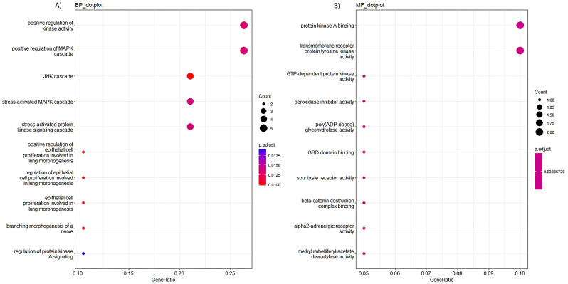
To explore the association between differential genes and CeAD, we included all independent SNPs with P value < 5 × 10− 7 and annotated them to nearby function genes within 1-Mb window. A total of 28 independent SNPs located in 20 protein-coding genes were included in enrichment analysis. We found that significant pathways were associated with biological process (BP) and molecular function (MF) in GO enrichment analysis (Fig. 2). Top 3 pathways in our gene set were GO:0033674, GO:0043410, and GO:0007254 for biological process, and GO:0051018, GO:0004714, and GO:0034211 for molecular function, respectively (Table 2). No significant cluster (P < 0.05) was found in KEGG pathway analysis.
Fig. 2.
GO enrichment analysis results of differential genes associated with CeAD in biological process (A) and molecular function (B)
Table 2.
Summary of enrichment pathways associated with CeAD
| ID | Description | Gene Ratio | P Value | P Adjust | Q Value | Gene ID |
|---|---|---|---|---|---|---|
| BP | ||||||
| GO:0033674 | positive regulation of kinase activity | 5/20 | 8.83E-05 | 0.0135 | 0.0079 | CDC42/ADRA2B/EPHB6/FGFR2/LRRK2 |
| GO:0043410 | positive regulation of MAPK cascade | 5/20 | 0.0001 | 0.0135 | 0.0079 | CDC42/ADRA2B/PJA2/FGFR2/LRRK2 |
| GO:0007254 | JNK cascade | 4/20 | 2.52E-05 | 0.0098 | 0.0057 | CDC42/PJA2/LRRK2/NPHS1 |
| GO:0051403 | stress-activated MAPK cascade | 4/20 | 9.35E-05 | 0.0135 | 0.0079 | CDC42/PJA2/LRRK2/NPHS1 |
| GO:0031098 | stress-activated protein kinase signaling cascade | 4/20 | 0.0001 | 0.0135 | 0.0079 | CDC42/PJA2/LRRK2/NPHS1 |
| GO:0060501 | positive regulation of epithelial cell proliferation involved in lung morphogenesis | 2/20 | 9.66E-06 | 0.0098 | 0.0057 | CDC42/FGFR2 |
| GO:2,000,794 | regulation of epithelial cell proliferation involved in lung morphogenesis | 2/20 | 2.03E-05 | 0.0098 | 0.0057 | CDC42/FGFR2 |
| GO:0060502 | epithelial cell proliferation involved in lung morphogenesis | 2/20 | 3.47E-05 | 0.0101 | 0.0059 | CDC42/FGFR2 |
| GO:0048755 | branching morphogenesis of a nerve | 2/20 | 4.33E-05 | 0.0101 | 0.0059 | FGFR2/LRRK2 |
| GO:0010738 | regulation of protein kinase A signaling | 2/20 | 0.0002 | 0.0191 | 0.0111 | PJA2/LRRK2 |
| MF | ||||||
| GO:0051018 | protein kinase A binding | 2/20 | 0.0013 | 0.0339 | 0.0173 | PJA2/LRRK2 |
| GO:0004714 | transmembrane receptor protein tyrosine kinase activity | 2/20 | 0.0019 | 0.0339 | 0.0173 | EPHB6/FGFR2 |
| GO:0034211 | GTP-dependent protein kinase activity | 1/20 | 0.0011 | 0.0339 | 0.0173 | LRRK2 |
| GO:0036479 | peroxidase inhibitor activity | 1/20 | 0.0011 | 0.0339 | 0.0173 | LRRK2 |
| GO:0004649 | poly (ADP-ribose) glycohydrolase activity | 1/20 | 0.0022 | 0.0339 | 0.0173 | PARG |
| GO:0032427 | GBD domain binding | 1/20 | 0.0022 | 0.0339 | 0.0173 | CDC42 |
| GO:0033040 | sour taste receptor activity | 1/20 | 0.0022 | 0.0339 | 0.0173 | PKD1L3 |
| GO:1,904,713 | beta-catenin destruction complex binding | 1/20 | 0.0022 | 0.0339 | 0.0173 | LRRK2 |
| GO:0004938 | alpha2-adrenergic receptor activity | 1/20 | 0.0032 | 0.0339 | 0.0173 | ADRA2B |
| GO:0047374 | methylumbelliferyl-acetate deacetylase activity | 1/20 | 0.0032 | 0.0339 | 0.0173 | CES1 |
Predictive model and validation
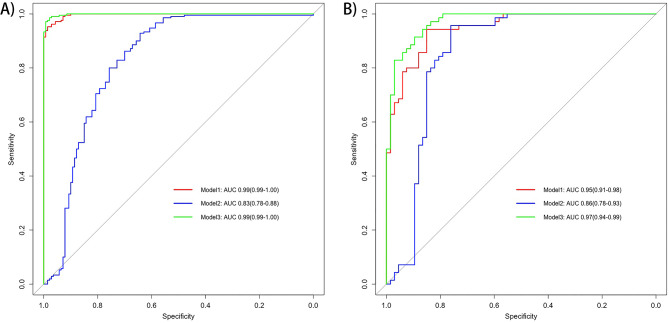
We set P value 5 × 10− 5 as threshold to construct PRS including 32 independent SNPs. Then we constructed predictive model for CeAD by using age, sex and PRS as predictors in the training set. The predictive ability of the model was estimated in validation set using area under the receiving operator characteristic curve (AUC). All three models could accurately predict the occurrence of CeAD both in training test and additional validation (Fig. 3). The AUC of Model 1 including only PRS reached 99% and 95% in training test and additional validation, respectively. The AUC of Model 2, including age and sex, reached 83% and 86% in training test and additional validation, respectively. The AUC of Model 3 including age, sex, and PRS together reached 97% (95%CI: 94 − 99%) in validation dataset.
Fig. 3.
The receiving operator characteristic curve of three different models in training test (A) and validation (B) set
Discussion
To our knowledge, this is the first study to investigate the genetic predisposition of CeAD in Chinese population. We identified ten leadind SNPs located in ten protein-coding genes for CeAD. Then, we applied a PRS by capturing all independent GWAS SNPs and demonstrated a significant association of the PRS with CeAD incidence after adjusting for age and sex both in training test and extra validation.
The present results showed that CeAD heritability is polygenic and yielded the following two findings. First, patients with CeAD were more likely to carry the variations in the genes associated with protein kinase pathways. Among them, rs34508376 (OR2L13) was a suggestive role in CeAD pathophysiology which was in line with the previous observations in aortic aneurysms [20]. The other nine genes were first-time associations in CeAD cases. Second, the PRS derived from GWAS risk variants predicted occurrence of CeAD with high stability and consistency.
It is believed that CeAD is a complex disease due to various factors [21]. According to previous studies, history of migraine, mechanical trauma and preceding infection are commonly reported in CeAD [4, 22]. However, these factors were absent in more than half of the patients [23]. Some inherited connective tissue diseases are also associated with CeAD, such as Fibromuscular dysplasia (FMD), Ehlers-Danlos syndrome (EDS) and Marfan syndrome (MFS) [24], suggesting that genetic predisposition contributed to the occurrence of CeAD. However, these monogenic disorders cannot explain the genetic involvement in the remaining sporadic cases. Thus, large-scale genetic sequencing analyses are needed to explore more general genetic variants in CeAD patients. Through GWAS analysis of enrolled 140 cases and 210 controls in the training dataset, we identified 13 leading SNPs to be associated with CeAD at a genome-wide significance level of P < 5 × 10− 8. Among them, 10 SNPs were annotated on functional genes. These ten functional genes have known roles in 20 important GO terms clustered into BP and MF. Top 3 terms in our gene set were associated with protein kinase pathways. It was in line with the previous studies [25]. Other studies have shown that the main pathophysiologic features of arterial dissection are the impairment of vessel wall, especially the disruption of medial layer. Thus, it was previously thought that genes which encoded proteins involved in the structure or function of the vascular smooth muscle cells (VSMC) elastin-contractile unit were altered to cause aortic aneurysms and dissection [26]. Recent reports indicated that macrophage metabolic reprogramming and hyper-eosinophilic inflammation are involved in the aortic dissection. To be specific, macrophages in aortic dissection had higher levels of several glycolytic intermediates and tricarboxylic acid cycle (TAC) intermediates than control, which leaded to the secretion of inflammatory factors damaging the vessel wall [27]. In addition, the accumulation of eosinophils in the arterial wall released cytotoxic products and induced inflammatory response [28, 29]. These results were in accord with the risk genes in our genetic analysis that the arterial dissection is a complex disease involving many biological processes besides the impairment of VSMC unit.
The latest study did find evidence for olfactory receptor 2L13 (OR2L13) in growth of aortic aneurysms (AAA) [20]. OR2L13 regulates the platelet activation in AAA. Platelets from patients with AAA and murine models of AAA demonstrated increased OR2L13 expression. Due to the same pathogenesis of splitting up of the arterial wall, the histological hallmark of AAA and CeAD are similar. In addition to OR2L13, several other genes in our study were also linked to vascular disease. EPHB6 encodes the receptor tyrosine kinase that was proved to regulate the vascular smooth muscle cell contraction and endothelial cell, and contribute to the development of atherosclerosis [30, 31]. Although LRRK2 is a gene associated with Parkinson’s disease, it has also been shown to regulate the function of vascular endothelial cells and promote inflammation in endothelial cells [32]. Thus, with the identified genes in our study, more research is needed to uncover the pathogenesis of CeAD.
Due to the limited sample size, we applied a series of strict filters in the above GWAS analysis to ensure the reliability of the results. However, strict filter criteria will cause some rare variants to be excluded from the analysis. Some rare genetic variants were identified to be responsible for the spontaneous coronary artery dissection [33]. Therefore, it is important to use new methods, such as gene-level collapsing analysis [34, 35], to gain insight into the genetic architecture of CeAD in the future.
Polygenic risk scores (PRS) aggregate many genetic variants across the human genome into a single score and have predictive value for multiple common diseases [18]. In recent years, genome-wide association studies (GWASs) have revealed numerous susceptible genes and loci for CeAD, indicating a more efficient predication role of PRS in CeAD [36]. However, all published genomics studies were conducted in European-ancestry populations, with few studies in other populations. We set P value 5 × 10− 5 as threshold to construct PRS including 32 independent SNPs in our cohort and constructed predictive model for CeAD by using age, sex and PRS as predictors both in internal and external verification. The predictive ability of PRS model for CeAD was stronger than age and sex. These results provide evidence that CeAD is a disease with a genetic background. Analyses that include these risk SNPs will be effective in identifying new associations with CeAD.
However, several limitations of the present study merit consideration. First, although our research had the largest sample size so far in China, the relatively small number of participants may lead to weak statistical power for evaluating the relationships. Second, some clinical risk factors are not included in our study, and the genetic data were from different platform. However, we have carried out more stringent quality control and manual check. Longitudinal and multicenter studies with large sample sizes are needed to investigate the genetic predisposition of CeAD. Third, the study focuses on the Chinese population, which is valuable, but also means that the findings might not be generalizable to other ethnic groups, given that most published genomic studies have been conducted in European-ancestry populations. However, we supplemented the results on the Chinese population so that we can compare the findings from different populations to reveal potential population-specific genetic variations and contribute to a more comprehensive understanding of CeAD susceptibility across ethnicities.
Electronic supplementary material
Below is the link to the electronic supplementary material.
Author contributions
Shufan Zhang and Dongliang Zhu made the formal analysis and wrote the main manuscript text. Zhengyu Wu wrote the review and edited the manuscript. Shilin Yang and Yuanzeng Liu made the study design. Shilin Yang and Zhu Zhu edited the draft. Xiaocui Kang and Xingdong Chen collected the resources. Qiang Dong, Chen Suo and Xiang Han did the project administration and supervised the project.
Funding
The funding of this work was from the following projects: (1) National Natural Science Foundation of China (No. 8227052180). (2) State Key Laboratory of Medical Neurobiology and MOE Frontiers Center for Brain Science, Fudan university. (3) The Cerebrovascular Disease Management Project of Sailing Foundation of China Stroke Association. (4) Heart and Brain Health Public Welfare Project of Buchang Zhiyuan Foundation (NO. HIGHER2022107). (5) Shanghai Municipal Health Commission’s TCM research project (No. 2024BJ005).
Data availability
The datasets generated in the current study are available in the figshare, DOI: 10.6084/m9.figshare.25424824.
Declarations
Ethics approval and consent to participate
Prior to the start of the research, all enrolled individuals signed informed consent. The study conformed to the principles of the declaration of Helsinki and was approved by the Internal Review Board of Huashan Hospital (HIRB) affiliated to Fudan University. The reference number was 2021-040.
Consent for publication
Not applicable.
Competing interests
The authors declare no competing interests.
Footnotes
Publisher’s Note
Springer Nature remains neutral with regard to jurisdictional claims in published maps and institutional affiliations.
Shufan Zhang, Dongliang Zhu and Zhengyu Wu contributed equally to this article.
Contributor Information
Qiang Dong, Email: dong_qiang@fudan.edu.cn.
Chen Suo, Email: suochen@fudan.edu.cn.
Xiang Han, Email: hansletter@fudan.edu.cn.
References
- 1.Ferro JM, Massaro AR, Mas JL. Aetiological diagnosis of ischaemic stroke in young adults. Lancet Neurol. 2010;9(11):1085–96. 10.1016/S1474-4422(10)70251-9 [DOI] [PubMed] [Google Scholar]
- 2.McCarty JL, et al. Ischemic infarction in young adults: a review for radiologists. Radiographics. 2019;39(6):1629–48. 10.1148/rg.2019190033 [DOI] [PubMed] [Google Scholar]
- 3.Benjamin EJ, et al. Heart Disease and Stroke Statistics-2018 update: a Report from the American Heart Association. Circulation. 2018;137(12):e67–492. 10.1161/CIR.0000000000000558 [DOI] [PubMed] [Google Scholar]
- 4.Engelter ST, et al. Cervical artery dissection: trauma and other potential mechanical trigger events. Neurology. 2013;80(21):1950–7. 10.1212/WNL.0b013e318293e2eb [DOI] [PubMed] [Google Scholar]
- 5.Renard M, et al. Clinical validity of genes for heritable thoracic aortic aneurysm and dissection. J Am Coll Cardiol. 2018;72(6):605–15. 10.1016/j.jacc.2018.04.089 [DOI] [PMC free article] [PubMed] [Google Scholar]
- 6.Pinard A, Jones GT, Milewicz DM. Genetics of thoracic and abdominal aortic diseases. Circ Res. 2019;124(4):588–606. 10.1161/CIRCRESAHA.118.312436 [DOI] [PMC free article] [PubMed] [Google Scholar]
- 7.Pfeiffer D, et al. Genetic imbalance is Associated with Functional Outcome after ischemic stroke. Stroke. 2019;50(2):298–304. 10.1161/STROKEAHA.118.021856 [DOI] [PMC free article] [PubMed] [Google Scholar]
- 8.Grond-Ginsbach C, et al. Genetic imbalance in patients with cervical artery dissection. Curr Genomics. 2017;18(2):206–13. 10.2174/1389202917666160805152627 [DOI] [PMC free article] [PubMed] [Google Scholar]
- 9.Debette S, et al. Common variation in PHACTR1 is associated with susceptibility to cervical artery dissection. Nat Genet. 2015;47(1):78–83. 10.1038/ng.3154 [DOI] [PMC free article] [PubMed] [Google Scholar]
- 10.Zhu Z, et al. Chinese cervicocephalic artery dissection study (CCADS): rationale and protocol for a multicenter prospective cohort study. BMC Neurol. 2018;18(1):6. 10.1186/s12883-018-1011-x [DOI] [PMC free article] [PubMed] [Google Scholar]
- 11.Li H, Durbin R. Fast and accurate short read alignment with Burrows-Wheeler transform. Bioinformatics. 2009;25(14):1754–60. 10.1093/bioinformatics/btp324 [DOI] [PMC free article] [PubMed] [Google Scholar]
- 12.Danecek P et al. Twelve years of SAMtools and BCFtools. Gigascience, 2021. 10(2). [DOI] [PMC free article] [PubMed]
- 13.Backman JD, et al. Exome sequencing and analysis of 454,787 UK Biobank participants. Nature. 2021;599(7886):628–34. 10.1038/s41586-021-04103-z [DOI] [PMC free article] [PubMed] [Google Scholar]
- 14.Purcell S, et al. PLINK: a tool set for whole-genome association and population-based linkage analyses. Am J Hum Genet. 2007;81(3):559–75. 10.1086/519795 [DOI] [PMC free article] [PubMed] [Google Scholar]
- 15.Howie BN, Donnelly P, Marchini J. A flexible and accurate genotype imputation method for the next generation of genome-wide association studies. PLoS Genet. 2009;5(6):e1000529. 10.1371/journal.pgen.1000529 [DOI] [PMC free article] [PubMed] [Google Scholar]
- 16.Marees AT, et al. A tutorial on conducting genome-wide association studies: quality control and statistical analysis. Int J Methods Psychiatr Res. 2018;27(2):e1608. 10.1002/mpr.1608 [DOI] [PMC free article] [PubMed] [Google Scholar]
- 17.Lee S, Wright FA, Zou F. Control of population stratification by correlation-selected principal components. Biometrics. 2011;67(3):967–74. 10.1111/j.1541-0420.2010.01520.x [DOI] [PMC free article] [PubMed] [Google Scholar]
- 18.Lambert SA, Abraham G, Inouye M. Towards clinical utility of polygenic risk scores. Hum Mol Genet. 2019;28(R2):R133–42. 10.1093/hmg/ddz187 [DOI] [PubMed] [Google Scholar]
- 19.LEWIS C M. Polygenic risk scores: from research tools to clinical instruments[J]. Genome Med. 2020;12(1):44. 10.1186/s13073-020-00742-5 [DOI] [PMC free article] [PubMed] [Google Scholar]
- 20.Morrell CN et al. Platelet olfactory receptor activation limits platelet reactivity and growth of aortic aneurysms. J Clin Invest, 2022. 132(9). [DOI] [PMC free article] [PubMed]
- 21.Salehi OS. Cervical artery dissection. Continuum (Minneap Minn). 2023;29(2):540–65. [DOI] [PubMed] [Google Scholar]
- 22.Kloss M, et al. Towards understanding seasonal variability in cervical artery dissection (CeAD). J Neurol. 2012;259(8):1662–7. 10.1007/s00415-011-6395-0 [DOI] [PubMed] [Google Scholar]
- 23.Metso TM, et al. Migraine in cervical artery dissection and ischemic stroke patients. Neurology. 2012;78(16):1221–8. 10.1212/WNL.0b013e318251595f [DOI] [PubMed] [Google Scholar]
- 24.Grond-Ginsbach C, Debette S. The association of connective tissue disorders with cervical artery dissections. Curr Mol Med. 2009;9(2):210–4. 10.2174/156652409787581547 [DOI] [PubMed] [Google Scholar]
- 25.Zeigler SM, Sloan B, Jones JA. Pathophysiology and Pathogenesis of Marfan Syndrome. Adv Exp Med Biol. 2021;1348:185–206. 10.1007/978-3-030-80614-9_8 [DOI] [PMC free article] [PubMed] [Google Scholar]
- 26.Karimi A, Milewicz DM. Structure of the elastin-contractile units in the thoracic aorta and how genes that cause thoracic aortic aneurysms and dissections disrupt this structure. Can J Cardiol. 2016;32(1):26–34. 10.1016/j.cjca.2015.11.004 [DOI] [PMC free article] [PubMed] [Google Scholar]
- 27.Lian G, et al. Macrophage metabolic reprogramming aggravates aortic dissection through the HIF1α-ADAM17 pathway(). EBioMedicine. 2019;49:291–304. 10.1016/j.ebiom.2019.09.041 [DOI] [PMC free article] [PubMed] [Google Scholar]
- 28.Pitliya A, et al. Eosinophilic inflammation in spontaneous coronary artery dissection: a potential therapeutic target? Med Hypotheses. 2018;121:91–4. 10.1016/j.mehy.2018.09.039 [DOI] [PubMed] [Google Scholar]
- 29.van Gaalen J, et al. Dissection of the posterior inferior cerebellar artery in the hypereosinophilic syndrome. J Neurol. 2011;258(12):2278–80. 10.1007/s00415-011-6089-7 [DOI] [PMC free article] [PubMed] [Google Scholar]
- 30.Luo H, et al. Receptor tyrosine kinase Ephb6 regulates vascular smooth muscle contractility and modulates blood pressure in concert with sex hormones. J Biol Chem. 2012;287(9):6819–29. 10.1074/jbc.M111.293365 [DOI] [PMC free article] [PubMed] [Google Scholar]
- 31.Sakamoto A, et al. Expression profiling of the ephrin (EFN) and eph receptor (EPH) family of genes in atherosclerosis-related human cells. J Int Med Res. 2011;39(2):522–7. 10.1177/147323001103900220 [DOI] [PubMed] [Google Scholar]
- 32.Hongge L, et al. The role of LRRK2 in the regulation of monocyte adhesion to endothelial cells. J Mol Neurosci. 2015;55(1):233–9. 10.1007/s12031-014-0312-9 [DOI] [PubMed] [Google Scholar]
- 33.Carss KJ, et al. Spontaneous coronary artery dissection: insights on Rare genetic variation from genome sequencing. Circ Genom Precis Med. 2020;13(6):e003030. 10.1161/CIRCGEN.120.003030 [DOI] [PMC free article] [PubMed] [Google Scholar]
- 34.Cameron-Christie S, et al. Exome-based rare-variant analyses in CKD. J Am Soc Nephrol. 2019;30(6):1109–22. 10.1681/ASN.2018090909 [DOI] [PMC free article] [PubMed] [Google Scholar]
- 35.Petrovski S, et al. An exome sequencing study to assess the role of Rare Genetic Variation in Pulmonary Fibrosis. Am J Respir Crit Care Med. 2017;196(1):82–93. 10.1164/rccm.201610-2088OC [DOI] [PMC free article] [PubMed] [Google Scholar]
- 36.Bakker MK, et al. Genome-wide association study of intracranial aneurysms identifies 17 risk loci and genetic overlap with clinical risk factors. Nat Genet. 2020;52(12):1303–13. 10.1038/s41588-020-00725-7 [DOI] [PMC free article] [PubMed] [Google Scholar]
Associated Data
This section collects any data citations, data availability statements, or supplementary materials included in this article.
Supplementary Materials
Data Availability Statement
The datasets generated in the current study are available in the figshare, DOI: 10.6084/m9.figshare.25424824.


