Abstract
Background:
Device-related thrombosis (DRT) after successful closure implantation on left atrial appendage (LAA) was considered as a major challenge and optimal strategy on antithrombotic therapy remains to be solved. This study was performed to compare the clinical effectiveness and safety of reduced rivaroxaban dose (RRD) and dual antiplatelet therapy (DAPT) after left atrial appendage closure (LAAC) implantation with the Watchman device.
Methods:
After successful LAAC, consecutive participants were medicated with a standard DAPT or RRD. The primary endpoints included DRT, thrombosis events (TE), and bleeding events that were documented during a 12-month follow-up period.
Results:
767 patients (DAPT: n = 140; RRD: n = 627) were initially included. After propensity score matching (PSM), 140 patients treated with DAPT and 280 patients with RRD were included in each group with similar baseline information, thromboembolic and bleeding risk factors, cardiovascular risk factors and concomitant medication. In the RRD group, 193 patients were on rivaroxaban 15 mg () and 47 received rivaroxaban 10 mg (). The incidence of DRT was documented in 12 (9.3%) patients in the DAPT group and 3 (6.3%) in and 7 (3.0%) in (log-rank p = 0.050). DAPT subgroups were more likely to experience shorter time to DRT as compared to ( vs. DAPT hazard ratio (HR) = 0.334, p = 0.015, 95% CI: 0.131–0.850). The median length of DRT in the group was significantly lower than that of the DAPT group (1.721 [1.610–1.818] mm vs. 1.820 [1.725–1.925] mm, p = 0.029). Compared with the unadjusted estimated rates of ischemic events for patients with similar congestive heart failure, hypertension, age 75 years, diabetes mellitus, prior stroke or transient ischemic attack or thromboembolism, vascular disease, age 65–74 years, sex category (-) scores, a significant decrease of 68.6% in ischemic stroke rates was noted in the group, which contributed to a 54.9% reduction of overall thromboembolic events. The overall minor bleeding was not significantly different amongst the three groups (p = 0.944). Procedural bleeding was more common in the DAPT group, as compared with the and groups.
Conclusions:
After successful closure implantation, long-term RRD significantly reduced the DRT and TE occurrence compared with DAPT.
Keywords: clinical effectiveness and safety, reduced rivaroxaban dose, dual antiplatelet therapy, nonvalvular atrial fibrillation, left atrial appendage closure
1. Introduction
Left atrial appendage closure (LAAC) has been currently proven to be effective and safe in stroke prevention among patients with non-valvular atrial fibrillation (NVAF) [1, 2]. Long-term follow-up revealed that LAAC significantly reduced the mortality of cardiovascular disease and all-cause mortality [3]. LAAC was regarded as an effective and safe alternative to oral anticoagulation (OAC) in thromboembolic (TE) prevention related to NVAF among patients contraindicated to long-term anticoagulation [4, 5]. Nowadays, thrombus development on the device after successful device insertion was considered as a major challenge with a reported incidence ranging from 3% to 5% of cases, which was considered as an increased thrombotic risk [6, 7].
Several antithrombotic strategies had been adopted for thrombus prevention after LAAC while endothelialization of the device is achieved. Current guidelines recommended a 45-day period of anticoagulation with a direct oral anticoagulant (DOAC) or warfarin after LAAC followed by dual antiplatelet therapy (DAPT) up to 6 months and then aspirin (100 mg qd) alone for life [8]. Nonetheless, concerns regarding bleeding risks and delayed device-related thrombosis (DRT) have prompted interest in exploring alternative antithrombotic regimens [9, 10].
Reduced rivaroxaban dose (RRD) has gained increasing attention as a potential alternative anticoagulation strategy for patients undergoing LAAC [11]. RRD, which involves a lower dose of rivaroxaban than typically used for anticoagulation, has shown promise as a potential alternative for reduction of DRT and thrombosis events (TE) without increasing bleeding risks [12, 13]. Clinical trials indicated that RRD has been proposed as a potentially effective approach to reduce the incidence of DRT without compromising safety [14, 15]. In the sub-analysis of J-ROCKET AF, the thrombotic and bleeding occurrence of RRD (10 mg) was consistent in patients with preserved renal function and moderate renal impairment, which confirmed the validity of RRD (10 mg) once daily for east Asia population with moderate renal impairment [16]. However, the efficacy and safety of RRD as a post-LAAC anticoagulation strategy have not been well studied.
Therefore, the main purpose of this study was to investigate the effectiveness and safety of RRD as a post-LAAC anticoagulation strategy for DRT and TE prevention without an increased bleeding risk during 1-year follow-up. The findings of this study may provide insights into the potential benefits and limitations of RRD as an antithrombotic regimen for LAAC.
2. Methods
2.1 Study Population and Design
This was a prospective, observational and single center study including consecutive eligible participants following percutaneous LAAC between September 2016 and September 2020. Ethics approval of antithrombotic protocols was granted by the Ethics Committee of Zhongshan Hospital, Fudan University. Patients eligible for Watchman (Boston Scientific, Natick, MA, USA) implantation met the following inclusion criteria: (1) age 18; (2) diagnosis as NVAF; (3) the potential ischemic stroke score () 2 or a congestive heart failure, hypertension, age 75 years, diabetes mellitus, prior stroke or transient ischemic attack or thromboembolism, vascular disease, age 65–74 years, sex category (-) score 3; (4) intolerant of long-term anticoagulants or at higher risk for bleeding. Participants who met the following criteria were excluded: (1) receiving long-term DAPT prior to Watchman implantation; (2) participants who were transferred to surgery due to the complications of LAAC procedures; (3) AF ablation planned during the follow-up.
There is currently concern regarding post-LAAC antithrombotic regimens. However, there is limited evidence on DRT and bleeding prevention. Therefore, this observational study sought to provide further data regarding the antithrombotic strategy among Chinese patients following LAAC. The study was not randomized; instead, the antithrombotic protocol was determined by the implanting physicians’ judgment. Subsequently, the patients were divided into three groups based on their prescribed antithrombotic plans, which were at the discretion of the physician. The RRD group comprised participants in the rivaroxaban 10 mg () or 15 mg (), who were initially medicated with 45 days of rivaroxaban 10 mg or 15 mg after operation. Subsequently these participants were switched to DAPT (aspirin 100 mg plus clopidogrel 75 mg) after confirming the adequate closure stability and no significant peridevice leak at 45-day trans-esophageal echocardiography (TEE) examination. After 6-month following TEE confirmation, mono-antiplatelet was continued indefinitely. Another group comprised patients with DAPT, who were prescribed aspirin (100 mg) and clopidogrel (75 mg) for 180 days after closure implantation and then long-term aspirin therapy. If the 45-day TEE revealed DRT, the antithrombotic strategy was switched to full dose rivaroxaban (20 mg qd) until the second TEE confirmation a total elimination of DRT.
2.2 Device Implantation Procedure
The LAAC device implantation had been described in detail [17]. The procedure was performed under general anesthesia with fluoroscopy and intracardiac echocardiography guidance. The post-implant anti-thrombotic regimen was individualized, and left to physician discretion. Participants were discharged after observation with no periprocedural complication. A routine TEE examination was performed 45 days after device implantation to determine the presence of significant residual flow (5 mm) or DRT.
2.3 In- and Out-of-Hospital Follow-Up
Detailed demographic and baseline clinical parameters were recorded from hospital information systems (HIS). - and hypertension, abnormal renal or liver function, stroke, bleeding, labile international normalized ratio, elderly, drugs or alcohol (HAS-BLED) score were determined in each patient for risk stratification of potential thromboembolism and bleeding risks. Laboratory parameters including liver, renal function and coagulation were also recorded. TEE was conducted to rule out a cardiac effusion post procedure.
Routine outpatient follow-ups performed for each enrolled participant included up to 3 repeated TEE examinations scheduled approximately 45 days, 180 days and yearly post procedure for the presence of DRT. Out-patient visits and trans-telephonic clinical evaluations were conducted every 3 months during the 1-year follow-up. All the follow-up TEE images and recordings were reviewed by one physician and participants who did not complete the follow-up examination were excluded from the final analysis.
2.4 Clinical Endpoints
The primary clinical endpoint was a composite of effective and safety characteristics of each strategy. The efficacy endpoints were as followings: (1) DRT defined as a well-circumscribed and uniformly echo-dense mass lying on the closure, measured by TEEs, (2) TE events including stroke or transient ischemic attack (TIA) determined on magnetic resonance imaging (MRI) or computed tomography (CT), peripheral thromboembolism, pulmonary embolism and venous thromboembolism. The safety endpoints included clinical major and non-major bleeding complications defined according to the guidance of the International Society on Thrombosis and Haemostasis [18]. The definition of major bleeding in the study involved a decrease in the hemoglobin level of no less than 20 g/L, transfusion of two or more units of blood, or symptomatic bleeding that affected a critical organ. Clinically significant non-major bleeding was defined as bleeding that necessitated medical attention from a healthcare professional, a higher level of intensive care, or an on-site evaluation.
2.5 Sample Size
The sample size was calculated based on the lower hospitalization rate of confirmed DRT and TE with long-term RRD compared with a standard antiplatelet therapy. Using PASS statistical software (version 11.0; NCSS, LLC. Kaysville, UT, USA), a class I error rate () of 0.05 and a statistical power of 90% (class II error rate = 0.1) were selected. To account for a 10% attrition rate, the study sought to enroll a minimum of 240 eligible participants.
2.6 Statistical Analyses
Continuous variables were presented as mean standard deviation (SD) and compared by the Mann-Whitney U tests or Student t-tests between the two groups mainly dependent on the normal distribution. Categorical variables were presented as frequencies or percentages n (%) and analyzed using 2 or Fisher’s precision probability tests.
The baseline characteristics comparison between groups was conducted using appropriate statistical tests, such as t-tests and 2 tests/Fisher’s precision probability test.
The primary efficacy and safety variables were the cumulative occurrence of confirmed DRT, TE, and bleeding complications, for each enrolled patient during the follow-up period. Kaplan-Meier curves were performed to illustrate the time-to-first thrombosis or bleeding, and log-rank tests were used to compare these curves. Statistical analysis was performed using SPSS software (version 22.0; IBM Corp., Armonk, NY, USA), and a p value of 0.05 was considered statistically significant.
3 Results
3.1 Baseline Characteristics of Study Populations
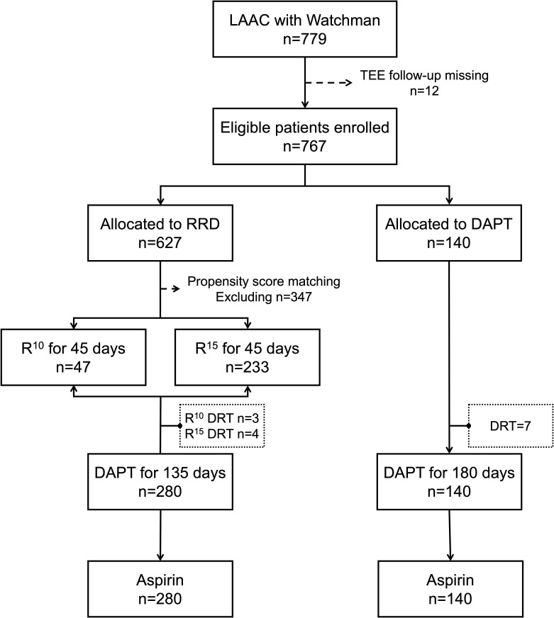
This cohort study initially enrolled 779 patients following successful Watchman implantation between December 2017 and December 2021. A total of 12 participants (DAPT group: n = 6 [0.8%]; RRD group: n = 6 [0.8%]) were finally excluded from the study because their follow-up TEEs were completed at a different clinical institution and no images were provided for review. Ultimately, 767 patients (DAPT group: n = 140; RRD group: n = 627; Mean age 68.0 9.0, male 478 (62.3%), Median - score 4; Median HAS-BLED score 2) were included in the analysis and followed for 12 months. Among participants in the RRD group, the anticoagulant regimen was rivaroxaban 15 mg once a day in 551 (87.9%) patients and 10 mg once a day in 76 (12.1%). The progression of anticoagulation therapy for post-LAAC operation is summarized in Fig. 1.
Fig. 1.
Enrollment flow chart of patients. LAAC, left atrial appendage closure; TEE, trans-esophageal echocardiography; RRD, reduced rivaroxaban doses; DAPT, dual antiplatelet therapy; , rivaroxaban 10 mg; , rivaroxaban 15 mg; DRT, device-related thrombosis.
Baseline information, cardiovascular risk factors, potential thromboembolic and bleeding risks, and concomitant medication are presented in Table 1. There was a higher percentage of higher HAS-BLED score in participants taking DAPT. After propensity score matching (PSM) with 1:2 ratio (140 patients for DAPT and 280 patients for RRD), the two subgroups were not significantly different in baseline information, cardiovascular risk factors, laboratory indicators and predetermined stroke and bleeding risk.
Table 1.
Baseline demographic and clinical characteristics between RRD and DAPT groups.
| Variables | Before matching | After matching | |||||||
| All (N = 767) | RRD (n = 627) | DAPT (n = 140) | p value | All (N = 420) | RRD (n = 280) | DAPT (n = 140) | p value | ||
| Age, y | 68.0 9.0 | 67.9 9.2 | 68.3 8.3 | 0.639 | 68.8 8.3 | 69.0 8.3 | 68.3 8.3 | 0.418 | |
| Male | 478 (62.3) | 391 (62.4) | 87 (62.1) | 0.962 | 267 (63.6) | 180 (64.3) | 87 (62.1) | 0.667 | |
| - score | 3.5 1.8 | 3.5 1.8 | 3.7 2.0 | 0.355 | 3.7 1.9 | 3.7 1.9 | 3.7 2.0 | 0.844 | |
| 3 | 372 (48.5) | 309 (49.3) | 63 (45.0) | 0.359 | 190 (45.2) | 127 (45.4) | 63 (45.0) | 0.945 | |
| 4 | 169 (22.0) | 141 (22.5) | 28 (20.0) | 0.521 | 86 (20.5) | 58 (20.7) | 28 (20.0) | 0.864 | |
| 5 | 226 (29.5) | 177 (28.2) | 49 (35.0) | 0.112 | 144 (34.3) | 95 (33.9) | 49 (35.0) | 0.827 | |
| HAS-BLED score | 2.5 1.2 | 2.4 1.2 | 3.2 1.2 | 0.001 | 3.1 1.2 | 3.0 1.2 | 3.2 1.2 | 0.092 | |
| 2 | 391 (51.0) | 352 (56.1) | 39 (27.9) | 0.001 | 115 (27.4) | 76 (27.1) | 39 (27.9) | 0.877 | |
| 3 | 217 (28.3) | 170 (27.1) | 47 (33.6) | 0.125 | 154 (36.7) | 107 (38.2) | 47 (33.6) | 0.352 | |
| 4 | 121 (15.8) | 81 (12.9) | 40 (28.6) | 0.001 | 113 (26.9) | 73 (26.1) | 40 (28.6) | 0.586 | |
| 5 | 38 (5.0) | 224 (3.8) | 14 (10.0) | 0.002 | 38 (9.0) | 24 (8.6) | 14 (10.0) | 0.630 | |
| Risk factors for stroke and bleeding | |||||||||
| CHF | 10 (1.3) | 8 (1.3) | 2 (1.4) | 1 | 5 (1.2) | 3 (1.1) | 2 (1.4) | 1.000 | |
| Hypertension | 488 (63.6) | 398 (63.5) | 90 (64.3) | 0.857 | 293 (69.8) | 203 (72.5) | 90 (64.3) | 0.084 | |
| 75 years of age | 179 (23.3) | 144 (23.0) | 35 (25.0) | 0.607 | 104 (24.8) | 69 (24.6) | 35 (25.0) | 0.936 | |
| 65–74 years of age | 345 (45.0) | 283 (45.1) | 62 (44.3) | 0.855 | 202 (48.1) | 140 (50.0) | 62 (44.3) | 0.269 | |
| Diabetes mellitus | 160 (20.9) | 124 (19.8) | 36 (25.7) | 0.118 | 79 (18.8) | 50 (17.9) | 29 (20.7) | 0.480 | |
| History of stroke/TIA | 326 (42.5) | 266 (42.4) | 60 (42.9) | 0.925 | 186 (44.3) | 126 (45.0) | 60 (42.9) | 0.677 | |
| Stroke | 282 (36.8) | 223 (35.6) | 59 (42.1) | 0.145 | 179 (42.6) | 120 (42.9) | 59 (42.1) | 0.889 | |
| TIA | 49 (6.4) | 43 (6.9) | 6 (4.3) | 0.26 | 17 (4.0) | 8 (2.9) | 9 (6.4) | 0.080 | |
| Vascular disease | 407 (53.1) | 329 (52.5) | 78 (55.7) | 0.487 | 228 (54.3) | 150 (53.6) | 78 (55.7) | 0.678 | |
| Renal Dysfunction | 44 (5.7) | 32 (5.1) | 12 (8.6) | 0.111 | 37 (8.8) | 25 (8.9) | 12 (8.6) | 0.903 | |
| Liver Dysfunction | 71 (9.3) | 55 (8.8) | 16 (11.4) | 0.327 | 53 (12.6) | 37 (13.2) | 16 (11.4) | 0.603 | |
| History of major bleeding | 56 (7.3) | 41 (6.5) | 15 (10.7) | 0.086 | 50 (11.9) | 35 (12.5) | 15 (10.7) | 0.594 | |
| Intracranial bleeding | 33 (4.3) | 25 (4.0) | 8 (5.7) | 0.363 | 30 (7.1) | 22 (7.9) | 8 (5.7) | 0.421 | |
| GI bleeding | 13 (1.7) | 10 (1.6) | 3 (2.1) | 0.715 | 10 (2.4) | 7 (2.5) | 3 (2.1) | 1.000 | |
| Other | 11 (1.4) | 7 (1.1) | 4 (2.9) | 0.123 | 11 (2.6) | 7 (2.5) | 4 (2.9) | 1.000 | |
| History of minor bleeding | 21 (2.7) | 14 (2.2) | 7 (5.0) | 0.07 | 17 (4.0) | 10 (3.6) | 7 (5.0) | 0.484 | |
| GI bleeding | 4 (0.5) | 3 (0.5) | 1 (0.7) | 0.554 | 3 (0.7) | 2 (0.7) | 1 (0.7) | 1.000 | |
| Epistaxis | 3 (0.4) | 2 (0.3) | 1 (0.7) | 0.454 | 2 (0.5) | 1 (0.4) | 1 (0.7) | 1.000 | |
| Other | 13 (1.7) | 9 (1.4) | 4 (2.9) | 0.271 | 6 (2.1) | 2 (1.4) | 4 (2.9) | 0.684 | |
| Labile INR | 36 (4.7) | 29 (4.6) | 7 (5.0) | 0.85 | 32 (7.6) | 25 (8.9) | 7 (5.0) | 0.153 | |
| Alcohol | 43 (5.6) | 38 (6.1) | 5 (3.6) | 0.247 | 33 (7.9) | 27 (9.6) | 6 (4.3) | 0.054 | |
| CAD | 122 (15.9) | 93 (14.9) | 29 (20.7) | 0.085 | 68 (16.2) | 39 (13.9) | 29 (20.7) | 0.075 | |
| LVEF, % | 63.1 6.8 | 63.1 6.8 | 62.7 6.8 | 0.471 | 63.0 6.9 | 63.1 7.1 | 62.7 6.7 | 0.575 | |
Values are mean SD, n (%). RRD, rivaroxaban dose; DAPT, dual antiplatelet therapy; CAD, coronary artery disease; -, congestive heart failure, hypertension, age 75 years, diabetes mellitus, prior stroke or transient ischemic attack or thromboembolism, vascular disease, age 65–74 years, sex category; CHF, congestive heart failure; INR, international normalized ratio; LVEF, left ventricular ejection fraction; GI, gastrointestinal; HAS-BLED, hypertension, abnormal renal or liver function, stroke, bleeding, labile international normalized ratio, elderly, drugs or alcohol; TIA, transient ischemic attack.
3.2 Efficacy Endpoints Evaluation
The RRD group had a 12-month median (interquartile range (IQR): 11–14) follow-up and the DAPT group had a 13-month (IQR: 11–15) median follow-up. In the RRD group, 193 were on and 47 on . In the first TEEs (within 2 days after LAAC), peri-device leaks 5 mm were recorded in 4 cases ( (n = 1), (n = 1) and DAPT (n = 2)). A second TEE was performed to confirm the presence of leaks among these patients, who were subsequently scheduled for percutaneous LAA leak closure. The primary efficacy endpoints were defined as a function of both the presence of DRT as well as the occurrence of thrombosis events. Early-phase formation of DRT was investigated, as reflected by the incidence of DRT and thrombus size. These patients were prescribed full-dose rivaroxaban (20 mg qd).
The incidence of DRT was documented in 12 (9.3%) patients in DAPT, 3 (6.3%) in and 7 (3.0%) in (log-rank p = 0.050), which indicated a higher DRT incidence in DAPT than that of and . A clear causal relationship between the TE and DRT could be established in 3 cases which were identified as ischemic strokes. As a result of switching to rivaroxaban therapy at full dose (20 mg), the DRTs were successfully managed. In the whole cohort of antithrombotic treated patients, DAPT subgroups were more likely to experience shorter time to DRT ( vs. DAPT, HR = 0.716, p = 0.603, 95% CI: 0.226–2.275; vs. DAPT HR = 0.334, p = 0.015, 95% CI: 0.131–0.850), as demonstrated in Fig. 2A.
Fig. 2.
Time to clinical events in antithrombotic treated patients, stratified into three subgroups (DAPT, and ) according to the different antithrombotic strategy. (A) referred as the comparison between DAPT and . (B) referred as the comparison between (C) DAPT and . (A) Kaplan-Meier survival curve of device-related thrombus (DRT), (B) Kaplan-Meier survival curve of thromboembolic (TE) events, (C) Kaplan-Meier survival curve of bleeding events. DAPT, dual antiplatelet therapy; , rivaroxaban 10 mg; , rivaroxaban 15 mg; HR, hazard ratio.
In the DAPT group, a total of 14 patients (10.0%) experienced TEs in terms of ischemic stroke or systemic embolism during the follow-up period compared with 6 patients (12.7%) in the and 8 (3.4%) in the matched group. Based on Kaplan-Meier survival analysis, TE reduction was significantly more favorable in the group ( vs. DAPT HR = 0.362, p = 0.018, 95% CI: 0.149–0.878, Fig. 2B).
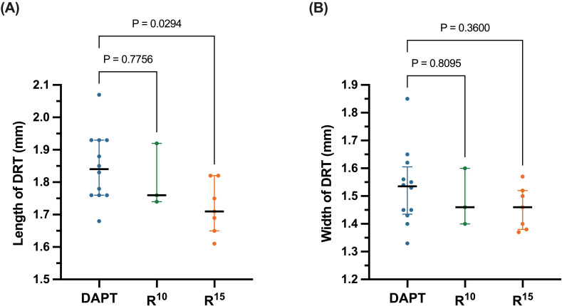
The median length of DRT in the group was significantly lower than that of the DAPT group (1.721 [1.610–1.818] mm vs. 1.820 [1.725–1.925] mm, p = 0.029), while no significant difference was detected between and DAPT (1.806 [1.740–1.924] mm vs. 1.820 [1.725–1.925] mm, p = 0.775), as shown in Fig. 3A. No significant difference in DRT width was observed among the three groups ( 1.486 (1.402–1.620) vs. DAPT 1.520 [1.400–1.610], p = 0.809; 1.457 (1.372–1.578) vs. DAPT 1.520 [1.400–1.610], p = 0.360, Fig. 3B).
Fig. 3.
(A) Length and (B) width of device-related thrombus (DRT) evaluated with transesophageal echocardiography. The solid black lines medians of each subgroup, while the error bars represent the interquartile range. DAPT, dual antiplatelet therapy; , rivaroxaban 10 mg; , rivaroxaban 15 mg.
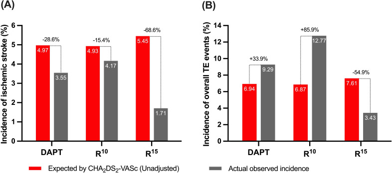
Compared with the unadjusted estimated rates of ischemic events for patients with similar - scores, a significant decrease of 68.6% in ischemic stroke rates was noted in the group, while slight insignificant reductions of 28.6% and 15.4% was observed in the DAPT and groups (Fig. 4A). In the whole cohort study, contributed a 54.9% reduction of overall TE events, as shown in Fig. 4B.
Fig. 4.
(A) Annualized ischemic stroke and (B) TE event rates after implantation vs expected (unadjusted) rates estimated based on the - (congestive heart failure, hypertension, age 75 years, diabetes mellitus, prior stroke or transient ischemic attack or thromboembolism, vascular disease, age 65–74 years, sex category) score of the 3 study groups. DAPT, dual antiplatelet therapy; , rivaroxaban 10 mg; , rivaroxaban 15 mg; TE, thromboembolic.
3.3 Safety Endpoints Evaluation
Details of the bleeding events in the entire patient cohort are reported in Table 2. No major bleeding was documented throughout the 12-month follow-up period. The rate of overall minor bleeding was not significantly different amongst the groups (8.5% among patients, 9.9% among patients and 10.7 among DAPT patients, (p = 0.944).
Table 2.
Bleeding complications comparison among , and DAPT groups.
| Bleeding complications, n (%) | DAPT | p value | |||
| Overall bleeding events | 4 (8.5%) | 23 (9.9%) | 15 (10.7%) | 0.944 | |
| GI bleeding | 1 (2.1%) | 5 (2.1%) | 3 (2.1%) | 1.000 | |
| Hematuria | 1 (2.1%) | 4 (1.7%) | 1 (0.7%) | 0.586 | |
| Operation site hemorrhage | 0 (0.0%) | 4 (1.7%) | 2 (1.4%) | 1.000 | |
| Bleeding gums | 1 (2.1%) | 6 (2.6%) | 4 (2.9%) | 1.000 | |
| Skin ecchymosis | 1 (2.1%) | 4 (1.7%) | 5 (3.6%) | 0.520 | |
| PLT 125 × /L | 4 (8.5%) | 10 (8.1%) | 15 (10.7%) | 0.745 | |
| Male: Hb 120 g/L | 5 (10.6%) | 11 (7.3%) | 16 (11.4%) | 0.748 | |
| Female: Hb 110 g/L | |||||
| PT 13 s | 12 (25.5%) | 62 (26.6%) | 32 (22.8%) | 0.856 | |
GI, Gastrointestinal; PLT, platelet; Hb, hemoglobin; PT, prothrombin time; DAPT, dual antiplatelet therapy; , rivaroxaban 10 mg; , rivaroxaban 15 mg. p-value represented with interaction.
Table 2 shows the accumulated anticoagulation-related complications and coagulation function tests among the groups. There was no significant reduction in levels of platelet (PLT), hemoglobin (Hb), or prothrombin time (PT) among , and DAPT groups (p 0.05). Based on Kaplan-Meier survival curve analysis, procedural bleeding was more common in the DAPT group, as compared with the and groups (Fig. 2), however, there was no significant difference among three groups.
4. Discussion
In this study, we prospectively investigated the clinical efficacy and safety between RRD and DAPT after successful LAAC implantation. The following are the main findings of the study. First, anticoagulation long term RRD led to a significant reduction of DRT and TE compared to DAPT. Second, long-term rivaroxaban provided more effective and safer thrombus prevention, when compared with unadjusted, estimated rates of ischemic stroke and TE for patients with similar -.
Similar to all medical devices implanted into the body, a longer implantation time beyond 90 days is expected to enable complete endothelialization for occluders post LAAC when exposed to circulating blood [19, 20]. Thrombosis formation might occur on the exposed device and adequate antithrombotic regimens are required for DRT prevention. Currently, pharmacological regimens following successful LAAC implantation were mainly dependent on strategies from recent clinical studies [21, 22]. Previous studies indicated short-term DAPT adoption followed by long-term aspirin could prevent DRT and TE events [23, 24]. Although antiplatelet therapy has confirmed the efficacy for thrombosis prevention after stent implantation, substantial variation still remains for the selection of the appropriate antithrombotic strategy for LAAC implantation. In this study we sought to compare the clinical efficacy and safety between RRD and standard antiplatelet strategy following LAAC. Some novel observations could be made based on the data derived from this study.
It is important to identify anticoagulants that can prevent the occurrence of DRT and TE. Currently, there is limited data regarding the correlation between antithrombotic strategies and thrombus on closure devices. In this study, the scheduled out-patient visits and trans-telephonic clinical evaluations were frequently conducted 6 months after discharge to help reduce the occurrence of thromboembolic events. Our results documented a lower DRT rate in RRD as compared to DAPT, which was similar to those in previously published studies from other groups [14, 25]. In one propensity matched comparison with Watchman closure implantation patients, the 6-month cumulative DRT occurrence was lower in DAPT as compared to half-Dose DOAC (3.4% vs. 0.0%), which was similar with our findings [14]. Another multicenter study with patients undergoing LAAC implantation indicated that DOACs proved to be a feasible and safe alternative antithrombotic regimen to warfarin for DRT and thromboembolic prevention after LAAC implantation, without increasing the risk of bleeding [14].
Activation of the coagulation system and enhanced thrombin generation without platelet aggregation were associated with DRT formation within days after LAAC [26]. In our results, persistent elevation of thrombosis size was also detected in the DAPT group after the procedure, which was consistent with the result of previous prospective studies. A randomized pilot study documented that circulating prothrombin fragments and thrombin-antithrombin complex were numerically lower after rivaroxaban treatment than that with DAPT, which might explain the lower rate of thrombosis and TE following medication with rivaroxaban after successful LAAC [27].
Another important factor for consideration is the safety for long-time anticoagulation among different antithrombotic regimens in patients undergoing LAAC. Our results indicated decreased bleeding occurrence after the initial follow-up, which might be related to more scheduled out-patient visits and clinical evaluations 6 months after discharge. Of note, a reduced DOAC dose was associated with lower bleeding for NVAF patients compared with vitamin K antagonists (VKA) in the east Asia population [28]. One large cohort of east Asia patients showed lower post-extraction bleeding rates with DOAC compared with warfarin (HR: 0.84; 95% CI: 0.54–1.31) [29]. In our study, there was no significant difference in coagulation function tests between the RRD and DAPT groups. Furthermore, our findings suggest that RRD may be a safe alternative to DAPT for short-term antithrombotic therapy after LAAC, without a statistically significant difference observed in bleeding events. However, the optimal antithrombotic regimen for long-term anticoagulation after LAAC remains uncertain, and individualized treatment plans should be developed based on patient-specific factors, such as bleeding risk, thromboembolic risk, comorbidities, and medication interactions. Although the overall minor bleeding rate was 10.0% in our study, the risk for postoperative bleeding was increased in these patients who underwent a percutaneous strategy and were exposed to anticoagulation therapy. Given the concern for bleeding and the need for TE prophylaxis, a minimally invasive surgical strategy such as epicardial LAA occlusion with no further anticoagulation is more favorable to patients at higher risk for bleeding and thrombosis. The rationale for this practice is inferred from the Left Atrial Appendage Occlusion Study (LAAOS III) trial, which demonstrated that LAA resection for AF patients during chest cardiac surgery contributed to a reduction of thrombosis and bleeding risks [30, 31]. As for NVAF patients with end-stage renal failure contraindicated to DOAC merging with high bleeding risk, epicardial LAA occlusion for such specific population might offer a clinical benefit [32]. Multi-disciplinary teams involving anesthesiologists, cardiologists and cardiac surgeons will need to determine which patients can reliably and safely undergo epicardial LAA closure in those patients with a contraindication for anticoagulation or those with anatomical abnormalities which are not conducive to percutaneous LAAO.
Limitations
The present study has many limitations. Firstly, our study is non-randomized and conducted at a single center, which might restrict the generalizability of the results to other cardiovascular centers and healthcare systems. Secondly, the sample size was relatively small and the follow-up duration of only 12 months may not be sufficient to determine the long-term efficacy and safety of different anticoagulation regimens, which potentially led to a low incidence of DRT. The exclusion of 12 participants due to incomplete follow-up TEEs and failure to provide images for review may have introduced selection bias. Thirdly, our study was followed up only by the cardiology department. Finally, the study only enrolled participants receiving RRD and DAPT, which may limit the generalizability of the findings to other anticoagulants. In conclusion, these limitations highlight the need for further studies with larger sample sizes, more frequent monitoring, and more diverse patient populations to confirm the conclusions and establish the clinical efficacy and safety of different antithrombotic strategies.
5. Conclusions
Based on the evidence presented in this cohort study, antithrombotic therapy with RRD may be a promising option for DAPT for reducing the risk of DRT and composite endpoints in patients following successful Watchman implantation. Further randomized controlled trials conducted at multiple centers are needed to compare the safety and efficacy of different antithrombotic regimens in these patients.
Acknowledgment
Not applicable.
Footnotes
Publisher’s Note: IMR Press stays neutral with regard to jurisdictional claims in published maps and institutional affiliations.
Contributor Information
Xiaochun Zhang, Email: zhang.xiaochun@zs-hospital.sh.cn.
Qianzhou Lv, Email: 13916088938@163.com.
Availability of Data and Materials
The datasets used and analyzed during the current study are available from the corresponding author on reasonable request.
Author Contributions
XL, YY, QJ, XZ and QL contributed to the conception and design, or acquisition of data, or analysis and interpretation of data, and completed the writing of the paper. XL, QJ, XZ and QL were involved in drafting the manuscript or revising it critically for important intellectual content. XL, YY, XZ and QL were responsible for the revision of the paper. All authors confirmed the final version of the paper. All authors have participated sufficiently in the work and agreed to be accountable for all aspects of the work.
Ethics Approval and Consent to Participate
The studies involving human participants were reviewed and approved by the Institutional Review Committee of Zhongshan Hospital, Fudan University (No: B2020-288R). The patients/participants provided their written informed consent to participate in this study.
Funding
This research received no external funding.
Conflict of Interest
The authors declare no conflict of interest.
References
- [1].Ueno H, Imamura T, Tanaka S, Fukuda N, Kinugawa K. Left atrial appendage closure for stroke prevention in nonvalvular atrial fibrillation: A current overview. Journal of Cardiology . 2023;81:420–428. doi: 10.1016/j.jjcc.2022.11.006. [DOI] [PubMed] [Google Scholar]
- [2].Wazni OM, Boersma L, Healey JS, Mansour M, Tondo C, Phillips K, et al. Comparison of anticoagulation with left atrial appendage closure after atrial fibrillation ablation: Rationale and design of the OPTION randomized trial. American Heart Journal . 2022;251:35–42. doi: 10.1016/j.ahj.2022.05.003. [DOI] [PubMed] [Google Scholar]
- [3].Osmancik P, Herman D, Neuzil P, Hala P, Taborsky M, Kala P, et al. Left Atrial Appendage Closure Versus Direct Oral Anticoagulants in High-Risk Patients With Atrial Fibrillation. Journal of the American College of Cardiology . 2020;75:3122–3135. doi: 10.1016/j.jacc.2020.04.067. [DOI] [PubMed] [Google Scholar]
- [4].Reddy VY, Doshi SK, Kar S, Gibson DN, Price MJ, Huber K, et al. 5-Year Outcomes After Left Atrial Appendage Closure: From the PREVAIL and PROTECT AF Trials. Journal of the American College of Cardiology . 2017;70:2964–2975. doi: 10.1016/j.jacc.2017.10.021. [DOI] [PubMed] [Google Scholar]
- [5].Chen M, Sun J, Wang QS, Zhang PP, Li W, Zhang R, et al. Long-term outcome of combined catheter ablation and left atrial appendage closure in atrial fibrillation patients. International Journal of Cardiology . 2022;368:41–48. doi: 10.1016/j.ijcard.2022.08.007. [DOI] [PubMed] [Google Scholar]
- [6].Kramer AD, Korsholm K, Jensen JM, Nørgaard BL, Peelukhana S, Herbst T, et al. Cardiac computed tomography following Watchman FLX implantation: device-related thrombus or device healing. European Heart Journal. Cardiovascular Imaging . 2023;24:250–259. doi: 10.1093/ehjci/jeac222. [DOI] [PMC free article] [PubMed] [Google Scholar]
- [7].Sedaghat A, Nickenig G, Schrickel JW, Ince H, Schmidt B, Protopopov AV, et al. Incidence, predictors and outcomes of device-related thrombus after left atrial appendage closure with the WATCHMAN device-Insights from the EWOLUTION real world registry. Catheterization and Cardiovascular Interventions . 2021;97:E1019–E1024. doi: 10.1002/ccd.29458. [DOI] [PubMed] [Google Scholar]
- [8].Saw J, Holmes DR, Cavalcante JL, Freeman JV, Goldsweig AM, Kavinsky CJ, et al. SCAI/HRS expert consensus statement on transcatheter left atrial appendage closure. Heart Rhythm . 2023;20:e1–e16. doi: 10.1016/j.hrthm.2023.01.007. [DOI] [PubMed] [Google Scholar]
- [9].Flores-Umanzor E, Cepas-Guillen P, Regueiro A, Sanchis L, Unigarro F, Brugaletta S, et al. Treatment of Device-Related Thrombosis After Left Atrial Appendage Occlusion: Initial Experience With Low-Dose Apixaban. Cardiovascular Revascularization Medicine: Including Molecular Interventions . 2022;40:201–203. doi: 10.1016/j.carrev.2021.11.007. [DOI] [PubMed] [Google Scholar]
- [10].Saw J, Nielsen-Kudsk JE, Bergmann M, Daniels MJ, Tzikas A, Reisman M, et al. Antithrombotic Therapy and Device-Related Thrombosis Following Endovascular Left Atrial Appendage Closure. JACC. Cardiovascular Interventions . 2019;12:1067–1076. doi: 10.1016/j.jcin.2018.11.001. [DOI] [PubMed] [Google Scholar]
- [11].Albertsen IE, Jensen M, Abdelgawwad K, Søgaard M, Larsen TB, Nielsen PB. Characteristics of patients receiving extended treatment after incident venous thromboembolism. Basic & Clinical Pharmacology & Toxicology . 2021;129:332–342. doi: 10.1111/bcpt.13637. [DOI] [PubMed] [Google Scholar]
- [12].Ye C, Han X, Chen Y, Xu D. Early myocardial injury and coagulation activation in thoracoscopic left atrial appendage clipping. Journal of Cardiac Surgery . 2022;37:4919–4927. doi: 10.1111/jocs.17169. [DOI] [PubMed] [Google Scholar]
- [13].Rodés-Cabau J, O’Hara G, Paradis JM, Bernier M, Rodriguez-Gabella T, Regueiro A, et al. Changes in Coagulation and Platelet Activation Markers Following Transcatheter Left Atrial Appendage Closure. American Journal of Cardiology . 2017;120:87–91. doi: 10.1016/j.amjcard.2017.03.253. [DOI] [PubMed] [Google Scholar]
- [14].Della Rocca DG, Magnocavallo M, Di Biase L, Mohanty S, Trivedi C, Tarantino N, et al. Half-Dose Direct Oral Anticoagulation Versus Standard Antithrombotic Therapy After Left Atrial Appendage Occlusion. JACC. Cardiovascular Interventions . 2021;14:2353–2364. doi: 10.1016/j.jcin.2021.07.031. [DOI] [PubMed] [Google Scholar]
- [15].Asmarats L, O’Hara G, Champagne J, Paradis JM, Bernier M, O’Connor K, et al. Short-Term Oral Anticoagulation Versus Antiplatelet Therapy Following Transcatheter Left Atrial Appendage Closure. CirculationCardiovascular Interventions . 2020;13:e009039. doi: 10.1161/CIRCINTERVENTIONS.120.009039. [DOI] [PubMed] [Google Scholar]
- [16].Hori M, Matsumoto M, Tanahashi N, Momomura SI, Uchiyama S, Goto S, et al. Safety and efficacy of adjusted dose of rivaroxaban in Japanese patients with non-valvular atrial fibrillation: subanalysis of J-ROCKET AF for patients with moderate renal impairment. Circulation Journal . 2013;77:632–638. doi: 10.1253/circj.cj-12-0899. [DOI] [PubMed] [Google Scholar]
- [17].Turagam MK, Neuzil P, Hala P, Mraz T, Dukkipati SR, Reddy VY. Intracardiac Echocardiography-Guided Left Atrial Appendage Closure With a Novel Foam-Based Conformable Device: Safety and 1-Year Outcomes. JACC. Clinical Electrophysiology . 2022;8:197–207. doi: 10.1016/j.jacep.2021.10.001. [DOI] [PubMed] [Google Scholar]
- [18].Spyropoulos AC, Brohi K, Caprini J, Samama CM, Siegal D, Tafur A, et al. Scientific and Standardization Committee Communication: Guidance document on the periprocedural management of patients on chronic oral anticoagulant therapy: Recommendations for standardized reporting of procedural/surgical bleed risk and patient-specific thromboembolic risk. Journal of Thrombosis and Haemostasis . 2019;17:1966–1972. doi: 10.1111/jth.14598. [DOI] [PubMed] [Google Scholar]
- [19].Liu T, Liu S, Zhang K, Chen J, Huang N. Endothelialization of implanted cardiovascular biomaterial surfaces: the development from in vitro to in vivo. Journal of Biomedical Materials Research. Part A . 2014;102:3754–3772. doi: 10.1002/jbm.a.35025. [DOI] [PubMed] [Google Scholar]
- [20].Edlinger C, Paar V, Kheder SH, Krizanic F, Lalou E, Boxhammer E, et al. Endothelialization and Inflammatory Reactions After Intracardiac Device Implantation. Advances in Experimental Medicine and Biology . 2022;1401:1–22. doi: 10.1007/5584_2022_712. [DOI] [PubMed] [Google Scholar]
- [21].Aarnink EW, Huijboom MFM, Bor WL, Maarse M, Zheng KL, Ten Cate H, et al. Hemostatic biomarkers and antithrombotic strategy in percutaneous left atrial interventions: State-of-the-art review. Thrombosis Research . 2022;215:41–51. doi: 10.1016/j.thromres.2022.05.009. [DOI] [PubMed] [Google Scholar]
- [22].Calabrò P, Gragnano F, Niccoli G, Marcucci R, Zimarino M, Spaccarotella C, et al. Antithrombotic Therapy in Patients Undergoing Transcatheter Interventions for Structural Heart Disease. Circulation . 2021;144:1323–1343. doi: 10.1161/CIRCULATIONAHA.121.054305. [DOI] [PubMed] [Google Scholar]
- [23].Weise FK, Bordignon S, Perrotta L, Konstantinou A, Bologna F, Nagase T, et al. Short-term dual antiplatelet therapy after interventional left atrial appendage closure with different devices. EuroIntervention . 2018;13:e2138–e2146. doi: 10.4244/EIJ-D-17-00901. [DOI] [PubMed] [Google Scholar]
- [24].Jalal Z, Dinet ML, Combes N, Pillois X, Renou P, Sibon I, et al. Percutaneous left atrial appendage closure followed by single antiplatelet therapy: Short- and mid-term outcomes. Archives of Cardiovascular Diseases . 2017;110:242–249. doi: 10.1016/j.acvd.2016.09.006. [DOI] [PubMed] [Google Scholar]
- [25].Vignali L, Gurgoglione FL, Barocelli F, Cattabiani MA, Solinas E, Maini A, et al. Looking for optimal antithrombotic strategy after transcatheter left atrial appendage occlusion: a real-world comparison of different antiplatelet regimens. International Journal of Cardiology . 2023;371:92–99. doi: 10.1016/j.ijcard.2022.09.066. [DOI] [PubMed] [Google Scholar]
- [26].Li X, Zhang X, Jin Q, Li Y, Zhou D, Lv Q, et al. The Impact of Dabigatran and Rivaroxaban on Variation of Platelet Activation Biomarkers and DRT Following Percutaneous Left Atrial Appendage Closure. Frontiers in Pharmacology . 2021;12:723905. doi: 10.3389/fphar.2021.723905. [DOI] [PMC free article] [PubMed] [Google Scholar]
- [27].Duthoit G, Silvain J, Marijon E, Ducrocq G, Lepillier A, Frere C, et al. Reduced Rivaroxaban Dose Versus Dual Antiplatelet Therapy After Left Atrial Appendage Closure: ADRIFT a Randomized Pilot Study. Circulation. Cardiovascular Interventions . 2020;13:e008481. doi: 10.1161/CIRCINTERVENTIONS.119.008481. [DOI] [PubMed] [Google Scholar]
- [28].Lau WCY, Torre CO, Man KKC, Stewart HM, Seager S, Van Zandt M, et al. Comparative Effectiveness and Safety Between Apixaban, Dabigatran, Edoxaban, and Rivaroxaban Among Patients With Atrial Fibrillation: A Multinational Population-Based Cohort Study. Annals of Internal Medicine . 2022;175:1515–1524. doi: 10.7326/M22-0511. [DOI] [PubMed] [Google Scholar]
- [29].Ono S, Ishimaru M, Yokota I, Konishi T, Okada A, Ono Y, et al. Risk of post-extraction bleeding with direct oral anticoagulant compared with warfarin: Retrospective cohort study using large scale claims data in Japan. Thrombosis Research . 2023;222:24–30. doi: 10.1016/j.thromres.2022.12.007. [DOI] [PubMed] [Google Scholar]
- [30].Bisleri G, Pandey AK, Verma S, Ali Hassan SM, Yanagawa B, Khandaker M, et al. Combined Minimally Invasive Surgical and Percutaneous Catheter Ablation of Atrial Fibrillation: JACC Review Topic of the Week. Journal of the American College of Cardiology . 2023;81:606–619. doi: 10.1016/j.jacc.2022.11.039. [DOI] [PubMed] [Google Scholar]
- [31].Verma S, Bhatt DL, Tseng EE. Time to Remove the Left Atrial Appendage at Surgery: LAAOS III in Perspective. Circulation . 2021;144:1088–1090. doi: 10.1161/CIRCULATIONAHA.121.055825. [DOI] [PubMed] [Google Scholar]
- [32].Rosati F, de Maat GE, Valente MAE, Mariani MA, Benussi S. Surgical clip closure of the left atrial appendage. Journal of Cardiovascular Electrophysiology . 2021;32:2865–2872. doi: 10.1111/jce.15181. [DOI] [PMC free article] [PubMed] [Google Scholar]
Associated Data
This section collects any data citations, data availability statements, or supplementary materials included in this article.
Data Availability Statement
The datasets used and analyzed during the current study are available from the corresponding author on reasonable request.


