Abstract
Circulating nucleosome levels are commonly elevated in physiological and pathological conditions. Their potential as biomarkers for diagnosing and prognosticating sepsis remains uncertain due, in part, to technical limitations in existing detection methods. This scoping review explores the possible role of nucleosome concentrations in the diagnosis, prognosis, and therapeutic management of sepsis. A comprehensive literature search of the Cochrane and Medline libraries from 1996 to 1 February 2024 identified 110 potentially eligible studies, of which 19 met the inclusion criteria, encompassing a total of 39 SIRS patients, 893 sepsis patients, 280 septic shock patients, 117 other ICU control patients, and 345 healthy volunteers. The enzyme-linked immunosorbent assay [ELISA] was the primary method of nucleosome measurement. Studies consistently reported significant correlations between nucleosome levels and other NET biomarkers. Nucleosome levels were higher in patients with sepsis than in healthy volunteers and associated with disease severity, as indicated by SOFA and APACHE II scores. Non-survivors had higher nucleosome levels than survivors. Circulating nucleosome levels, therefore, show promise as early markers of NETosis in sepsis, with moderate diagnostic accuracy and strong correlations with disease severity and prognosis. However, the available evidence is drawn mainly from single-center, observational studies with small sample sizes and varied detection methods, warranting further investigation.
Keywords: nucleosome, NETosis, sepsis, infection, sepsis diagnosis, prognosis
1. Introduction
Sepsis remains a significant global health concern, responsible for approximately 20% of deaths worldwide, with septic shock mortality rates reaching close to 60% [1]. Biomarkers can be used to indicate the presence of sepsis, its severity, and its response to treatment [2,3]. Despite the identification of more than 250 potential biomarkers of sepsis [4], only two, the host-response markers C-reactive protein (CRP) and procalcitonin (PCT), are widely used in clinical practice and these are not specific for sepsis as levels can be raised in other conditions in critically ill patients; as such, serial values are more useful than single measurements. The complexity of sepsis makes it unlikely that a single biomarker will be relevant for all patients at all times and further evaluation and validation is needed to determine the clinical utility of individual biomarkers for specific purposes, including diagnosis, prognosis, and therapeutic guidance. Here, we will discuss the potential role of circulating nucleosomes as biomarkers of sepsis.
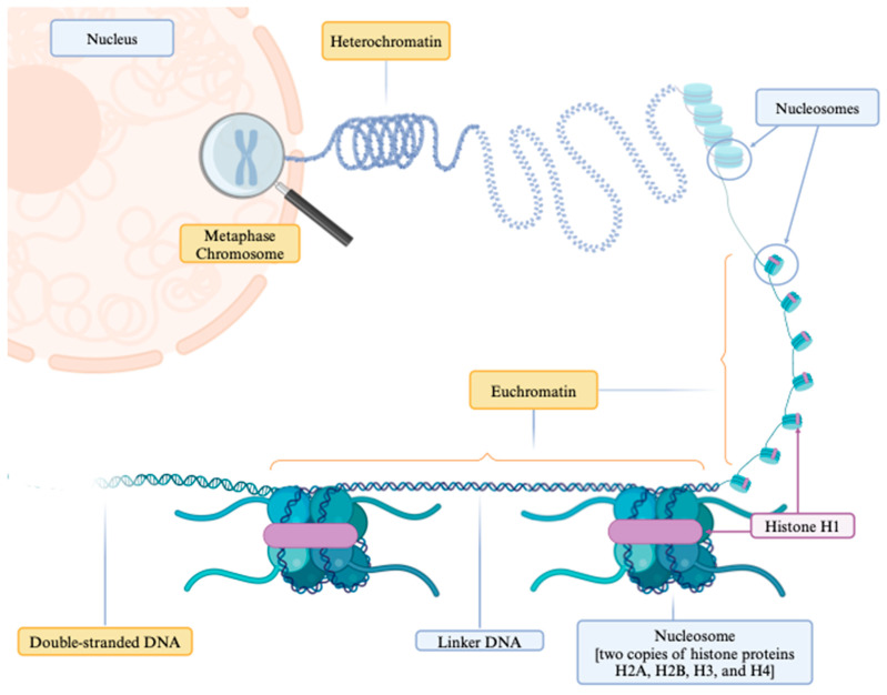
Chromatin is a substance composed of DNA and proteins that form chromosomes within the nucleus of cells. The fundamental structural unit of chromatin in all eukaryotic cells is the nucleosome, comprising the following two main components: histones and DNA, as depicted in Figure 1. The histone portion offers structural support and consists of two copies each of the histone proteins H2A, H2B, H3, and H4, encircling approximately 145–147 base pairs of DNA for about 1.65 times. These nucleosomes are interconnected by “linker DNA” segments, approximately 20–80 base pairs long, and bound by histone H1 [5]. This intricate structure regulates chromatin compaction/condensation and governs transcriptional access to the nucleosome [6]. Biochemical and structural studies have elucidated the dynamic nature of nucleosomes, which are essential for gene transcription regulation, DNA replication, repair processes, and efficient higher-order chromatin compaction [5,7,8]. Various molecules and mechanisms, including DNA breathing [7], post-translational histone modifications, histone chaperones, histone variants, and chromatin remodelers, contribute to this dynamic regulation [9,10].
Figure 1.
The structure of chromatin within a cell nucleus. This illustration depicts chromatin unfolding to expose euchromatin regions, characterized by a relaxed structure conducive to transcriptional processes. Nucleosomes are complexes of DNA coiled about 1.65 times around core histone proteins: two copies each of the histone proteins H2A, H2B, H3, and H4, bound together and secured by the histone H1. These nucleosomes are interconnected by segments of DNA known as “linker DNA”. The images were created using https://www.biorender.com (accessed on 10 April 2024).
Nucleosomes are released during cellular damage and cell death [11], and circulating levels are increased in physiological conditions, including aging [12], physical exercise [13], and stress [14], as well as pathological conditions, such as aging-related degenerative disorders [15], inflammatory responses, autoimmune diseases, ischemic stroke, trauma, and malignancies [12]. These nucleosomes have a short half-life and are typically removed from circulation within 10 min, primarily degraded by endonucleases in the blood, metabolized in the liver, or eliminated by macrophages and immune cells [16,17,18]. Notably, circulating nucleosomes, along with post-translational histone modifications and specific tumor markers, can help in the diagnosis of certain cancers [19], and monitoring their changes during cancer treatment can be useful in assessing therapeutic efficacy [19].
Studies in animal models of sepsis [18] and trials with healthy volunteers receiving lipopolysaccharides [LPSs] [20] have observed increased nucleosome concentrations, indicative of nucleosome release in response to innate immune cell activation. In sepsis, neutrophil activation triggers the release of granule proteins and chromatin, forming neutrophil extracellular traps [NETs] through a process termed NETosis [21]. This process, along with other forms of increased cell death, such as apoptosis [22], necrosis [23], and pyroptosis [24,25], contribute to nucleosome release into the extracellular space. Core histone proteins, including H3, H4, H2A, and H2B, have been identified as major components of NETs, emphasizing their significance in septic pathophysiology [26].
Circulating nucleosome levels may increase in sepsis due to several factors as follows: (a) increased release of circulating free DNA [cfDNA] leading to a biphasic nucleosome release. The first phase is marked by the contribution of cell death within hematopoietic cells, and the second phase by release from non-hematopoietic cells, such as epithelial and endothelial cells [11,27,28]. Indeed, immune [e.g., neutrophils, monocytes, macrophages, mast cells, dendritic cells, eosinophils, basophils] and parenchymal [e.g., endothelial cells] cell death is reported in sepsis [29,30,31]; (b) insufficient clearance due to suppressed or decreased deoxyribonuclease activity [32,33] and the diminished phagocytosis of apoptotic cells [34]; (c) the release of histones, which induces direct cellular toxicity, prompting immune responses, inflammation, and further cellular injury and death, leading to the amplification of nucleosome cascades [35]; and (d) the binding of acute phase proteins, such as CRP, to positively charged histone components, impeding the removal of circulating nucleosomes [36].
2. Immunostimulatory Role of Nucleosomes
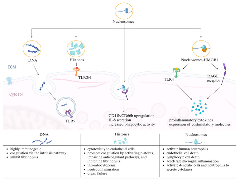
Circulating nucleosomes are potent triggers of immune responses [37]. Both components of the nucleosome–double-stranded DNA [dsDNA] and histones–exhibit diverse immunostimulatory effects, both in vitro and in vivo. Histones induce cytotoxicity and proinflammatory signaling through Toll-like receptors [TLRs] 2 and 4, whereas DNA triggers signaling through TLR9 and intracellular nucleic acid sensing mechanisms [38]. Histones are cytotoxic to endothelial cells and promote coagulation by activating platelets, impairing anticoagulant pathways, and inhibiting fibrinolysis [39,40]. The administration of purified histones in mice mirrors human sepsis, with thrombocytopenia, neutrophil migration, and organ failure developing [41]. DNA, which is highly immunogenic, represents a crucial pathogen-associated molecular pattern [PAMP] during infection [42]. It can initiate coagulation via the intrinsic pathway and inhibit fibrinolysis [43,44]. However, the in vivo administration of DNA to healthy or septic mice showed no harmful effects [45].
In some clinical studies, the terms histones and nucleosomes are used interchangeably because of detection method limitations [38]. However, in this review, we focus specifically on studies that measure nucleosome concentrations in sepsis. Following their release into the extracellular space, nucleosomes are internalized by various mammalian cells through multiple endocytic pathways [46]. Cellular uptake is facilitated by electrostatic interactions between histone N-terminal tails and cell surface ligands, followed by clathrin- and caveolae-dependent endocytosis [46]. Nucleosomes induce the direct activation of human neutrophils, triggering CD11b/CD66b upregulation, interleukin [IL]-8 secretion, and increased phagocytic activity, independent of the TLR2/TLR4 pathway [47,48,49]. Studies on the immunostimulatory role of nucleosomes suggest cell-type dependence. Purified nucleosomes at physiological concentrations activate human neutrophils [37], induce endothelial [38] and lymphocyte cell death [50], accelerate microglial inflammation [51], and activate dendritic cells and neutrophils to secrete cytokines [52,53]. Additionally, nucleosome high-mobility group box 1 protein [HMGB1] complexes stimulate immune responses via TLR4 and the receptor for advanced glycation end-products [RAGEs], inducing the secretion of proinflammatory cytokines and expression of costimulatory molecules [54,55]. These findings underscore the diverse and intricate immunostimulatory effects of circulating nucleosomes in various cellular contexts. The involvement of nucleosomes in the processes of inflammation and sepsis is illustrated in Figure 2.
Figure 2.
Visualization of the role of nucleosomes in the pathogenesis of inflammation and sepsis DNA, histones, and the nucleosome complex with the high-mobility group box 1 protein [HMGB1] and engaging with cell surface receptors like Toll-like receptor [TLR]2/4 and the receptor for advanced glycation end-products [RAGEs], as well as intracellular receptors such as TLR9, initiating signaling cascades. The consequences of these interactions include the up-regulation of CD11b/CD66b, secretion of interleukin [IL]-8, increased phagocytic activity, and the release of proinflammatory cytokines. These events contribute to several effects, as described at the bottom of the figure. The images were created using https://www.biorender.com (accessed on 10 April 2024).
3. Nucleosome Administration in Sepsis
Despite their immunostimulatory role, the administration of nucleosomes in sepsis does not appear to be toxic [16]. No cytotoxicity was observed, even at high doses when nucleosomes were injected into healthy or septic mice [15,45]. Intact nucleosomes themselves are not procoagulant, unlike the individual purified components of DNA and histones, which exhibit procoagulant properties [14].
Given the lack of direct toxicity associated with nucleosomes, targeting nucleosomes themselves may not be a valid therapeutic focus in sepsis. Instead, therapeutic strategies typically target histones or related signaling pathways [56]. These strategies include inhibiting histone release or NETosis [57], neutralizing histones with anti-histone monoclonal antibodies [58], or blocking signaling pathways, such as TLRs [59]. Targeting histones could potentially decrease circulating nucleosome levels by mitigating the amplification cascade effect and leading to decreased cell death. A translational study in an ewe septic shock model demonstrated that targeting histones using the histone-neutralizing polyanion sodium-β-O-methyl cellobioside sulfate [mCBS] resulted in decreased circulating nucleosome levels [60].
Elevated circulating nucleosome levels have been observed to correlate with sepsis severity and outcome. In liver transplant patients, nucleosome levels were associated with the occurrence of acute kidney injury, early allograft dysfunction, and early mortality after transplantation [61]. Similarly, raised nucleosome levels after graft reperfusion were associated with the occurrence of systemic inflammatory response syndrome [61].
4. Unanswered Questions
Despite the known increase in circulating nucleosome levels in sepsis, several questions remain unanswered as follows:
Can circulating nucleosome concentrations serve as a biomarker for NET formation in sepsis?
Are circulating nucleosome concentrations diagnostic biomarkers for sepsis?
Can circulating nucleosome concentrations serve as markers of organ dysfunction or disease severity in sepsis?
Are circulating nucleosome concentrations prognostic biomarkers in sepsis?
Can nucleosome levels be used to guide sepsis therapy?
We conducted a scoping review to address these unanswered questions and provide insights into the utility of circulating nucleosomes in the context of sepsis diagnosis, prognosis, and therapy.
5. Methods
The Preferred Reporting Items for Systematic Reviews and Meta-Analyses [PRISMA 5.15] guidelines [62] were employed for this review, with an extension for scoping reviews [PRISMA-ScR] [63]. A systematic search of the Cochrane and Medline libraries from 1996 to 1 February 2024 was performed to identify studies that investigated the role of circulating nucleosomes in NETosis in differentiating patients with sepsis from healthy volunteers, those with systemic inflammatory response syndrome [SIRS] without infection, or other ICU patients, or assessed associations between circulating nucleosome levels and disease severity or prognosis. The search used the following keywords: nucleosome AND/OR infection AND/OR sepsis AND/OR septic shock AND/OR NETosis. Studies involving animals, patients without probable infection, and healthy volunteers receiving lipopolysaccharide [LPS] were excluded. Studies in children younger than 28 days were also excluded due to variations in etiology and prognosis between early- and late-onset sepsis in neonates compared to adults [64]. Additionally, studies in adults and children with acute respiratory distress syndrome [ARDS] were excluded because ARDS can be sepsis- or non-sepsis-related [65].
Two independent investigators [FS, AM] extracted patient and study characteristics, and any discrepancies were resolved through consensus.
6. Results
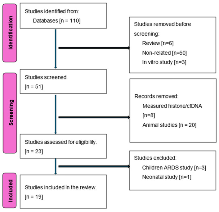
Our initial search yielded 110 studies, of which 19 met the inclusion criteria, including data from a total of 39 patients with SIRS, 893 patients with sepsis, 280 patients with septic shock, another 117 ICU patients, and 345 healthy volunteers [18,66,67,68,69,70,71,72,73,74,75,76,77,78,79,80,81,82,83]. The study selection flowchart is shown in Figure 3. Eleven studies were prospective, and two were multicenter (Table 1).
Figure 3.
Flowchart of included studies.
Table 1.
Studies included in the current review.
| Author [Ref.] | Year | Study Type | Sample | Methods | Catching Ab Detection Ab |
Patient Population | Range |
|---|---|---|---|---|---|---|---|
| Haem Rahimi et al. [66] | 2023 | Retrospective Monocenter |
Plasma | Chemiluminescence immunoassay [Volition] | Nucleosomes H3.1 [H2A, H3B, H3, H4 + DNA] | 50 healthy volunteers 151 septic shock |
Median 15.4 ng/mL Day 1–2 median 1515 ng/mL |
| Eichhorn et al. [67] | 2023 | Prospective Monocenter |
Plasma | ELISA ROCHE |
Mono anti-histone Mono anti-DNA-POD |
25 healthy volunteers 78 sepsis patients [14 with COVID] |
0.01 [0.01; 0.02] AU 0.09 [0.05; 0.11] AU without COVID 0.11 [0.04; 0.15] AU with COVID |
| Rai et al. [68] | 2022 | Prospective Monocenter |
Serum | ELISA [Orgentec] | Unknown | 80 sepsis | male: 209.8 [68.0–1263.0] ρg/μL female: 248.7 [65.0–1721.0] ρg/μL |
| Morimont et al. [69] | 2022 | Retrospective Monocenter |
Plasma | ELISA [Volition] | Nucleosomes H3.1 [H2A, H3B, H3, H4 + DNA] | 48 control patients 46 septic shock 22 critical COVID-19 |
24.6 [12.2–61.7] ng/mL 862 [252–9398] ng/mL |
| Beltrán-García et al. [70] | 2021 | Retrospective Monocenter |
Plasma | ELISA Kit 1 [home made] Kit2 [Roche] |
Mono anti-histone Mono anti-DNA-POD |
17 healthy volunteers 9 ICU control patients 10 septic ICU patients 17 septic shock |
70.66 ± 42.22 ng/mL (kit 1); 0.083 ± 0.04 AU (kit 2) 56.97 ± 25.76 ng/mL (kit 1); 0.080 ± 0.01 AU (kit 2) 111.8 ± 74.50 ng/mL (kit 1); 0.130 ± 0.08 AU (kit 2) 152.7 ± 74.93 ng/mL (kit 1); 0.216 ± 0.17 AU (kit 2) |
| van der Meer et al. [18] | 2019 | Retrospective Monocenter |
Plasma | ELISA | Mono anti-histone mAb CLB-ANA/58 |
20 sepsis patients | NR [only figure available] |
| Patel et al. [71] | 2019 | Retrospective Monocenter |
Plasma | ELISA [Roche] | Mono anti-histone Mono anti-DNA-POD |
50 healthy volunteers 20 sepsis + no DIC 59 sepsis + non-overt DIC 24 sepsis + overt DIC |
<10AU <10 AU 10–15 AU 20–30 AU |
| Lee et al. [72] | 2018 | Prospective Monocenter |
Plasma | ELISA [Roche] | Mono anti-histone Mono anti-DNA-POD | 21 sepsis patients 23 healthy volunteers |
0.3 ± 0.08 U/L 0.1 ± 0.03 U/L |
| Duplessis et al. [73] | 2018 | Retrospective Multicenter [4 in USA] |
Plasma | ELISA [Roche] | Mono anti-histone Mono anti-DNA-POD | 24 non-infectious SIRS 4 uncomplicated sepsis 127 severe sepsis 35 septic shock |
1.1 ± 1.7 µg/mL 1.7 ± 1.9 µg/mL 3.0 ± 9.4 µg/mL 5.5 ± 10.9 µg/mL |
| Kaufman et al. [74] | 2017 | Prospective Monocenter |
Plasma | ELISA [Roche] | Mono anti-histone Mono anti-DNA-POD |
30 healthy volunteers 24 sepsis |
0 [0–0.1] µg/mL 0.35 [0–1.9] µg/mL |
| Delabranche et al. [75] | 2017 | Prospective Monocenter |
Plasma | ELISA [Roche] | Mono anti-histone Mono anti-DNA-POD | 20 septic shock [10 with DIC vs. 10 without] | Higher in patients with DIC |
| Raffray et al. [76] | 2015 | Prospective Monocenter |
Plasma | ELISA [Roche] | Mono anti-histone Mono anti-DNA-POD | 17 healthy volunteers 49 septic shock 22 cardiogenic shock |
NR [only figure available] |
| Miki et al. [77] | 2015 | Prospective Monocenter |
Plasma | ELISA [Roche] | Mono anti-histone Mono anti-DNA-POD |
5 healthy volunteers 30 sepsis patients [20 survivors, 10 non-survivors] |
NR [only figure available] |
| Huson et al. [78] | 2015 | Prospective Monocenter |
Plasma | ELISA [Sanquin] | Monoclonal antibody H3 Monoclonal antibody nucleosome |
35 healthy controls 105 sepsis patients 60 asymptomatic HIV patients 126 patients with malaria |
NR 64 U/mL NR 175 U/mL |
| de Jong et al. [79] | 2014 | Prospective Monocenter |
Plasma | ELISA | H3 H2A, H 2B and dsDNA |
82 healthy controls 44 sepsis [12 non-survivors] |
Survivors 33.6 ± 4 U/mL * Non-survivors 192.3 ± 5 U/mL * |
| Zeerleder et al. [80] | 2012 | Retrospective Monocenter |
Plasma | ELISA | H3 H2A, H 2B and dsDNA |
38 children with meningococcal sepsis | 47–8638 U/mL |
| Chen et al. [81] | 2012 | Prospective Multicenter [2 hospitals in China] |
Plasma | ELISA [Roche] | Mono anti-histone Mono anti-DNA-POD | Medical: 45 sepsis vs. 29 controls [no sepsis] Post-surgery: 70 sepsis vs. 21 controls [no sepsis] |
2.98 [0.30–12.60] vs. 1.29 [0.11–9.86] AU 1.86 [0.40–10.27] vs. 0.78 [0.35–9.69] AU |
| Weber et al. [82] | 2008 | Prospective Monocenter |
Serum | ELISA [Roche] | Mono anti-histone Mono anti-DNA-POD |
11 healthy volunteers 16 severe sepsis patients 10 ICU patients without sepsis |
0.118 ± 0.036 AU 0.356 ± 0.057 AU 0.149 ± 0.026 AU |
| Zeerleder et al. [83] | 2003 | Retrospective Monocenter |
Plasma | ELISA | H3 H2A, H 2B and dsDNA,x |
14 fever 15 SIRS 32 severe sepsis 8 septic shock |
38 [<35–285] units/mL 53 [<35–793] units/mL 269 [<35–1947] units/mL 814 [52–1979] units/mL |
* Values on day 7; NR: not reported.
The enzyme-linked immunosorbent assay (ELISA) was the primary method for measuring plasma/serum nucleosome levels; commercial and homemade kits were used with the Cell Death Detection ELISA kit (Roche, Mannheim, Germany) most commonly employed. This kit does not offer a standard curve for objective nucleosome quantification, so the results are expressed in arbitrary units [AU].
Among the nineteen studies, eight investigated the correlation between admission nucleosome levels and markers of NET formation [18,69,73,74,75,78,79,80]. Seven of the eight studies reported a positive association [69,73,74,75,78,79,80] (Table 2). Various NET markers were studied, including citrullinated H3R8-nucleosomes [69], citrullinated histones [69], human neutrophil elastase DNA [74], elastase–α1-antitrypsin complexes [18,78,80], neutrophil elastase [NE] [69,79] and myeloperoxidase [MPO] [69,75].
Table 2.
Correlation of nucleosome levels with markers of neutrophil extracellular traps [NETs].
| Author [Ref.] | NET Biomarker Utilized | Reported Correlation |
|---|---|---|
| van der Meer et al. [18] | elastase-a1-antitrypsin | r = 0.155 (p = 0.1295) |
| Morimont et al. [69] | citrullinated H3R8-nucleosomes, free citrullinated histones, NE and MPO | NE: Pearson r = 0.719 |
| Duplessis et al. [73] | cfDNA | r = 0.41 |
| Kaufman et al. [74] | human neutrophil elastase DNA | r2 = 0.3962 (p = 0.0499) |
| Delabranche et al. [75] | DNA-bound MPO | r2 = 0.397 (p = 0.004) |
| Huson et al. [78] | elastase-α1antitrypsin | r = 0.41 (p < 0.0001) |
| de Jong et al. [79] | neutrophil elastase | r = 0.84 (p < 0.001) |
| Zeerleder et al. [80] | elastase–α1antitrypsin complexes | r = 0.206 (p = 0.200) |
NE: neutrophil elastase; MPO: myeloperoxidase.
Thirteen studies explored nucleosomes as a potential diagnostic marker in sepsis [66,67,70,71,72,73,74,77,78,79,81,82,83]; all except one [72] reported significant differences in circulating nucleosome levels on admission between septic patients and healthy volunteers, patients with SIRS, or other ICU patients. Three studies reported areas under the receiver operating characteristic curves [AUCs] for diagnosing sepsis and comparing sepsis patients with non-septic control patients, with values ranging from 0.63 to 0.88 [70,73,81]. One study reported a specificity of 100% and sensitivity of 67–78% [70], and a second study reported a sensitivity of 64% and specificity of 76% with the best nucleosome cut-off of 2.09 AU [81].
Among the nine studies that measured nucleosome levels over time [66,68,69,73,74,77,80,81,83], six reported correlations with disease severity, as measured using the sequential organ failure assessment [SOFA] and/or Acute Physiology, Age and Chronic Health Evaluation [APACHE II] scores [Table 3] [66,69,73,80,81,83]; three studies reported no association [68,74,77].
Table 3.
Nucleosome levels and sepsis severity.
| Author [Ref.] | Sample Collection Time | Nucleosome Levels Predict Sepsis Severity | Severity Score | ||
|---|---|---|---|---|---|
| SOFA | APACHE II | SAPS II | |||
| Haem Rahimi et al. [66] | Daily (D1–D8) | Yes | r = 0.4 (p < 0.0001) |
NR | r = 0.2 (p = 0.008) |
| Rai et al. [68] | Single (within 24 h of diagnosis) | No | 0.08 (p = 0.46) |
0.10 (p = 0.34) |
NR |
| Morimont et al. [69] | Single (admission) | Yes | 0.61 | 0.47 | NR |
| Duplessis et al. [73] | Daily (T0 and T24) | Yes (a small correlation) | NR | 0.24 | NR |
| Kaufman et al. [74] | Within 24 h of admission and on day 4 | No | R2 = 0.195 (p = 0.0362) |
NR | NR |
| Miki et al. [77] | Days 0, 1, 3, 7 | No | NR | NR | NR |
| Zeerleder et al. [80] | Days 0–8 | Yes | R = 0.44 (p = 0.008) |
NR | NR |
| Chen et al. [81] | Admission, days 1, 3, 5, 7 | Yes | Admission r = 0.21 (p = 0.03) | Admission r = 0.24 (p = 0.01) | NR |
| Zeerleder et al. [83] | Admission | Yes | NR | NR | NR |
SOFA: sequential organ failure assessment, and NR: not reported.
Thirteen studies reported early differences in circulating nucleosome concentrations in survivors and non-survivors [66,67,68,69,73,74,76,77,78,79,80,81,83], eight of which showed significant differences in admission nucleosome levels between the groups [66,68,73,74,76,78,79,80] (Table 4). Three studies reported AUCs for predicting mortality, ranging from 0.63 to 0.75 [66,68,73] (Table 4).
Table 4.
Nucleosome levels and sepsis prognosis.
| Author [Ref.] | Survivors | Non-Survivors |
p Value [Survivors vs. Non-Survivors] |
Prediction of Mortality |
|---|---|---|---|---|
| Haem Rahimi et al. [66] | 1333.14 [385.14–3637.92] ng/mL | 1919 [880.75–12,098.9] ng/mL | 0.006 | Cut-off 4639 ng/mL AUC 0.63 |
| Eichhorn et al. [67] | 0.09 [0.05; 0.11] AU | 0.11 [0.05; 0.2] AU | NS | NR |
| Rai et al. [68] | 185.0 [68.0–1721.0] pg/µL | 345.0 [65.0–1584.2] pg/µL | 0.004 | Cut-off 215.0 [pg/μL] AUC: 0.68 [95% CI 0.56–0.80] Odds ratio 3.42 [1.35–8.68] |
| Morimont et al. [69] | 785.2 [173.4–3076.1] ng/mL | 901.6 [402.7–16,032.5] ng/mL | 0.0664 | NR |
| Duplessis et al. [73] | 3.2 ± 9.1 µg/mL | 5.0 ± 4.9 µg/mL | 0.007 | T0: AUC: 0.75 T24: AUC: 0.67 |
| Kaufman et al. [74] | NR | NR | NR | Yes, Wald = 5.31 |
| Raffray et al. [76] | 9.2 AU | 71.8 AU | p < 0.0001 | NR [available from figure] |
| Miki et al. [77] | NR | NR | NS | Day 7 AUC: 0.57 |
| Huson et al. [78] | 60 [25–135] AU/mL | 333 [298–456] AU/mL | 0.0002 | NR |
| de Jong et al. [79] | 33.6 ± 4 AU/mL | 192.3 ± 5 AU/mL | 0.001 | NR |
| Zeerleder et al. [80] | 583 (47–2329) AU/mL | 2244 [610–8638] AU/mL | 0.0061 | NR |
| Chen et al. [81] | 1.97 AU | 2.58 AU | 0.06 | NR |
| Zeerleder et al. [83] | 276 [35–1947] AU/mL | 628 [35–1979] AU/mL | 0.333 | NR |
AUC: area under the receiver operating characteristic curve; AU: artificial unit; NR: not reported; and NS: not significant.
No study specifically investigated the use of circulating nucleosome levels to guide sepsis therapy.
7. Discussion
The main findings, in relation to the study questions listed earlier, can be summarized as follows:
Can circulating nucleosome concentrations serve as a biomarker for NET formation in sepsis? Circulating nucleosome concentrations are reliable biomarkers for NETosis in sepsis, reflecting the release of chromatin into the extracellular space as part of the immune response.
-
Are circulating nucleosome concentrations diagnostic biomarkers for sepsis?
Nucleosome concentrations have moderate utility as diagnostic biomarkers for sepsis, with limited sensitivity and specificity.
-
Can circulating nucleosome concentrations serve as markers of organ dysfunction or disease severity in sepsis?
There is a correlation between circulating nucleosome levels and the severity of sepsis, making them a potential biomarker for the stratification of disease severity.
-
Are circulating nucleosome concentrations prognostic biomarkers in sepsis?
Nucleosome concentrations on admission serve as good prognostic biomarkers for predicting mortality within 28 to 30 days.
-
Can nucleosome levels be used to guide sepsis therapy?
Currently, there is no evidence to support the use of circulating nucleosomes to guide therapy in sepsis.
The activation of neutrophils and the subsequent release of chromatin into the extracellular space as NETs plays a crucial role in immune response during sepsis. NETosis is triggered by pathogen-associated molecular patterns [PAMPs] from microbes or damage-associated molecular patterns [DAMPs] from damaged tissues. NETs can help trap and kill bacteria and can also contribute to tissue damage and organ dysfunction by activating blood coagulation, impairing fibrinolysis, and injuring endothelial cells. Markers of NET release include various molecules, such as cell-free DNA [cf-DNA], nucleosomes, histones, neutrophil elastase, myeloperoxidase, calprotectin, cathelicidins, defensins, and actin. Among these markers, circulating nucleosome levels have been of particular interest due to their association with NETosis. In healthy individuals, an intravenous injection of Escherichia coli LPS resulted in a sharp increase in circulating nucleosome levels, peaking approximately 3 h post-injection before rapidly decreasing [18,20]. In septic patients, one small retrospective study showed that nucleosome concentrations were similar in those with [>100 ng/mL, n = 8] and without [<100 ng/mL, n = 12] neutrophil activation on admission, with no correlation between the nucleosome and elastase–a1-antitrypsin complex levels [18]. However, in the same study, nucleosome levels correlated positively with elastase–a1-antitrypsin complex levels in LPS-challenged healthy volunteers [18], suggesting that nucleosome levels’ ability to detect NETosis may depend on the duration and severity of the sepsis. Nevertheless, all the other seven studies [69,73,74,75,78,79,80] that compared the admission of nucleosome concentrations and markers of NET formation in patients with sepsis reported a positive correlation. Larger-scale studies are needed to provide further insight into the relationship between circulating nucleosome levels and NETosis markers in patients with sepsis.
The search for reliable biomarkers for sepsis has been challenging among the studies included in our analysis, with 13 compared admission circulating nucleosome levels in healthy volunteers or control ICU patients and patients with sepsis. All except one biomarker showed a significant difference in nucleosome concentrations in the two groups of subjects, with three studies reporting AUCs for sepsis diagnosis between 0.63 and 0.88 [70,73,81], indicating moderate diagnostic accuracy.
Despite the limited specificity and sensitivity of nucleosome concentrations as a diagnostic biomarker for sepsis, they may still serve as a useful stratification tool in sepsis. The studies that monitored nucleosome levels over time found a strong correlation between nucleosome concentrations and sepsis severity [66,69,80,81]. In an observational study of 50 healthy volunteers and 151 patients with sepsis, the presence of combined high nucleosome and IL-6 values at admission identified a subset of patients who died rapidly [66], suggesting that high nucleosome levels may indicate a hyper-inflammatory response; such patients may need more aggressive anti-inflammatory treatments. In summary, although nucleosome concentrations may not be ideal as a standalone diagnostic biomarker for sepsis because of their limited specificity and sensitivity, they hold promise as stratification markers and, thus, can possibly help guide sepsis treatment. Further research with larger studies is needed to explore the potential of circulating nucleosome levels for guiding treatment strategies in septic patients.
The potential benefit of targeting circulating nucleosomes in sepsis therapeutics remains uncertain, as evidenced by conflicting findings from in vivo studies [45,60]. The administration of histones to mice with sepsis led to increased levels of markers of inflammation and coagulation, suggesting a potentially detrimental effect [41]. However, similar effects were not observed in sham or septic mice who were administered DNA or nucleosomes, suggesting that the harmful effects may be specific to histones [45]. Moreover, a recent translational study conducted in an ovine septic shock model reported promising results regarding the neutralization of circulating histones [60]; in this study, the neutralization of circulating histones appeared to interrupt the harmful amplification cycle induced by increased histone levels. This intervention resulted in decreased inflammation, reduced vasopressor requirements, improved tissue perfusion, mitigated multi-organ dysfunction, and ultimately increased survival rates. These findings suggest that while circulating nucleosomes, particularly histones, may contribute to the pathogenesis of sepsis, targeted interventions aimed at neutralizing or reducing their levels could potentially offer therapeutic benefits.
The variation in methods and kits for measuring nucleosome levels across centers and studies poses challenges in reaching a consensus regarding thresholds for quantifying the presence of NETs in sepsis and septic shock. Some tests have been unable to differentiate between SIRS and sepsis, highlighting the need for standardized and reliable assays. The Cell Death Detection ELISAPLUS kit [Roche] was widely used in the studies in this review, although it was initially designed for apoptosis assessment [84] and only later modified for nucleosome evaluation [85]. Additionally, serum nucleosome levels have been reported to be higher than plasma levels with ELISA tests [86], potentially due to the clotting process, although there were no direct comparisons in the included studies. Newer commercial kits using a chemiluminescence immunoassay performed on an automated immunoanalyzer offer several advantages. They provide rapid and reliable quantification, enabling circulating nucleosomes and their post-translational modifications to be monitored in real-time or near-real-time. Standardized and automated methods can facilitate large-scale routine testing in clinical practice, potentially leading to more accurate, reliable, and homogeneous data across different patient populations. We showed, using an ovine septic shock model [60], that the Nu.Q H3.1 ELISA sandwich assay [Volition, Isnes, Belgium] performed better than the Cell Death Detection ELISAPLUS kit [Roche], providing more reliable measurements: the co-efficiency correlation between the two kits was 0.33 [95% CI: 0.19 to 0.47 p < 0.0001].
Our study has several limitations that should be considered when interpreting the results. First, all the studies included in our analysis were observational with limited patient cohort sizes, and most were conducted at single centers, leading inherently to potential bias and limiting the generalizability of the findings to broader populations. Second, most studies had small sample sizes, which may limit the statistical power and generalizability of the results. Larger sample sizes are necessary to obtain more robust and reliable findings. Third, different methods were used to measure nucleosome levels across the studies, making it challenging to make comparisons and impossible to conduct formal meta-analyses. This heterogeneity complicates the interpretation of our results. To address these limitations and provide more conclusive evidence, future research should aim for prospective, large-scale, multicenter clinical studies with standardized measurement methods. Such studies would offer greater statistical power, increased generalizability, and more homogenous findings, ultimately advancing our understanding of the role of circulating nucleosomes in sepsis and their potential clinical implications.
8. Conclusions
Circulating nucleosome levels show promise as a biomarker for NETosis in sepsis, particularly in the early stages of the condition. While their diagnostic performance for sepsis is only moderate, they exhibit stronger correlations with sepsis severity and prognosis. Moreover, circulating nucleosome levels hold potential as a target for personalized treatment in sepsis management. However, further evaluation through large-scale randomized clinical trials is necessary to validate these findings and definitively determine their clinical utility.
Author Contributions
F.S. conceived the manuscript; F.S. and A.M. conducted the scoping review; M.S. (Michele Salvagno) drew the figures. F.S. wrote the first draft of the manuscript. All authors provided substantial intellectual contribution, participated in the modification of the first draft, and approved the final version of this manuscript. All authors have read and agreed to the published version of the manuscript.
Conflicts of Interest
The other authors have no conflicts of interest to declare.
Funding Statement
This research received no external funding.
Footnotes
Disclaimer/Publisher’s Note: The statements, opinions and data contained in all publications are solely those of the individual author(s) and contributor(s) and not of MDPI and/or the editor(s). MDPI and/or the editor(s) disclaim responsibility for any injury to people or property resulting from any ideas, methods, instructions or products referred to in the content.
References
- 1.Jarczak D., Kluge S., Nierhaus A. Sepsis—Pathophysiology and Therapeutic Concepts. Front. Med. 2021;8:628302. doi: 10.3389/fmed.2021.628302. [DOI] [PMC free article] [PubMed] [Google Scholar]
- 2.Barichello T., Generoso J.S., Singer M., Dal-Pizzol F. Biomarkers for sepsis: More than just fever and leukocytosis—A narrative review. Crit. Care. 2022;26:14. doi: 10.1186/s13054-021-03862-5. [DOI] [PMC free article] [PubMed] [Google Scholar]
- 3.Rello J., Van Engelen T.S.R., Alp E., Calandra T., Cattoir V., Kern W.V., Netea M.G., Nseir S., Opal S.M., van de Veerdonk F.L., et al. Towards precision medicine in sepsis: A position paper from the European Society of Clinical Microbiology and Infectious Diseases. Clin. Microbiol. Infect. 2018;24:1264–1272. doi: 10.1016/j.cmi.2018.03.011. [DOI] [PubMed] [Google Scholar]
- 4.Pierrakos C., Velissaris D., Bisdorff M., Marshall J.C., Vincent J.L. Biomarkers of sepsis: Time for a reappraisal. Crit. Care. 2020;24:1–15. doi: 10.1186/s13054-020-02993-5. [DOI] [PMC free article] [PubMed] [Google Scholar]
- 5.Cutter A., Hayes J.J. A brief review of nucleosome structure. FEBS Lett. 2015;589:2914–2922. doi: 10.1016/j.febslet.2015.05.016. [DOI] [PMC free article] [PubMed] [Google Scholar]
- 6.Clausell J., Happel N., Hale T.K., Doenecke D., Beato M. Histone H1 subtypes differentially modulate chromatin condensation without preventing ATP-dependent remodeling by SWI/SNF or NURF. PLoS ONE. 2009;4:e0007243. doi: 10.1371/journal.pone.0007243. [DOI] [PMC free article] [PubMed] [Google Scholar]
- 7.Polach K.J., Widom J. Mechanism of protein access to specific DNA sequences in chromatin: A dynamic equilibrium model for gene regulation. J. Mol. Biol. 1995;254:130–149. doi: 10.1006/jmbi.1995.0606. [DOI] [PubMed] [Google Scholar]
- 8.Kujirai T., Ehara H., Sekine S.I., Kurumizaka H. Structural transition of the nucleosome during transcription elongation. Cells. 2023;12:1388. doi: 10.3390/cells12101388. [DOI] [PMC free article] [PubMed] [Google Scholar]
- 9.Gansen A., Felekyan S., Kühnemuth R., Lehmann K., Tóth K., Seidel C.A.M., Langowski J. High precision FRET studies reveal reversible transitions in nucleosomes between microseconds and minutes. Nat. Commun. 2018;9:4628. doi: 10.1038/s41467-018-06758-1. [DOI] [PMC free article] [PubMed] [Google Scholar]
- 10.Sehrawat P., Shobhawat R., Kumar A. Catching nucleosome by its decorated tails determines its functional states. Front. Genet. 2022;13:903923. doi: 10.3389/fgene.2022.903923. [DOI] [PMC free article] [PubMed] [Google Scholar]
- 11.Gao X., Hao S., Yan H., Ding W., Li K., Li J. Neutrophil extracellular traps contribute to the intestine damage in endotoxemic rats. J. Surg. Res. 2015;195:211–218. doi: 10.1016/j.jss.2014.12.019. [DOI] [PubMed] [Google Scholar]
- 12.Church M.C., Workman J.L., Suganuma T. Macrophages, metabolites, and nucleosomes: Chromatin at the intersection between aging and inflammation. Int. J. Mol. Sci. 2021;22:10274. doi: 10.3390/ijms221910274. [DOI] [PMC free article] [PubMed] [Google Scholar]
- 13.Schoenfeld J., Roeh A., Holdenrieder S., von Korn P., Haller B., Krueger K., Falkai P., Halle M., Hasan A., Scherr J. High-mobility group box 1 protein, receptor for advanced glycation end products and nucleosomes increases after marathon. Front. Physiol. 2023;14:1118127. doi: 10.3389/fphys.2023.1118127. [DOI] [PMC free article] [PubMed] [Google Scholar]
- 14.Bahceci B., Kokacya M.H., Copoglu U.S., Bahceci I., Sahin K., Bagcioglu E., Dokuyucu R. Elevated nucleosome level and oxidative stress in schizophrenia patients. Bratisl. Lek. Listy. 2015;116:587–590. doi: 10.4149/BLL_2015_114. [DOI] [PubMed] [Google Scholar]
- 15.Nishimoto S., Fukuda D., Higashikuni Y., Tanaka K., Hirata Y., Murata C., Kim-Kaneyama J.-R., Sato F., Bando M., Yagi S., et al. Obesity-induced DNA released from adipocytes stimulates chronic adipose tissue inflammation and insulin resistance. Sci. Adv. 2016;2:e1501332. doi: 10.1126/sciadv.1501332. [DOI] [PMC free article] [PubMed] [Google Scholar]
- 16.Gauthier V.J., Tyler L.N., Mannik M. Blood clearance kinetics and liver uptake of mononucleosomes in mice. J. Immunol. 1996;156:1151–1156. doi: 10.4049/jimmunol.156.3.1151. [DOI] [PubMed] [Google Scholar]
- 17.Peitsch M.C., Hesterkamp T., Polzar B., Mannherz H.G., Tschopp J. Functional characterisation of serum DNase I in MRL-lpr/lpr mice. Biochem. Biophys. Res. Commun. 1992;186:739–745. doi: 10.1016/0006-291X(92)90808-X. [DOI] [PubMed] [Google Scholar]
- 18.van der Meer A.J., Kroeze A., Hoogendijk A.J., Soussan A.A., Ellen van der Schoot C., Wuillemin W.A., Voermans C., van der Poll T., Zeerleder S. Systemic inflammation induces release of cell-free DNA from hematopoietic and parenchymal cells in mice and humans. Blood Adv. 2019;3:724–728. doi: 10.1182/bloodadvances.2018018895. [DOI] [PMC free article] [PubMed] [Google Scholar]
- 19.Wang H., Wang Y., Zhang D., Li P. Circulating nucleosomes as potential biomarkers for cancer diagnosis and treatment monitoring. Int. J. Biol. Macromol. 2024;262:130005. doi: 10.1016/j.ijbiomac.2024.130005. [DOI] [PubMed] [Google Scholar]
- 20.Perlee D., Van Vught L.A., Scicluna B.P., Maag A., Lutter R., Kemper E.M., Veer C.V., Punchard M.A., González J., Richard M.P., et al. Intravenous infusion of human adipose mesenchymal stem cells modifies the host response to lipopolysaccharide in humans: A randomized, single-blind, parallel group, placebo controlled trial. Stem Cells. 2018;36:1778–1788. doi: 10.1002/stem.2891. [DOI] [PubMed] [Google Scholar]
- 21.Brinkmann V., Reichard U., Goosmann C., Fauler B., Uhlemann Y., Weiss D.S., Weinrauch Y., Zychlinsky A. Neutrophil extracellular traps kill bacteria. Science. 2004;303:1532–1535. doi: 10.1126/science.1092385. [DOI] [PubMed] [Google Scholar]
- 22.Gabler C., Blank N., Hieronymus T., Schiller M., Berden J., Kalden J., Lorenz H.-M. Extranuclear detection of histones and nucleosomes in activated human lymphoblasts as an early event in apoptosis. Ann. Rheum. Dis. 2004;63:1135–1144. doi: 10.1136/ard.2003.011452. [DOI] [PMC free article] [PubMed] [Google Scholar]
- 23.Ungerer V., Bronkhorst A.J., Van den Ackerveken P., Herzog M., Holdenrieder S. Serial profiling of cell-free DNA and nucleosome histone modifications in cell cultures. Sci. Rep. 2021;11:9460. doi: 10.1038/s41598-021-88866-5. [DOI] [PMC free article] [PubMed] [Google Scholar]
- 24.Magna M., Pisetsky D.S. The role of HMGB1 in the pathogenesis of inflammatory and autoimmune diseases. Mol. Med. 2014;20:138–146. doi: 10.2119/molmed.2013.00164. [DOI] [PMC free article] [PubMed] [Google Scholar]
- 25.Chen L., Zhao Y., Lai D., Zhang P., Yang Y., Li Y., Fei K., Jiang G., Fan J. Neutrophil extracellular traps promote macrophage pyroptosis in sepsis. Cell Death Dis. 2018;9:597. doi: 10.1038/s41419-018-0538-5. [DOI] [PMC free article] [PubMed] [Google Scholar]
- 26.Urban C.F., Ermert D., Schmid M., Abu-Abed U., Goosmann C., Nacken W., Brinkmann V., Jungblut P.R., Zychlinsky A. Neutrophil extracellular traps contain calprotectin, a cytosolic protein complex involved in host defense against Candida albicans. PLoS Pathog. 2009;5:e1000639. doi: 10.1371/journal.ppat.1000639. [DOI] [PMC free article] [PubMed] [Google Scholar]
- 27.Saffarzadeh M., Juenemann C., Queisser M.A., Lochnit G., Barreto G., Galuska S.P., Lohmeyer J., Preissner K.T. Neutrophil extracellular traps directly induce epithelial and endothelial cell death: A predominant role of histones. PLoS ONE. 2012;7:e32366. doi: 10.1371/journal.pone.0032366. [DOI] [PMC free article] [PubMed] [Google Scholar]
- 28.Hotchkiss R.S., Nicholson D.W. Apoptosis and caspases regulate death and inflammation in sepsis. Nat. Rev. Immunol. 2006;6:813–822. doi: 10.1038/nri1943. [DOI] [PubMed] [Google Scholar]
- 29.Nija R.J., Sanju S., Sidharthan N., Mony U. Extracellular trap by blood cells: Clinical implications. Tissue Eng. Regen. Med. 2020;17:141–153. doi: 10.1007/s13770-020-00241-z. [DOI] [PMC free article] [PubMed] [Google Scholar]
- 30.Aulik N.A., Hellenbrand K.M., Czuprynski C.J. Mannheimia haemolytica and Its leukotoxin cause macrophage extracellular trap formation by bovine macrophages. Infect. Immun. 2012;80:1923–1933. doi: 10.1128/IAI.06120-11. [DOI] [PMC free article] [PubMed] [Google Scholar]
- 31.Loureiro A., Pais C., Sampaio P. Relevance of macrophage extracellular traps in C. albicans killing. Front. Immunol. 2019;10:2767. doi: 10.3389/fimmu.2019.02767. [DOI] [PMC free article] [PubMed] [Google Scholar]
- 32.Koyama R., Arai T., Kijima M., Sato S., Miura S., Yuasa M., Kitamura D., Mizuta R. DNase γ, DNase I and caspase-activated DNase cooperate to degrade dead cells. Genes Cells. 2016;21:1150–1163. doi: 10.1111/gtc.12433. [DOI] [PubMed] [Google Scholar]
- 33.Janovičová Ľ., Čonka J., Lauková L., Celec P. Variability of endogenous deoxyribonuclease activity and its pathophysiological consequences. Mol. Cell. Probes. 2022;65:101844. doi: 10.1016/j.mcp.2022.101844. [DOI] [PubMed] [Google Scholar]
- 34.Sae-Khow K., Tachaboon S., Wright H.L., Edwards S.W., Srisawat N., Leelahavanichkul A., Chiewchengchol D. Defective neutrophil function in patients with sepsis is mostly restored by ex vivo ascorbate incubation. J. Inflamm. Res. 2020;13:263–274. doi: 10.2147/JIR.S252433. [DOI] [PMC free article] [PubMed] [Google Scholar]
- 35.Li Y., Wan D., Luo X., Song T., Wang Y., Yu Q., Jiang L., Liao R., Zhao W., Su B. Circulating histones in sepsis: Potential outcome predictors and therapeutic targets. Front. Immunol. 2021;12:650184. doi: 10.3389/fimmu.2021.650184. [DOI] [PMC free article] [PubMed] [Google Scholar]
- 36.Burlingame R.W., Volzer M.A., Harris J., Du Clos T.W. The effect of acute phase proteins on clearance of chromatin from the circulation of normal mice. J. Immunol. 1996;156:4783–4788. doi: 10.4049/jimmunol.156.12.4783. [DOI] [PubMed] [Google Scholar]
- 37.Rönnefarth V.M., Erbacher A.I.M., Lamkemeyer T., Madlung J., Nordheim A., Rammensee H.G., Decker P. TLR2/TLR4-independent neutrophil activation and recruitment upon endocytosis of nucleosomes reveals a new pathway of innate immunity in systemic lupus erythematosus. J. Immunol. 2006;177:7740–7749. doi: 10.4049/jimmunol.177.11.7740. [DOI] [PubMed] [Google Scholar]
- 38.Marsman G., Zeerleder S., Luken B.M. Extracellular histones, cell-free DNA, or nucleosomes: Differences in immunostimulation. Cell Death Dis. 2016;7:e2518. doi: 10.1038/cddis.2016.410. [DOI] [PMC free article] [PubMed] [Google Scholar]
- 39.Ammollo C.T., Semeraro F., Xu J., Esmon N.L., Esmon C.T. Extracellular histones increase plasma thrombin generation by impairing thrombomodulin-dependent protein C activation. J. Thromb. Haemost. 2011;9:1795–1803. doi: 10.1111/j.1538-7836.2011.04422.x. [DOI] [PubMed] [Google Scholar]
- 40.Sharma N., Haggstrom L., Sohrabipour S., Dwivedi D.J., Liaw P.C. Investigations of the effectiveness of heparin variants as inhibitors of histones. J. Thromb. Haemost. 2022;20:1485–1495. doi: 10.1111/jth.15706. [DOI] [PubMed] [Google Scholar]
- 41.Xu J., Zhang X., Pelayo R., Monestier M., Ammollo C.T., Semeraro F., Taylor F.B., Esmon N.L., Lupu F., Esmon C.T. Extracellular histones are major mediators of death in sepsis. Nat. Med. 2009;15:1318–1321. doi: 10.1038/nm.2053. [DOI] [PMC free article] [PubMed] [Google Scholar]
- 42.Ablasser A., Chen Z.J. cGAS in action: Expanding roles in immunity and inflammation. Science. 2019;363:eaat8657. doi: 10.1126/science.aat8657. [DOI] [PubMed] [Google Scholar]
- 43.Gould T.J., Lysov Z., Liaw P.C. Extracellular DNA and histones: Double-edged swords in immunothrombosis. J. Thromb. Haemost. 2015;13((Suppl. 1)):S82–S91. doi: 10.1111/jth.12977. [DOI] [PubMed] [Google Scholar]
- 44.Medeiros S.K., Zafar N., Liaw P.C., Kim P.Y. Purification of silica-free DNA and characterization of its role in coagulation. J. Thromb. Haemost. 2019;17:1860–1865. doi: 10.1111/jth.14565. [DOI] [PubMed] [Google Scholar]
- 45.Medeiros S.K., Sharma N., Dwivedi D., Cani E., Zhou J., Dwivedi N., Sohrabipour S., Liaw P.C. The effects of DNASE I and low-molecular-weight heparin in a murine model of polymicrobial abdominal sepsis. Shock. 2023;59:666–672. doi: 10.1097/SHK.0000000000002095. [DOI] [PubMed] [Google Scholar]
- 46.Wang H., Shan X., Ren M., Shang M., Zhou C. Nucleosomes enter cells by clathrin- and caveolin-dependent endocytosis. Nucleic Acids Res. 2021;49:12306–12319. doi: 10.1093/nar/gkab1121. [DOI] [PMC free article] [PubMed] [Google Scholar]
- 47.Watson K., Gooderham N.J., Davies D.S., Edwards R.J. Nucleosomes bind to cell surface proteoglycans. J. Biol. Chem. 1999;274:21707–21713. doi: 10.1074/jbc.274.31.21707. [DOI] [PubMed] [Google Scholar]
- 48.Sun L., Wu J., Du F., Chen X., Chen Z.J. Cyclic GMP-AMP synthase is a cytosolic DNA sensor that activates the type I interferon pathway. Science. 2013;339:786–791. doi: 10.1126/science.1232458. [DOI] [PMC free article] [PubMed] [Google Scholar]
- 49.Wang H., Zang C., Ren M., Shang M., Wang Z., Peng X., Zhang Q., Wen X., Xi Z., Zhou C. Cellular uptake of extracellular nucleosomes induces innate immune responses by binding and activating cGMP-AMP synthase [cGAS] Sci. Rep. 2020;10:15385. doi: 10.1038/s41598-020-72393-w. [DOI] [PMC free article] [PubMed] [Google Scholar]
- 50.Decker P., Wolburg H., Rammensee H.G. Nucleosomes induce lymphocyte necrosis. Eur. J. Immunol. 2003;33:1978–1987. doi: 10.1002/eji.200323703. [DOI] [PubMed] [Google Scholar]
- 51.Wu H., Bao H., Liu C., Zhang Q., Huang A., Quan M., Li C., Xiong Y., Chen G., Hou L. Extracellular nucleosomes accelerate microglial inflammation via C-type lectin receptor 2D and Toll-like receptor 9 in mPFC of mice with chronic stress. Front. Immunol. 2022;13:854202. doi: 10.3389/fimmu.2022.854202. [DOI] [PMC free article] [PubMed] [Google Scholar]
- 52.Gowda N.M., Wu X., Gowda D.C. The nucleosome [histone-DNA complex] is the TLR9-specific immunostimulatory component of Plasmodium falciparum that activates DCs. PLoS ONE. 2011;6:e20398. doi: 10.1371/journal.pone.0020398. [DOI] [PMC free article] [PubMed] [Google Scholar]
- 53.Decker P., Singh-Jasuja H., Haager S., Kötter I., Rammensee H.G. Nucleosome, the main autoantigen in systemic lupus erythematosus, induces direct dendritic cell activation via a MyD88-independent pathway: Consequences on inflammation. J. Immunol. 2005;174:3326–3334. doi: 10.4049/jimmunol.174.6.3326. [DOI] [PubMed] [Google Scholar]
- 54.Park J.S., Gamboni-Robertson F., He Q., Svetkauskaite D., Kim J.Y., Strassheim D., Kim J.-Y., Strassheim D., Sohn J.-W., Yamada S., et al. High mobility group box 1 protein interacts with multiple Toll-like receptors. Am. J. Physiol. Cell Physiol. 2006;290:C917–C924. doi: 10.1152/ajpcell.00401.2005. [DOI] [PubMed] [Google Scholar]
- 55.Urbonaviciute V., Fürnrohr B.G., Meister S., Munoz L., Heyder P., De Marchis F., Bianchi M.E., Kirschning C., Wagner H., Manfredi A.A., et al. Induction of inflammatory and immune responses by HMGB1-nucleosome complexes: Implications for the pathogenesis of SLE. J. Exp. Med. 2008;205:3007–3018. doi: 10.1084/jem.20081165. [DOI] [PMC free article] [PubMed] [Google Scholar]
- 56.Silk E., Zhao H., Weng H., Ma D. The role of extracellular histone in organ injury. Cell Death Dis. 2017;8:e2812. doi: 10.1038/cddis.2017.52. [DOI] [PMC free article] [PubMed] [Google Scholar]
- 57.Huang L.J., Chuang I.C., Dong H.P., Yang R.C. Hypoxia-inducible factor 1α regulates the expression of the mitochondrial ATPase inhibitor protein [IF1] in rat liver. Shock. 2011;36:90–96. doi: 10.1097/SHK.0b013e318219ff2a. [DOI] [PubMed] [Google Scholar]
- 58.Deng Q., Pan B., Alam H.B., Liang Y., Wu Z., Liu B., Mor-Vaknin N., Duan X., Williams A.M., Tian Y., et al. Citrullinated histone H3 as a therapeutic target for endotoxic shock in mice. Front. Immunol. 2020;10:2957. doi: 10.3389/fimmu.2019.02957. [DOI] [PMC free article] [PubMed] [Google Scholar]
- 59.Ramasubramanian B., Kim J., Ke Y., Li Y., Zhang C.O., Promnares K., Tanaka K.A., Birukov K.G., Karki P., Birukova A.A. Mechanisms of pulmonary endothelial permeability and inflammation caused by extracellular histone subunits H3 and H4. FASEB J. 2022;36:e22470. doi: 10.1096/fj.202200303RR. [DOI] [PubMed] [Google Scholar]
- 60.Garcia B., Su F., Dewachter L., Wang Y., Li N., Remmelink M., Van Eycken M., Khaldi A., Favory R., Herpain A., et al. Neutralization of extracellular histones by sodium-Β-O-methyl cellobioside sulfate in septic shock. Crit. Care. 2023;27:458. doi: 10.1186/s13054-023-04741-x. [DOI] [PMC free article] [PubMed] [Google Scholar]
- 61.Faria L.C., Andrade A.M.F., Trant C.G.M.C., Lima A.S., Machado P.A.B., Porto R.D., Andrade M.V.M. Circulating levels of high-mobility group box 1 protein and nucleosomes are associated with outcomes after liver transplant. Clin. Transplant. 2020;34:e13869. doi: 10.1111/ctr.13869. [DOI] [PubMed] [Google Scholar]
- 62.McInnes M.D., Moher D., Thombs B.D., McGrath T.A., Bossuyt P.M., Clifford T., Cohen J.F., Deeks J.J., Gatsonis C., Hooft L., et al. Preferred Reporting Items for a Systematic Review and Meta-analysis of Diagnostic Test Accuracy Studies: The PRISMA-DTA Statement. JAMA. 2018;319:388. doi: 10.1001/jama.2017.19163. [DOI] [PubMed] [Google Scholar]
- 63.Page M.J., Moher D., Bossuyt P.M., Boutron I., Hoffmann T.C., Mulrow C.D., Shamseer L., Tetzlaff J.M., Akl E.A., Brennan S.E., et al. PRISMA 2020 explanation and elaboration: Updated guidance and exemplars for reporting systematic reviews. BMJ. 2021;372:n160. doi: 10.1136/bmj.n160. [DOI] [PMC free article] [PubMed] [Google Scholar]
- 64.Lenz M., Maiberger T., Armbrust L., Kiwit A., Von der Wense A., Reinshagen K., Elrod J., Boettcher M. cfDNA and DNases: New biomarkers of sepsis in preterm neonates—A pilot study. Cells. 2022;11:192. doi: 10.3390/cells11020192. [DOI] [PMC free article] [PubMed] [Google Scholar]
- 65.Sheu C.C., Gong M.N., Zhai R., Chen F., Bajwa E.K., Clardy P.F., Gallagher D.C., Thompson B.T., Christiani D.C. Clinical characteristics and outcomes of sepsis-related vs non-sepsis-related ARDS. Chest. 2010;138:559–567. doi: 10.1378/chest.09-2933. [DOI] [PMC free article] [PubMed] [Google Scholar]
- 66.Haem Rahimi M., Bidar F., Lukaszewicz A.C., Garnier L., Payen-Gay L., Venet F., Monneret G. Association of pronounced elevation of NET formation and nucleosome biomarkers with mortality in patients with septic shock. Ann. Intensive Care. 2023;13:102. doi: 10.1186/s13613-023-01204-y. [DOI] [PMC free article] [PubMed] [Google Scholar]
- 67.Eichhorn T., Huber S., Weiss R., Ebeyer-Masotta M., Lauková L., Emprechtinger R., Bellmann-Weiler R., Lorenz I., Martini J., Pirklbauer M., et al. Infection with SARS-CoV-2 is associated with elevated levels of IP-10, MCP-1, and IL-13 in sepsis patients. Diagnostics. 2023;13:1069. doi: 10.3390/diagnostics13061069. [DOI] [PMC free article] [PubMed] [Google Scholar]
- 68.Rai N., Khanna P., Kashyap S., Kashyap L., Anand R.K., Kumar S. Comparison of serum nucleosomes and tissue inhibitor of metalloproteinase1 [timp1] in predicting mortality in adult critically ill patients in sepsis: Prospective observational study. Indian J. Crit. Care Med. 2022;26:804–810. doi: 10.5005/jp-journals-10071-24258. [DOI] [PMC free article] [PubMed] [Google Scholar]
- 69.Morimont L., Dechamps M., David C., Bouvy C., Gillot C., Haguet H., Favresse J., Ronvaux L., Candiracci J., Herzog M., et al. NETosis and nucleosome biomarkers in septic shock and critical COVID-19 patients: An observational study. Biomolecules. 2022;12:1038. doi: 10.3390/biom12081038. [DOI] [PMC free article] [PubMed] [Google Scholar]
- 70.Beltrán-García J., Manclús J.J., García-López E.M., Carbonell N., Ferreres J., Rodríguez-Gimillo M., Garcés C., Pallardó F.V., García-Giménez J.L., Montoya Á., et al. Comparative analysis of chromatin-delivered biomarkers in the monitoring of sepsis and septic shock: A pilot study. Int. J. Mol. Sci. 2021;22:9935. doi: 10.3390/ijms22189935. [DOI] [PMC free article] [PubMed] [Google Scholar]
- 71.Patel P., Walborn A., Rondina M., Fareed J., Hoppensteadt D. Markers of inflammation and infection in sepsis and disseminated intravascular coagulation. Clin. Appl. Thromb. Hemost. 2019;25:1076029619843338. doi: 10.1177/1076029619843338. [DOI] [PMC free article] [PubMed] [Google Scholar]
- 72.Lee K.H., Cavanaugh L., Leung H., Yan F., Ahmadi Z., Chong B.H., Passam F. Quantification of NETs-associated markers by flow cytometry and serum assays in patients with thrombosis and sepsis. Int. J. Lab. Hematol. 2018;40:392–399. doi: 10.1111/ijlh.12800. [DOI] [PubMed] [Google Scholar]
- 73.Duplessis C., Gregory M., Frey K., Bell M., Truong L., Schully K., Lawler J., Langley R.J., Kingsmore S.F., Woods C.W., et al. Evaluating the discriminating capacity of cell death [apoptotic] biomarkers in sepsis. J. Intensive Care. 2018;6:72. doi: 10.1186/s40560-018-0341-5. [DOI] [PMC free article] [PubMed] [Google Scholar]
- 74.Kaufman T., Magosevich D., Moreno M.C., Guzman M.A., D’Atri L.P., Carestia A., Fandiño M.E., Fondevila C., Schattner M. Nucleosomes and neutrophil extracellular traps in septic and burn patients. Clin. Immunol. 2017;183:254–262. doi: 10.1016/j.clim.2017.08.014. [DOI] [PubMed] [Google Scholar]
- 75.Delabranche X., Stiel L., Severac F., Galoisy A.C., Mauvieux L., Zobairi F., Lavigne T., Toti F., Anglès-Cano E., Meziani F., et al. Evidence of Netosis in septic shock-induced disseminated intravascular coagulation. Shock. 2017;47:313–317. doi: 10.1097/SHK.0000000000000719. [DOI] [PubMed] [Google Scholar]
- 76.Raffray L., Douchet I., Augusto J.F., Youssef J., Contin-Bordes C., Richez C., Duffau P., Truchetet M.-E., Moreau J.-F., Cazanave C., et al. Septic shock sera containing circulating histones induce dendritic cell–regulated necrosis in fatal septic shock patients. Crit. Care Med. 2015;43:e107–e116. doi: 10.1097/CCM.0000000000000879. [DOI] [PubMed] [Google Scholar]
- 77.Miki T., Iba T. Kinetics of circulating damage-associated molecular patterns in sepsis. J. Immunol. Res. 2015;2015:424575. doi: 10.1155/2015/424575. [DOI] [PMC free article] [PubMed] [Google Scholar]
- 78.Huson M.A.M., Zeerleder S.S., Van Mierlo G., Wouters D., Grobusch M.P., Van Der Poll T. HIV infection is associated with elevated nucleosomes in asymptomatic patients and during sepsis or malaria. J. Infect. 2015;71:266–269. doi: 10.1016/j.jinf.2015.03.008. [DOI] [PubMed] [Google Scholar]
- 79.de Jong H.K., Koh G.C., Achouiti A., van der Meer A.J., Bulder I., Stephan F., Roelofs J.J., Day N.P., Peacock S.J., Zeerleder S., et al. Neutrophil extracellular traps in the host defense against sepsis induced by Burkholderia pseudomallei [melioidosis] Intensive Care Med. Exp. 2014;2:21. doi: 10.1186/s40635-014-0021-2. [DOI] [PMC free article] [PubMed] [Google Scholar]
- 80.Zeerleder S., Stephan F., Emonts M., de Kleijn E.D., Esmon C.T., Varadi K., Hack C.E., Hazelzet J.A. Circulating nucleosomes and severity of illness in children suffering from meningococcal sepsis treated with protein C. Crit. Care Med. 2012;40:3224. doi: 10.1097/CCM.0b013e318265695f. [DOI] [PubMed] [Google Scholar]
- 81.Chen Q., Ye L., Jin Y., Zhang N., Lou T., Qiu Z., Jin Y., Cheng B., Fang X. Circulating nucleosomes as a predictor of sepsis and organ dysfunction in critically ill patients. Int. J. Infect. Dis. 2012;16:e558–e564. doi: 10.1016/j.ijid.2012.03.007. [DOI] [PubMed] [Google Scholar]
- 82.Weber S.U., Lehmann L.E., Schewe J., Schröder S., Book M., Hoeft A., Stüber F., Thiele J.T. Low serum α-tocopherol and selenium are associated with accelerated apoptosis in severe sepsis. BioFactors. 2008;33:107–119. doi: 10.1002/biof.5520330203. [DOI] [PubMed] [Google Scholar]
- 83.Zeerleder S., Zwart B., Wuillemin W.A., Aarden L.A., Groeneveld A.B.J., Caliezi C., van Nieuwenhuijze A.E.M., van Mierlo G.J., Eerenberg A.J.M., Lämmle B., et al. Elevated nucleosome levels in systemic inflammation and sepsis. Crit. Care Med. 2003;31:1947–1951. doi: 10.1097/01.CCM.0000074719.40109.95. [DOI] [PubMed] [Google Scholar]
- 84.Salgame P., Varadhachary A.S., Primiano L.L., Fincke J.E., Muller S., Monestier M. An ELISA for detection of apoptosis. Nucleic Acids Res. 1997;25:680–681. doi: 10.1093/nar/25.3.680. [DOI] [PMC free article] [PubMed] [Google Scholar]
- 85.Holdenrieder S., Stieber P., Bodenmüller H., Fertig G., Fürst H., Schmeller N., Untch M., Seidel D. Nucleosomes in serum as a marker for cell death. Clin. Chem. Lab. Med. 2001;39:596–605. doi: 10.1515/CCLM.2001.095. [DOI] [PubMed] [Google Scholar]
- 86.Mittra I., Nair N.K., Mishra P.K. Nucleic acids in circulation: Are they harmful to the host? J. Biosci. 2012;37:301–312. doi: 10.1007/s12038-012-9192-8. [DOI] [PubMed] [Google Scholar]


