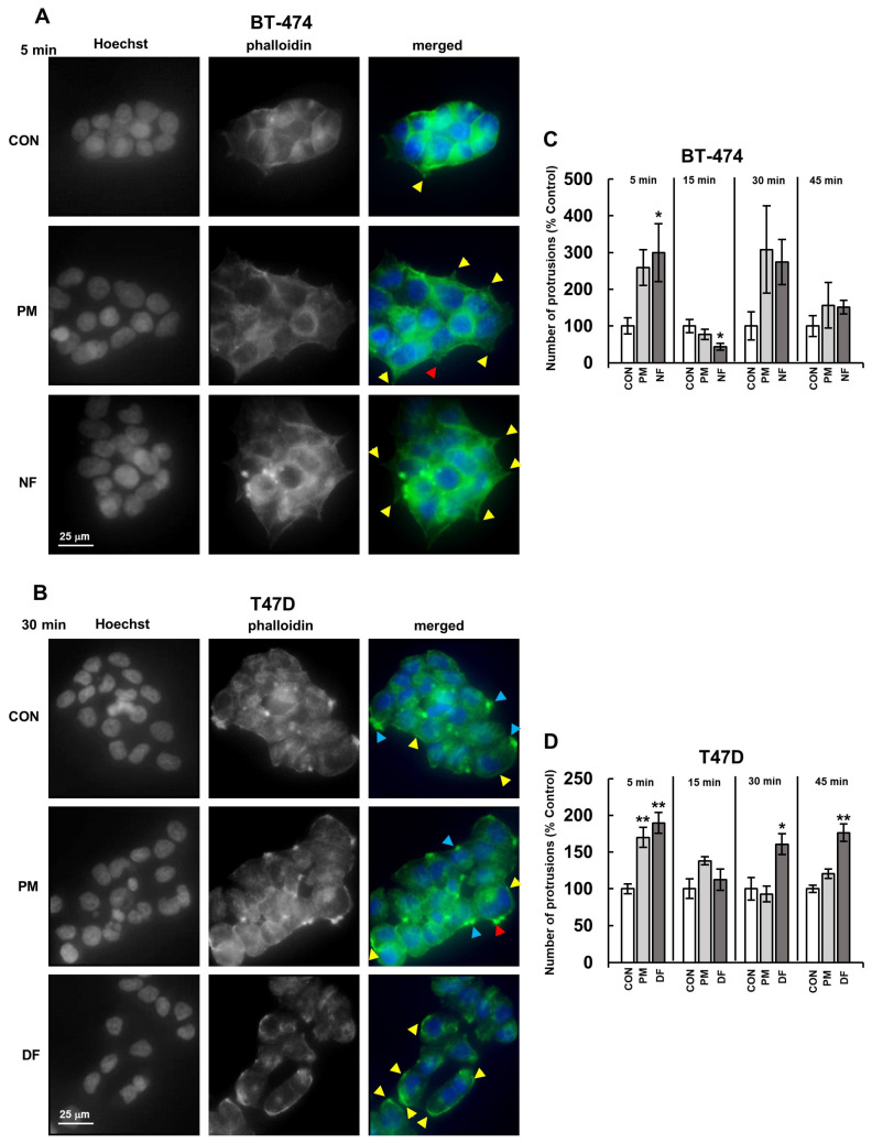
Figure 3.
DS rapidly influenced the actin cytoskeleton organization in luminal breast cancer cells. (A,B) Representative images showing the effects of the DS variants (PM–DS from porcine intestinal mucosa, NF–DS from normal human fascia, DF–DS from fibrosis-affected human fascia) at a concentration of 25 µg/mL on the formation of cellular protrusions in the BT-474 (A) or T-47D (B) cells that were incubated with the glycans for the indicated time periods and then stained with phalloidin-Alexa Fluor (actin filaments–green) and Hoechst (nuclei–blue). Yellow arrowheads (A,B) indicate lamellipodia-like processes, blue arrowheads (B)–large dot-like structures, and the red arrowhead (A,B) shows clusters of projections resembling filopodia. (C,D) The dynamics of the DS variant-mediated effects on the formation of lamellipodia-like protrusions in the BT-474 (C) and T-47D (D) cells. The cells were grown in the presence of the DS variants for the indicated periods of time. The number of protrusions (indicated by yellow or blue arrowheads in the BT-474 or T-47D cells, respectively) was counted for all of the visible cells in four non-overlapping fields from each of two independent experiments. The results are expressed as the percentage of the effect that was visible in the untreated controls and are presented as the mean ±SEM for all of the obtained images. */**—statistically significant differences (p < 0.05/p < 0.01, respectively) versus the control.

