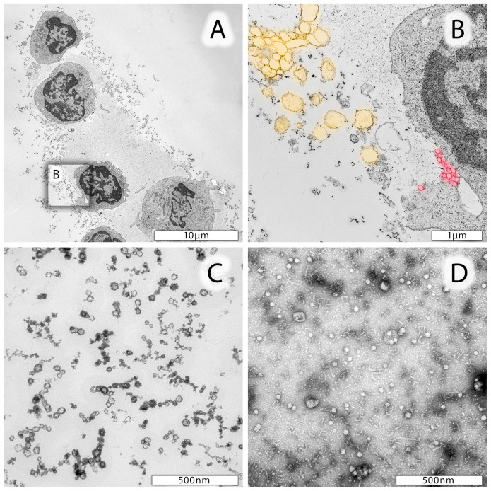
Figure 1.
Representative electron micrographs of leukocytes from CSF. (A) Lymphocytes and one monocyte in the lower part of the image. Cells are surrounded by a protein matrix and numerous vesicles differing in size. (B) Enlarged area of a lymphocyte marked in (A) with likely extruding microparticles highlighted in yellow and exosomes highlighted in red. (C) Smaller (<40 nm in diameter) particles in CSF specimen which may be due to proteins, oxidized phospholipids, and nucleic acids. (D) Negative staining preparation of mesenchymal stem-cell-derived exosomes.

