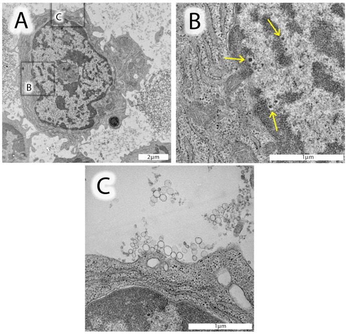
Figure 2.
Electron micrographs of leukocytes from a selected AF patient #15159. (A) Lymphocyte from the CSF with a well-structured, metabolically active nucleus, mitochondria, and endoplasmic reticulum. The nucleus of this lymphocyte contains homo- and heterochromatin and a well-identified nucleolus in the center. Distinct, highly electron-dense spots, structurally similar to herpesvirus nucleocapsids devoid of an envelope, can be identified in the distal area of the heterochromatin. (B) Enlarged insert of the nuclear area (black square in (A)) shows these likely herpesvirus nucleocapsids with a characteristically light halo surrounding the nucleocapsid (yellow arrows). (C) Enlarged insert area from (A) documenting the presence of exosomes (50–120 nm in diameter) close to lymphocyte’s cell surface. In the upper part of this image insert, very small vesicles can be identified, likely corresponding to proteins, oxidized phospholipids, and/or nucleic acid aggregates.

