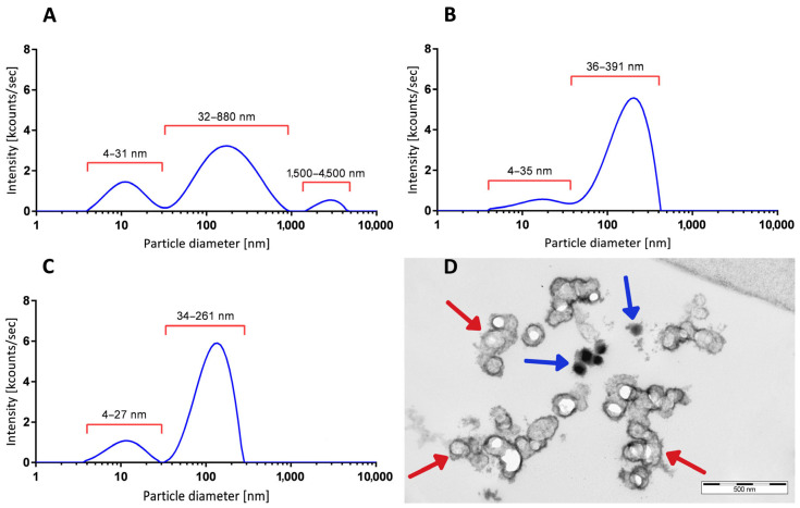
Figure 3.
Vesicle size distributions from CSF-derived particles (EVs). (A) DLS-generated size distribution profile of cell-free CSF after step 1 centrifugation from a representative AF patient, with peaks at 12.20 nm, 215.00 nm, and 2790 nm in diameter. (B) Size distribution profile of another patient’s preparation after step 3 ultracentrifugation. Enriched EVs show a major peak at 183.5 nm in diameter and a smaller peak fraction with 16.0 nm in diameter. (C) DLS-generated size distribution pattern of highly purified mesenchymal stem-cell-derived exosomes (XoGloR, Kimeralabs.com) showing a major peak at 124.0 nm and a smaller peak at 11.90 nm in diameter. For vesicle size distribution plots, the relative intensities (kcounts/s, y-axis) were plotted against the log10 values of particles’ diameters (x-axis). (D) Ultrastructure of cryopressure-processed, highly purified mesenchymal stem-cell-derived exosomes (XoGloR, Kimeralabs.com). Preparation contains lipid-bilayer-enclosed vesicles (red arrows) and lipid-bilayer-negative electron-dense aggregates (blue arrows).

