Abstract
Obesity is frequently accompanied by non-alcoholic fatty liver disease (NAFLD). These two diseases are associated with altered lipid metabolism, in which reverse cholesterol transport (LXRα/ABCA1/ABCG1) and leptin response (leptin receptor (Ob-Rb)/Sam68) are involved. The two pathways were evaluated in peripheral blood mononuclear cells (PBMCs) from 86 patients with morbid obesity (MO) before and six months after Roux-en-Y gastric bypass (RYGB) and 38 non-obese subjects. In the LXRα pathway, LXRα, ABCA1, and ABCG1 mRNA expressions were decreased in MO compared to non-obese subjects (p < 0.001, respectively). Ob-Rb was decreased (p < 0.001), whereas Sam68 was increased (p < 0.001) in MO. RYGB did not change mRNA gene expressions. In the MO group, the LXRα pathway (LXRα/ABCA1/ABCG1) negatively correlated with obesity-related variables (weight, body mass index, and hip), inflammation (C-reactive protein), and liver function (alanine-aminotransferase, alkaline phosphatase, and fatty liver index), and positively with serum albumin. In the Ob-R pathway, Ob-Rb and Sam68 negatively correlated with alanine-aminotransferase and positively with albumin. The alteration of LXRα and Ob-R pathways may play an important role in NAFLD development in MO. It is possible that MO patients may require more than 6 months following RYBGB to normalize gene expression related to reverse cholesterol transport or leptin responsiveness.
Keywords: obesity, bariatric surgery, Roux-en-Y gastric bypass, Ob-R, LXRα, ABCA1, ABCG1
1. Introduction
Obesity represents a significant public health problem in developed countries, with a multitude of obesity-related comorbidities. These include a chronic low-grade inflammatory state, insulin resistance, type 2 diabetes mellitus (T2DM), hypertension, hepatic steatosis, and abnormal lipid metabolism, including decreased high-density lipoprotein (HDL) levels. Bariatric surgical procedures, such as laparoscopic Roux-en-Y gastric bypass (RYGB), are recommended as the most effective treatment for morbid obesity (MO). Furthermore, obesity is also associated with high levels of leptin [1,2]. The action of leptin is mediated by the leptin receptor (Ob-R), a member of the class I type cytokine receptor family [3]. Ob-Rb is the long isoform of the leptin receptor, which allows for complete intracellular signaling. In the pathway of Ob-R signaling, the Src-associated mitosis 68 kDa protein (mostly known as Sam68), a member of the signal transduction and activation of RNA metabolism (STAR) family of RNA-binding proteins, has been previously implicated [4]. Sam68 mediates leptin effects in different diseases [4,5] and is involved in the regulation of hepatic gluconeogenesis [6], which is also associated with leptin effects [7].
Liver X receptor α (LXRα) is an oxysterol-activated nuclear receptor present in different human cells that regulates the expression of genes linked to cholesterol metabolism [8]. Previously, we demonstrated a downregulation of LXRα in visceral adipose tissue from patients with MO [9]. In peripheral blood cells, such as macrophages, this nuclear receptor modulates gene transcription involved in homeostasis cholesterol, such as the ATP-binding cassette (ABC) transporters (ABCA1 and ABCG1), lipogenesis, and the anti-inflammatory response [10]. In previous studies [11,12,13], we have demonstrated their presence in peripheral blood mononuclear cells (PBMCs) and the relevance of LXRα and ABCA1 and ABCG1 transporters in regulating cholesterol homeostasis in monocytes/macrophages and neutrophils. In addition to its pivotal role in cholesterol homeostasis, the nuclear receptor LXRα plays a fundamental role in regulating inflammation in these PBMCs.
Obesity is frequently accompanied by metabolic dysfunction-associated steatotic liver disease (MASLD) and sarcopenia, which are closely intertwined [14]. MASLD, formerly known as non-alcoholic fatty liver disease (NAFLD), is defined as the presence of hepatic steatosis, which is defined as the accumulation of neutral lipids such as triacylglycerol and cholesteryl esters within the liver. In MASLD, the role of metabolic syndrome, obesity, and T2DM is essential. Sarcopenia is defined as the loss of skeletal muscle mass and muscle function. These diseases, obesity, sarcopenia, and MASLD, are associated with an increase in insulin resistance and chronic low-grade inflammation. However, the current therapeutic approach for MO patients with MASLD is to ensure weight loss through bariatric surgery.
It is noteworthy that the Ob-R and LXRα pathways are also associated with MASLD. Leptin plays a critical role in the development of liver fibrosis [15]. Additionally, leptin reduced LXRα protein level and activity in hepatic stellate cells [16]. However, the role of LXRα in the liver is debated, with apparently contradictory results. LXRα activation has been associated with increased liver fat deposition and the development of hepatic steatosis [17]. However, it has also been demonstrated to possess anti-inflammatory properties [18]. Furthermore, the reduction of LXRα produced a depletion of hepatic cellular lipid content in a fatty liver mouse model [19]. In contrast, other studies found that LXR activation improved liver injury, as evidenced by reductions in alanine aminotransferase (ALT), aminotransferase (AST), and tumor necrosis factor (TNF)α levels [20].
The aim of this study was to identify biomarkers in peripheral blood (a tissue easily obtained with minimally invasive techniques), which will enable us to gain a deeper understanding of how metabolic changes following RYGB may influence the relationship between leptin/LXRα pathways and different fat and fibrosis indexes. To this end, we analyze the mRNA expression of Ob-R, Sam68, LXRα, ABCA1, and ABCG1 in PBMCs from a group of non-obese subjects and patients with MO before and 6 months after RYGB.
2. Results
2.1. Anthropometric Characteristics and Biochemical Study
Table 1 summarizes the anthropometric and biochemical variables of non-obese subjects and patients with MO before and 6 months after RYGB. As expected, most of the anthropometric and biochemical parameters were altered in the obese group, which improved after RYGB, including fat (fatty liver index (FLI)) and fibrosis (non-alcoholic fatty liver disease fibrosis score (NAFLD FS)) indexes. Meanwhile, 0% of non-obese subjects had metabolic syndrome. In the MO group, 82% had metabolic syndrome before RYGB and 62.9% after RYGB.
Table 1.
Anthropometric and biochemical variables of non-obese subjects and patients with morbid obesity (MO) before and six months after RYGB.
| Non-Obese Subjects | MO Pre-Surgery | MO Post-Surgery | |
|---|---|---|---|
| Sex (Men/Women) | 35 (12/23) | 87 (22/65) | - |
| Age (years) | 43.4 ± 13.9 | 42.5 ± 10.0 | - |
| %EWL | - | - | 36 ± 31.5 |
| %TWL | - | - | 17.3 ± 14.6 |
| Weight (kg) | 70.3 ± 13.0 | 135.3 ± 25.4 3 | 96.5 ± 18.4 3,b |
| BMI (kg/m2) | 26.1 ± 3.9 | 50.4 ± 7.6 3 | 36.1 ± 6.6 3,b |
| Waist (cm) | 92.5 ± 9.2 | 138.3 ± 16.2 3 | 110.6 ± 13.1 3,b |
| Hip (cm) | 99.7 ± 8.0 | 147.5 ± 18.5 3 | 121.6 ± 16.9 3,b |
| Glucose (mg/dL) | 85.7 ± 8.8 | 108.8 ± 49.6 3 | 81.1 ± 13.5 b |
| Cholesterol (mg/dL) | 204.9 ± 30.6 | 197.3 ± 35.7 | 183.6 ± 33.5 2,b |
| Triglycerides (mg/dL) | 109.2 ± 61.3 | 149.7 ± 80.2 2 | 109.9 ± 47.4 b |
| HDL (mg/dL) | 54.8 ± 12.1 | 47.2 ± 12.7 2 | 47.0 ± 8.9 2,a |
| LDL (mg/dL) | 128.2 ± 27.0 | 120.0 ± 31.1 | 114.2 ± 30.3 1,b |
| Insulin (µU/mL) | 10.1 ± 4.5 | 22.0 ± 13.8 3 | 8.8 ± 4.1 b |
| HOMA-IR | 2.17 ± 1.05 | 5.95 ± 4.5 3 | 1.81 ± 0.88 b |
| CRP (mg/dL) | 3.3 ± 3.3 | 11.4 ± 7.0 3 | 4.6 ± 4.1 b |
| Leptin (ng/mL) | 17.5 ± 12.7 | 71.1 ± 38.4 3 | 19.28 ± 13.9 b |
| Adiponectin (µg/mL) | 10.7 ± 6.8 | 6.5 ± 3.3 2 | 10.5 ± 5.5 c |
| AST (IU/L) | 22.0 ± 13.5 | 27.6 ± 18.6 | 20.9 ± 8.8 c |
| ALT (IU/L) | 25.5 ± 17.0 | 44.3 ± 33.1 3 | 27.5 ± 14.5 c |
| GGT (IU/L) | 31.8 ± 32.2 | 38.4 ± 31.4 | 24.6 ± 26.3 2,c |
| ALP (IU/L) | 64.4 ± 30.1 | 79.5 ± 24.1 1 | 75.1 ± 39.5 1 |
| Albumin (g/dL) | 4.7 ± 0.3 | 4.0 ± 0.4 3 | 4.0 ± 0.3 3,c |
| FLI | 53.5 ± 32.9 | 98.6 ± 2.9 3 | 72.0 ± 25.7 2 |
| NAFLD FS | −1.17 ± 1.47 | −0.69 ± 1.48 | −1.53 ± 1.31 c |
Results are expressed as mean ± SD. Significant differences with respect to healthy control group: 1 p < 0.05, 2 p < 0.01, 3 p < 0.001. Significant differences in patients with MO between before and six months after RYGB: a p < 0.05, b p < 0.01, c p < 0.001. BMI: body mass index; HDL: high-density lipoprotein; LDL: low-density lipoprotein; HOMA-IR: homeostasis model assessment of insulin resistance; AST: aspartate aminotransferase; ALT: alanine aminotransferase; GGT: gamma-glutamyltransferase; ALP: alkaline phosphatase; FLI: fatty liver index; NAFLD FS: non-alcoholic fatty liver disease fibrosis score.
2.2. LXRα, ABCA1, and ABCG1 Expression in PBMCs from Non-Obese Subjects and Patients with MO Pre- and Post-Surgery
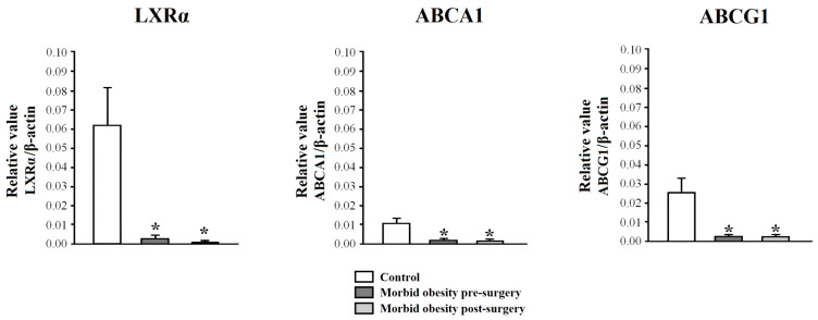
Figure 1 shows the mRNA expression in PBMCs from non-obese subjects and patients with MO pre- and post-surgery. The mRNA expression of LXRα, ABCA1, and ABCG1 (p < 0.001, respectively) in PBMCs from patients with MO was statistically lower than in non-obese subjects. RYGB did not restore this gene expression. There were no significant differences in LXRα, ABCA1, and ABCG1 according to the presence or absence of metabolic syndrome.
Figure 1.
LXRα, ABCA1, and ABCG1 mRNA expression in PBMCs from non-obese subjects (control) and patients with morbid obesity pre- and post-surgery. Data are represented as mean ± standard error of the mean. * p < 0.001: significant differences with regard to control subjects.
2.3. Ob-Rb and Sam68 Expression in PBMC from Non-Obese Subjects and Patients with MO Pre- and Post-Surgery
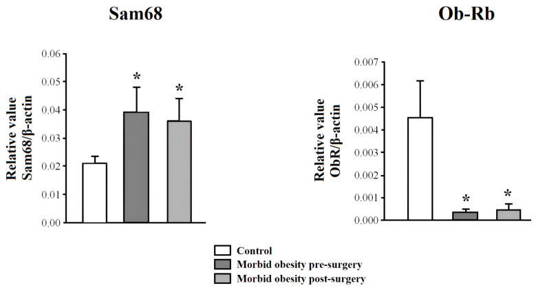
Figure 2 shows the mRNA expression in PBMCs from non-obese subjects and patients with MO pre- and post-surgery. Ob-Rb mRNA expression was significantly decreased (p < 0.001), and Sam68 expression was increased (p < 0.001) in PBMCs from MO patients compared to non-obese subjects. RYGB did not reverse this increased gene expression. There were no significant differences in Ob-Rb and Sam68 according to the presence or absence of metabolic syndrome.
Figure 2.
Ob-Rb and SAM68 mRNA expression in PBMCs from non-obese subjects (control) and patients with morbid obesity pre- and post-surgery. Data are represented as mean ± standard error of the mean. * p < 0.001: significant differences with regard to control subjects.
2.4. Association between mRNA Expression and Anthropometric/Biochemical Variables in Patients with MO Pre- and Post-Surgery
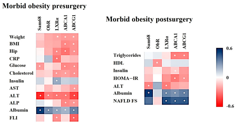
The mRNA expression in PBMCs from patients with MO before RYBGB was significantly correlated with several anthropometric (weight, BMI, and hip circumference) and biochemical variables (glucose, cholesterol, CRP, AST, ALT, alkaline phosphatase (ALP), albumin, insulin, and FLI) (Figure 3). Genes related to cholesterol transport, such as LXRα, ABCA1, and ABCG1, were those with a greater number of significant associations.
Figure 3.
Heat maps represent those significant correlations between Ob-Rb, SAM68, LXRα, ABCA1, and ABCG1 mRNA expression in PBMCs from patients with morbid obesity pre- and post-surgery with anthropometric and biochemical variables. The Spearman correlation is displayed on a color scale from blue (positive correlation) to red (negative correlation). * Significant correlations (p < 0.05). BMI: body mass index; CRP: C-reactive protein; HDL: high-density lipoprotein; HOMA-IR: homeostasis model assessment of insulin resistance; AST: aspartate aminotransferase; ALT: alanine aminotransferase; ALP: alkaline phosphatase; FLI: fatty liver index; NAFLD FS: non-alcoholic fatty liver disease fibrosis score.
The mRNA expression in PBMCs from patients with MO six months after RYGB was significantly correlated with several biochemical variables, including triglycerides, HDL, ALT, albumin, NAFLD FS, insulin, and HOMA-IR (Figure 3). ABCA1 and ABCG1 were the genes with a greater number of significant associations.
3. Discussion
In the present work, we have extended the knowledge of metabolic changes on reverse cholesterol transport mechanisms and leptin response before and six months after RYGB as expected and as we have already shown in previous studies [21]. RYGB resulted in a reduction in peripheral inflammation (e.g., CRP levels) and T2DM. However, the recovery of lipid metabolism is slower and less understood. Although there was an improvement in HDL, the level was still lower than in the control subjects. Similar results have been previously described, although HDL levels were recovered after 12 months [22]. This improvement in HDL levels and its properties would have beneficial effects on obesity-associated atherogenic disease.
The mechanisms involved in the regulation of the atherogenic profile in patients with MO could include the ABCA1 and ABCG1 transporters. However, there are few studies on PBMCs. We found that the expression of ABCA1 and ABCG1 was reduced in patients with MO, as shown in another study performed in visceral adipose tissue [23]. This reduction could play an important role in the downregulation of HDL levels in MO. These two proteins are involved in the generation and formation of HDL [24,25]. Although previous studies have described that RYGB induces an improvement in the atherogenic lipid profile by a shift toward a more cardioprotective HDL and an increase in plasma efflux capacity via ABCG1 six months after surgery [26,27], our study did not show this increase. Our data at 6 months after RYGB suggest that, although there is a slight improvement, more time would be needed to normalize the expression levels of these genes in PBMCs and thus to normalize HDL levels and functionality.
In this study, we showed that LXRα expression was downregulated in PBMCs from patients with MO and negatively correlated with CRP and total cholesterol. In addition, LXRα, ABCA1, and ABCG1 expressions were negatively correlated with several obesity-related variables, such as weight, BMI, and hip circumference, reinforcing their association with obesity. Their reduced expression could be due to the increased serum LPS levels found in obesity [28], which repress the expression of LXR and, consequently, of ABCA1/ABCG1 [29]. Although this result may suggest that an upregulation of LXRα would be beneficial to increase the ABCA1 and ABCG1 expression, promote the reverse cholesterol transport (less total cholesterol), and reduce inflammation (less CRP), precautions must be taken. It is true that LXRα activation may suppress inflammation and improve atherosclerosis, but it can also promote the development of obesity and liver steatosis [30]
As expected, the high leptin levels found in patients with MO decreased to normal levels after RYGB. Although leptin can be secreted into the blood by a wide variety of tissues, in the case of morbid obesity, the most important tissues are the stomach and adipose tissue. The reduction in leptin levels after bariatric surgery is a result of the surgery itself due to the removal of part of the stomach and the large loss of adipose tissue. In previous studies, serum leptin levels were associated with an increased risk of sarcopenic obesity [31], suggesting that leptin may also play a role in sarcopenic obesity. However, it can deteriorate further after bariatric surgery [32]. The effects of leptin are mediated by its receptor, Ob-R, which was decreased in PBMCs from MO patients. Ob-Rb is critical because it is the only leptin receptor with a long cytoplasmic tail, allowing complete intracellular signaling. It plays a key role in mediating the effects of leptin on appetite control and energy balance via the JAK-STAT signaling pathway [33].
A previous study also showed that children with obesity had significantly lower Ob-R levels and higher leptin levels [34]. This decreased Ob-Rb expression could be due to the leptin excess and the low-grade inflammation present in obesity [35]. Regarding the effect of RYGB, although bariatric surgery has recently been described to enhance the leptin signaling pathway due to the decreased inflammatory state [36], our data did not show changes in mRNA expression. However, its level increased one year after laparoscopic sleeve gastrectomy, although it was still lower than that of the control group [37]. In addition, the weight loss one year after adjustable gastric banding increased soluble leptin receptor levels [38]. Perhaps more time would be needed to normalize its expression level. In the leptin pathway, we found an increase in the expression of Sam68. Sam68 influences alternative splicing of several genes critical to processes such as adipogenesis [39]. However, its obesity-related function in PBMCs has not yet been defined. In relation to bariatric surgery, the Sam68 gene still maintains high expression levels 6 months after RYGB. Further studies are needed to clarify the role of Sam68 in MO patients.
Another interesting point found in this study was the relationship between the expression of these genes and different liver variables. Although the gold standard for liver tests is liver biopsy, we did not have access to it for ethical reasons, nor to liver ultrasound or transient elastography. Therefore, we have used indirect markers, such as liver enzymes, FLI, and NAFLD FS, to study liver status. LXRα, ABCA1, and ABCG1 were negatively associated with ALT levels, which is related to lipid metabolism and obesity and reflects hepatocellular injury in patients with NAFLD [40,41]. Greater inhibition of LXRα, ABCA1, and ABCG1 would be associated with greater inflammation (CRP) and worse liver function (high ALT and AP levels and lower albumin concentration) [42]. Moreover, our data also showed that LXRα was associated with FLI, an indirect marker of liver steatosis, as suggested by a previous study [43]. The accumulation of fatty acids in the liver, as evidenced by elevated FLI levels, is a defining feature of MASLD [44]. This ectopic fat deposition is a consequence of the increased insulin resistance observed in obesity. In other organs, such as skeletal muscle, this fat deposition results in sarcopenia [45]. Furthermore, the severity of hepatic fibrosis and steatosis increases with the prevalence of sarcopenia [46]. Moreover, the relationship between sarcopenia and cholesterol metabolism is further supported by another previous study in which two proteins, cholesteryl ester transfer protein and apolipoprotein A2, were identified as potential biomarkers for sarcopenia, thereby improving the diagnostic accuracy of this condition [47]. On the other hand, the association found between Ob-Rb with ALT and albumin levels suggests that a decrease in leptin signaling may be associated with a hepatic impairment. In this regard, the development of NAFLD is associated with increased leptin levels and leptin resistance (low Ob-R signaling) [48]. In this regard, the up-regulation of leptin receptor may result in increased leptin sensitivity, which would be sufficient to treat NAFLD [49].
However, a limitation of the present study is that we performed an evaluation only 6 months after RYGB. At that time, the patient was still adapting to a new lifestyle and had not achieved total weight loss. In addition, it would have been better to conduct the study with liver samples. However, given the ethical impossibility of taking liver biopsies, we have carried out this study in PBMCs with the aim of finding biomarkers in peripheral blood (a tissue easily obtained with minimally invasive techniques). Other studies have also shown that changes in PBMC gene expression could be used as early biomarkers for the diagnosis of metabolic disorders, could have clinical significance, and could ultimately provide surrogate transcriptional markers of biological efficacy in relevant tissues [50,51].
In conclusion, this is the first time that it has been described that the expression of LXRα, ABCA1, ABCG1, and Ob-Rb in PBMCs was decreased in patients with MO before and after RYGB and was associated with several variables related to obesity, inflammation, and liver function. This decrease may play an important role in the downregulation of HDL levels and in the development of MASLD in MO. Our data at six months after RYGB suggest that more time would be needed to normalize the expression levels of genes related to reverse cholesterol transport or leptin responsiveness in PBMCs and thus to normalize HDL levels and liver function. Further research in this area is highly needed using transcriptomics and proteomics to perform a more complete analysis of the up- and down-regulation of specific obesity-related genes in these conditions, including in liver samples biopsied at the time of bariatric surgery.
4. Methods
4.1. Subjects
The study included 86 patients with MO before and six months after RYGB and 38 healthy, non-obese subjects (body mass index (BMI) ˂ 30 kg/m2). Patients were excluded if they had acute inflammatory disease, infectious disease, or because of patient decision. Non-obese subjects were similar in age to the group with MO, had a stable body weight for at least 3 months before the study, and without acute inflammatory disease, infectious disease, and lipid and carbohydrate metabolism disorders. The percentage of total weight loss (%TWL) was calculated as 100 × (baseline weight − postoperative weight)/baseline weight. The percentage of excess weight loss (%EWL) was calculated as 100 × [(baseline weight) − (postoperative weight)]/[(baseline weight) − (ideal weight)], where ideal weight is defined by the weight corresponding to a BMI of 25 kg/m2. The patients included in the study were subjected to the International Diabetes Federation (IDF) metabolic syndrome classification criteria to determine the percentage of patients who met the definition of presence or absence of metabolic syndrome [52]. The study was conducted in accordance with the Code of Ethics of the World Medical Association (Declaration of Helsinki). All participants gave their written informed consent, and the study was reviewed and approved by the Malaga Provincial Research Ethics Committee (Malaga, Spain).
4.2. Sample Collection
Peripheral blood samples were collected from patients with MO before and six months after RYGB and from non-obese subjects to isolate the cellular fraction and serum [53]. Serum was separated and immediately frozen at −80 °C. PBMCs were isolated from 10 mL of blood in a Ficoll-Paque density gradient (GE Healthcare, Buckinghamshire, UK) [53]. Samples were processed and frozen immediately upon receipt at the Regional University Hospital Biobank (Andalusian Public Health System Biobank, Spain).
4.3. Laboratory Measurements
Serum biochemical parameters were measured in duplicate using an Advia Chemistry XPT autoanalyzer (Siemens Healthcare Diagnostics, Malvern, PA, USA). The low-density lipoprotein (LDL) fraction was calculated according to the Friedewald equation. Serum insulin levels were measured by immunoassay using an ADVIA Centaur autoanalyzer (Siemens Healthcare Diagnostics, Malvern, PA, USA). The determination of leptin and high-sensitivity C-reactive protein (CRP) were performed by commercial enzyme-linked immunosorbent assay (Mediagnost GmbH, Reutlingen, Germany, and DRG Instruments GmbH, Marburg, Germany, respectively). HOMA-IR was calculated with the following equation: HOMA-IR = fasting insulin (µIU/mL) × fasting glucose (mmol/L)/22.5. The fatty liver index (FLI) was used for assessing hepatic steatosis [54]. To evaluate liver fibrosis, the NAFLD fibrosis score (NAFLD FS) was used [55].
4.4. Real-Time Quantitative PCR of mRNA Levels
Purification of RNA from PBMCs was performed by the QIAamp RNA Blood Mini Kit (QIAGEN Science, Hilden, Germany). Total RNA was reverse-transcribed into cDNA, and RT-PCR was carried out in an ABI Prism 7300 Sequence Detection System (Applied Biosystems, Foster City, CA, USA). PCR reactions were performed in triplicate with SYBR Green PCR Master Mix (Applied Biosystems, Foster City, CA, USA) using the following primers: LXRα: forward, 5’-AAGCCCTGCATGCCTACGT-3′, reverse, 5′-TGCAGACGCAGTGCAAACA-3′; ABCA1: forward, 5′-CCCTGTGGAATGTACCTATGTG-3′, reverse, 5′-GAGGTGTCCCAAAGATGCAA-3′; ABCG1: forward,5′-CAGTCGCTCCTTAGCACCA-3′, reverse, 5′-TCCATGCTCGGACTCTCTG-3′; Ob-Rb: forward, 5′-ATAGTTCAGTCACCAAGTGC-3′, reverse, 5′-GTCCTGGAGAACTCTGATGTCC-3′; Sam68: forward, 5′-TTTGTGGGGAAGATTCTTGG-3′, reverse 5′-GGGGGTCCAAAGACTTCAAT-3′ and β-actin: forward, 5’-CCAGCTCACCATGGATGATG-3’, reverse, 5’-ATGCCGGAGCCGTTGTC-3’. Relative levels of transcripts above were quantified by the comparative threshold cycle (Ct) method as described in the ABI Prism 7300 User Bulletin 2 and normalized to β-actin mRNA levels.
4.5. Statistical Analysis
Statistical analysis was performed using the SPSS 26.0 software package (SPSS Inc., Chicago, IL, USA). Normal distribution was analyzed using the Kolmogorov–Smirnov test. Most of the parameters analyzed did not have a normal distribution, and nonparametric analyses were used. Differences between two related variables were analyzed using the Wilcoxon test. The Spearman correlation coefficients were calculated to estimate the correlations between variables. Statistically significant differences were considered when p ≤ 0.05. Results are presented as mean ± standard deviation.
Acknowledgments
CIBEREHD and CIBEROBN are ISCIII projects.
Author Contributions
Study concept and design: C.J.-C., S.L.-E., V.S.-M. and E.G.-F.; acquisition of data: G.A., C.S.-M., G.M.M.-N., F.J.M.-R., S.V., S.G.-S., C.R.-D., A.H.-P. and M.I.F.-R.; analysis and interpretation of data: C.J.-C., S.L.-E., G.A., L.G.-S. and E.G.-F.; drafting of the manuscript: C.J.-C., S.L.-E., G.A., L.G.-S., E.G.-F. and V.S.-M.; critical revision of the manuscript: E.G.-F. and V.S.-M.; statistical analysis: C.J.-C., S.L.-E., G.A., L.G.-S. and E.G.-F.; obtained funding: S.L.-E., V.S.-M. and E.G.-F.; study supervision: E.G.-F. and V.S.-M. All authors have read and agreed to the published version of the manuscript.
Institutional Review Board Statement
The study was conducted in accordance with the Declaration of Helsinki and approved by the Malaga Provincial Research Ethics Committee of the Biomedical Research Institute of Malaga (IBIMA) (protocol code: PI09/01016).
Informed Consent Statement
Informed consent was obtained from all subjects involved in the study.
Data Availability Statement
The datasets presented in this article are not readily available because they are part of an ongoing study. Requests to access the datasets should be directed to the corresponding author, [E.G.-F.], upon reasonable request.
Conflicts of Interest
The authors declare no conflict of interest.
Funding Statement
This study has been supported by the Instituto de Salud Carlos III (Spain) (PI09/01016) (“A way to make Europe”), the Consejería de Salud y Familias (Junta de Andalucía, Spain) (PE-0098-2019, PI-2013-575), and the Consejería de Universidad, Investigación e Innovación (Junta de Andalucía, Spain) (P10-CTS6928, P11-CTS8161 and P11-CTS8081, and CTS-151), and co-financed by the European Regional Development Fund (FEDER) (“Andalusia moves with Europe”). C.J.-C. is supported by the Margarita Salas Fellowship (University of Seville, Spain). C.R.-D. is supported by a grant from the Consejería de Transformación Económica, Industria, Conocimiento y Universidades de la Junta de Andalucía (Spain) (DOC_01610). E.G.-F. is supported by the Nicolas Monardes program from the Consejería de Salud de Andalucía (Spain) (RC-0005-2020).
Footnotes
Disclaimer/Publisher’s Note: The statements, opinions and data contained in all publications are solely those of the individual author(s) and contributor(s) and not of MDPI and/or the editor(s). MDPI and/or the editor(s) disclaim responsibility for any injury to people or property resulting from any ideas, methods, instructions or products referred to in the content.
References
- 1.Murri M., García-Fuentes E., García-Almeida J.M., Garrido-Sánchez L., Mayas M.D., Bernal R., Tinahones F.J. Changes in Oxidative Stress and Insulin Resistance in Morbidly Obese Patients After Bariatric Surgery. Obes. Surg. 2010;20:363–368. doi: 10.1007/s11695-009-0021-6. [DOI] [PubMed] [Google Scholar]
- 2.Pérez-Pérez A., Vilariño-García T., Fernández-Riejos P., Martín-González J., Segura-Egea J.J., Sánchez-Margalet V. Role of leptin as a link between metabolism and the immune system. Cytokine Growth Factor. Rev. 2017;35:71–84. doi: 10.1016/j.cytogfr.2017.03.001. [DOI] [PubMed] [Google Scholar]
- 3.Sánchez-Jiménez F., Pérez-Pérez A., González-Yanes C., Najib S., Varone C.L., Sánchez-Margalet V. Leptin receptor activation increases Sam68 tyrosine phosphorylation and expression in human trophoblastic cells. Mol. Cell Endocrinol. 2011;332:221–227. doi: 10.1016/j.mce.2010.10.014. [DOI] [PubMed] [Google Scholar]
- 4.Pérez-Pérez A., Sánchez-Jiménez F., Vilariño-García T., de la Cruz L., Virizuela J.A., Sánchez-Margalet V. Sam68 Mediates the Activation of Insulin and Leptin Signalling in Breast Cancer Cells. PLoS ONE. 2016;11:e0158218. doi: 10.1371/journal.pone.0158218. [DOI] [PMC free article] [PubMed] [Google Scholar]
- 5.Vilariño-García T., Pérez-Pérez A., Santamaría-López E., Prados N., Fernández-Sánchez M., Sánchez-Margalet V. Sam68 mediates leptin signaling and action in human granulosa cells: Possible role in leptin resistance in PCOS. Endocr. Connect. 2020;9:479–488. doi: 10.1530/EC-20-0062. [DOI] [PMC free article] [PubMed] [Google Scholar]
- 6.Qiao A., Zhou J., Xu S., Ma W., Boriboun C., Kim T., Yan B., Deng J., Yang L., Zhang E., et al. Sam68 promotes hepatic gluconeogenesis via CRTC2. Nat. Commun. 2021;12:3340. doi: 10.1038/s41467-021-23624-9. [DOI] [PMC free article] [PubMed] [Google Scholar]
- 7.Perry R.J., Petersen K.F., Shulman G.I. Pleotropic effects of leptin to reverse insulin resistance and diabetic ketoacidosis. Diabetologia. 2016;59:933–937. doi: 10.1007/s00125-016-3909-4. [DOI] [PMC free article] [PubMed] [Google Scholar]
- 8.Sáenz J., Alba G., Reyes-Quiroz M.E., Geniz I., Jiménez J., Sobrino F., Alba G. Grapefruit Flavonoid Naringenin Regulates the Expression of LXRα in THP-1 Macrophages by Modulating AMP-Activated Protein Kinase. Mol. Pharm. 2018;15:1735–1745. doi: 10.1021/acs.molpharmaceut.7b00797. [DOI] [PubMed] [Google Scholar]
- 9.Moreno-Santos I., Garcia-Serrano S., Boughanem H., Garrido-Sanchez L., Tinahones F.J., Garcia-Fuentes E., Macias-Gonzalez M. The Antagonist Effect of Arachidonic Acid on GLUT4 Gene Expression by Nuclear Receptor Type II Regulation. Int. J. Mol. Sci. 2019;20:963. doi: 10.3390/ijms20040963. [DOI] [PMC free article] [PubMed] [Google Scholar]
- 10.Zelcer N., Tontonoz P. Liver X receptors as integrators of metabolic and inflammatory signaling. J. Clin. Investig. 2006;116:607–614. doi: 10.1172/JCI27883. [DOI] [PMC free article] [PubMed] [Google Scholar]
- 11.Sáenz J., Alba G., Reyes-Quiroz M.E., Geniz I., Jiménez J., Sobrino F., Santa-María C. Curcumin enhances LXRα in an AMP-activated protein kinase-dependent manner in human macrophages. J. Nutr. Biochem. 2018;54:48–56. doi: 10.1016/j.jnutbio.2017.11.006. [DOI] [PubMed] [Google Scholar]
- 12.Reyes-Quiroz M.E., Alba G., Saenz J., Santa-María C., Geniz I., Jiménez J., Ramírez R., Martín-Nieto J., Pintado E., Sobrino F. Oleic acid modulates mRNA expression of liver X receptor (LXR) and its target genes ABCA1 and SREBP1c in human neutrophils. Eur. J. Nutr. 2014;53:1707–1717. doi: 10.1007/s00394-014-0677-0. [DOI] [PubMed] [Google Scholar]
- 13.Alba G., Reyes M.E., Santa-María C., Ramírez R., Geniz I., Jiménez J., Martín-Nieto J., Pintado E., Sobrino F. Transcription of liver X receptor is down-regulated by 15-deoxy-Δ(12,14)-prostaglandin J(2) through oxidative stress in human neutrophils. PLoS ONE. 2012;7:e42195. doi: 10.1371/journal.pone.0042195. [DOI] [PMC free article] [PubMed] [Google Scholar]
- 14.Wong R., Yuan L.Y. Sarcopenia and metabolic dysfunction associated steatotic liver disease: Time to address both. World J. Hepatol. 2024;16:871–877. doi: 10.4254/wjh.v16.i6.871. [DOI] [PMC free article] [PubMed] [Google Scholar]
- 15.García-Suárez C., Crespo J., Fernández-Gil P.L., Amado J.A., García-Unzueta M.T., Pons Romero F. Concentraciones plasmáticas de leptina en los pacientes con cirrosis biliar primaria y su relación con el grado de fibrosis [Plasma leptin levels in patients with primary biliary cirrhosis and their relationship with degree of fibrosis] Gastroenterol. Hepatol. 2004;27:47–50. doi: 10.1016/S0210-5705(03)79085-2. [DOI] [PubMed] [Google Scholar]
- 16.Saxena N.K., Ikeda K., Rockey D.C., Friedman S.L., Anania F.A. Leptin in hepatic fibrosis: Evidence for increased collagen production in stellate cells and lean littermates of ob/ob mice. Hepatology. 2002;35:762–771. doi: 10.1053/jhep.2002.32029. [DOI] [PMC free article] [PubMed] [Google Scholar]
- 17.Dixon E.D., Nardo A.D., Claudel T., Trauner M. The Role of Lipid Sensing Nuclear Receptors (PPARs and LXR) and Metabolic Lipases in Obesity, Diabetes and NAFLD. Genes. 2021;12:645. doi: 10.3390/genes12050645. [DOI] [PMC free article] [PubMed] [Google Scholar]
- 18.Körner A., Zhou E., Müller C., Mohammed Y., Herceg S., Bracher F., Rensen P.C.N., Wang Y., Mirakaj V., Giera M. Inhibition of Δ24-dehydrocholesterol reductase activates pro-resolving lipid mediator biosynthesis and inflammation resolution. Proc. Natl. Acad. Sci. USA. 2019;116:20623–20634. doi: 10.1073/pnas.1911992116. [DOI] [PMC free article] [PubMed] [Google Scholar]
- 19.Lin Y.N., Wang C.C.N., Chang H.Y., Chu F.Y., Hsu Y.A., Cheng W.K., Ma W.C., Chen C.J., Wan L., Lim Y.P. Ursolic Acid, a Novel Liver X Receptor α (LXRα) Antagonist Inhibiting Ligand-Induced Nonalcoholic Fatty Liver and Drug-Induced Lipogenesis. J. Agric. Food Chem. 2018;66:11647–11662. doi: 10.1021/acs.jafc.8b04116. [DOI] [PubMed] [Google Scholar]
- 20.Liu Y., Han X., Bian Z., Peng Y., You Z., Wang Q., Chen X., Qiu D., Ma X. Activation of liver X receptors attenuates endotoxin-induced liver injury in mice with nonalcoholic fatty liver disease. Dig. Dis. Sci. 2012;57:390–398. doi: 10.1007/s10620-011-1902-9. [DOI] [PubMed] [Google Scholar]
- 21.García-Fuentes E., García-Almeida J.M., García-Arnés J., Rivas-Marín J., Gallego-Perales J.L., González-Jiménez B., Cardona I., García-Serrano S., Garriga M.J., Gonzalo M., et al. Morbidly obese individuals with impaired fasting glucose have a specific pattern of insulin secretion and sensitivity: Effect of weight loss after bariatric surgery. Obes. Surg. 2006;16:1179–1188. doi: 10.1381/096089206778392383. [DOI] [PubMed] [Google Scholar]
- 22.Genua I., Puig N., Miñambres I., Benítez S., Gil P., Grau-Agramunt M., Rivas-Urbina A., Balagué C., Fernández-Alanin S., García-Osuna Á., et al. Changes in the composition and function of lipoproteins after bariatric surgery in patients with severe obesity. J. Clin. Med. 2021;10:1716. doi: 10.3390/jcm10081716. [DOI] [PMC free article] [PubMed] [Google Scholar]
- 23.Choromanska B., Mysliwiec P., Hady H.R., Dadan J., Mysliwiec H., Bonda T., Chabowski A., Miklosz A. The implication of adipocyte atp-binding cassette A1 and G1 transporters in metabolic complications of obesity. J. Physiol. Pharmacol. 2019;70:143–152. doi: 10.26402/jpp.2019.1.14. [DOI] [PubMed] [Google Scholar]
- 24.Kennedy M.A., Barrera G.C., Nakamura K., Baldán A., Tarr P., Fishbein M.C., Frank J., Francone O.L., Edwards P.A. ABCG1 has a critical role in mediating cholesterol efflux to HDL and preventing cellular lipid accumulation. Cell Metab. 2005;1:121–131. doi: 10.1016/j.cmet.2005.01.002. [DOI] [PubMed] [Google Scholar]
- 25.Wang S., Smith J.D. ABCA1 and nascent HDL biogenesis. Biofactors. 2014;40:547–554. doi: 10.1002/biof.1187. [DOI] [PMC free article] [PubMed] [Google Scholar]
- 26.Aron-Wisnewsky J., Julia Z., Poitou C., Bouillot J.L., Basdevant A., Chapman M.J., Clement K., Guerin M. Effect of bariatric surgery-induced weight loss on SR-BI-, ABCG1-, and ABCA1-mediated cellular cholesterol efflux in obese women. J. Clin. Endocrinol. Metab. 2011;96:1151–1159. doi: 10.1210/jc.2010-2378. [DOI] [PubMed] [Google Scholar]
- 27.Alaminos-Castillo M.Á., Ho-Plagaro A., García-Serrano S., Santiago-Fernandez C., Rodríguez-Pacheco F., Garrido-Sanchez L., Rodriguez C., Valdes S., Gonzalo M., Moreno-Ruiz F.J., et al. Increased PON lactonase activity in morbidly obese patients is associated with impaired lipid profile. Int. J. Clin. Pract. 2019;73:e13315. doi: 10.1111/ijcp.13315. [DOI] [PubMed] [Google Scholar]
- 28.Di Vincenzo F., Del Gaudio A., Petito V., Lopetuso L.R., Scaldaferri F. Gut microbiota, intestinal permeability, and systemic inflammation: A narrative review. Intern. Emerg. Med. 2023;19:275–293. doi: 10.1007/s11739-023-03374-w. [DOI] [PMC free article] [PubMed] [Google Scholar]
- 29.Castrillo A., Joseph S.B., Vaidya S.A., Haberland M., Fogelman A.M., Cheng G., Tontonoz P. Crosstalk between LXR and toll-like receptor signaling mediates bacterial and viral antagonism of cholesterol metabolism. Mol. Cell. 2003;12:805–816. doi: 10.1016/S1097-2765(03)00384-8. [DOI] [PubMed] [Google Scholar]
- 30.Tanaka N., Aoyama T., Kimura S., Gonzalez F.J. Targeting nuclear receptors for the treatment of fatty liver disease. Pharmacol. Ther. 2017;179:142–157. doi: 10.1016/j.pharmthera.2017.05.011. [DOI] [PMC free article] [PubMed] [Google Scholar]
- 31.Vassilev G., Galata C., Finze A., Weiss C., Otto M., Reissfelder C., Blank S. Sarcopenia after Roux-en-Y Gastric Bypass: Detection by Skeletal Muscle Mass Index vs. Bioelectrical Impedance Analysis. J. Clin. Med. 2022;11:1468. doi: 10.3390/jcm11061468. [DOI] [PMC free article] [PubMed] [Google Scholar]
- 32.Armandi A., Rosso C., Caviglia G.P., Bugianesi E. Insulin Resistance across the Spectrum of Nonalcoholic Fatty Liver Disease. Metabolites. 2021;11:155. doi: 10.3390/metabo11030155. [DOI] [PMC free article] [PubMed] [Google Scholar]
- 33.Boyle C.A., Kola P.K., Oraegbuna C.S., Lei S. Leptin excites basolateral amygdala principal neurons and reduces food intake by LepRb-JAK2-PI3K-dependent depression of GIRK channels. J. Cell Physiol. 2024;239:e31117. doi: 10.1002/jcp.31117. [DOI] [PMC free article] [PubMed] [Google Scholar]
- 34.Reinehr T., Kratzsch J., Kiess W., Andler W. Circulating soluble leptin receptor, leptin, and insulin resistance before and after weight loss in obese children. Int. J. Obes. 2005;29:1230–1235. doi: 10.1038/sj.ijo.0803027. [DOI] [PubMed] [Google Scholar]
- 35.Zhang Y., Jin W., Zhang D., Lin C., He H., Xie F., Gan L., Fu W., Wu L., Wu Y. TNF-α Antagonizes the Effect of Leptin on Insulin Secretion through FOXO1-Dependent Transcriptional Suppression of LepRb in INS-1 Cells. Oxid. Med. Cell Longev. 2022;2022:9142798. doi: 10.1155/2022/9142798. [DOI] [PMC free article] [PubMed] [Google Scholar]
- 36.Nozari Y., Park C., Brietzke E., Iacobucci M., Gill H., McIntyre R. Correlation between improved leptin signaling and cognitive function post bariatric surgery. J. Affect. Disord. 2023;326:225–231. doi: 10.1016/j.jad.2023.01.100. [DOI] [PubMed] [Google Scholar]
- 37.Mazahreh T.S., Alfaqih M., Saadeh R., Al-Zoubi N.A., Hatamleh M., Alqudah A., Aleshawi A.J., Alzoubi A. The effects of laparoscopic sleeve gastrectomy on the parameters of leptin resistance in obesity. Biomolecules. 2019;9:533. doi: 10.3390/biom9100533. [DOI] [PMC free article] [PubMed] [Google Scholar]
- 38.Laimer M., Ebenbichler C.F., Kaser S., Sandhofer A., Weiss H., Nehoda H., Aigner F., Patsch J.R. Weight loss increases soluble leptin receptor levels and the soluble receptor bound fraction of leptin. Obes. Res. 2002;10:597–601. doi: 10.1038/oby.2002.81. [DOI] [PubMed] [Google Scholar]
- 39.Zhang P., Wu W., Ma C., Du C., Huang Y., Xu H., Li C., Cheng X., Hao R., Xu Y. RNA-Binding Proteins in the Regulation of Adipogenesis and Adipose Function. Cells. 2022;11:2357. doi: 10.3390/cells11152357. [DOI] [PMC free article] [PubMed] [Google Scholar]
- 40.Loomba R., Hwang S.J., O’Donnell C.J., Ellison R.C., Vasan R.S., D’Agostino R.B., Sr., Liang T.J., Fox C.S. Parental obesity and offspring serum alanine and aspartate aminotransferase levels: The Framingham heart study. Gastroenterology. 2008;134:953–959. doi: 10.1053/j.gastro.2008.01.037. [DOI] [PMC free article] [PubMed] [Google Scholar]
- 41.Ruhl C.E., Everhart J.E. Determinants of the association of overweight with elevated serum alanine aminotransferase activity in the United States. Gastroenterology. 2003;124:71–79. doi: 10.1053/gast.2003.50004. [DOI] [PubMed] [Google Scholar]
- 42.Kawaguchi K., Sakai Y., Terashima T., Shimode T., Seki A., Orita N., Takeshita Y., Shimakami T., Takatori H., Arai K., et al. Decline in serum albumin concentration is a predictor of serious events in nonalcoholic fatty liver disease. Medicine. 2021;100:e26835. doi: 10.1097/MD.0000000000026835. [DOI] [PMC free article] [PubMed] [Google Scholar]
- 43.Ahn S.B., Jang K., Jun D.W., Lee B.H., Shin K.J. Expression of liver X receptor correlates with intrahepatic inflammation and fibrosis in patients with nonalcoholic fatty liver disease. Dig. Dis. Sci. 2014;59:2975–2982. doi: 10.1007/s10620-014-3289-x. [DOI] [PubMed] [Google Scholar]
- 44.Yang Z.Y., Chen W.L. Examining the Association Between Serum Leptin and Sarcopenic Obesity. J. Inflamm. Res. 2021;14:3481–3487. doi: 10.2147/JIR.S320445. [DOI] [PMC free article] [PubMed] [Google Scholar]
- 45.Abdul-Ghani M.A., DeFronzo R.A. Pathogenesis of insulin resistance in skeletal muscle. J. Biomed. Biotechnol. 2010;2010:476279. doi: 10.1155/2010/476279. [DOI] [PMC free article] [PubMed] [Google Scholar]
- 46.Petta S., Ciminnisi S., Di Marco V., Cabibi D., Cammà C., Licata A., Marchesini G., Craxì A. Sarcopenia is associated with severe liver fibrosis in patients with non-alcoholic fatty liver disease. Aliment. Pharmacol. Ther. 2017;45:510–518. doi: 10.1111/apt.13889. [DOI] [PubMed] [Google Scholar]
- 47.Wu J., Cao L., Wang J., Wang Y., Hao H., Huang L. Characterization of serum protein expression profiles in the early sarcopenia older adults with low grip strength: A cross-sectional study. BMC Musculoskelet. Disord. 2022;23:894. doi: 10.1186/s12891-022-05844-2. [DOI] [PMC free article] [PubMed] [Google Scholar]
- 48.Casado M.E., Collado-Pérez R., Frago L.M., Barrios V. Recent Advances in the Knowledge of the Mechanisms of Leptin Physiology and Actions in Neurological and Metabolic Pathologies. Int. J. Mol. Sci. 2023;24:1422. doi: 10.3390/ijms24021422. [DOI] [PMC free article] [PubMed] [Google Scholar]
- 49.Nason S.R., Kim T., Antipenko J.P., Finan B., DiMarchi R., Hunter C.S., Habegger K.M. Glucagon-Receptor Signaling Reverses Hepatic Steatosis Independent of Leptin Receptor Expression. Endocrinology. 2020;161:bqz013. doi: 10.1210/endocr/bqz013. [DOI] [PMC free article] [PubMed] [Google Scholar]
- 50.Jung U.J., Seo Y.R., Ryu R., Choi M.S. Differences in metabolic biomarkers in the blood and gene expression profiles of peripheral blood mononuclear cells among normal weight, mildly obese and moderately obese subjects. Br. J. Nutr. 2016;116:1022–1032. doi: 10.1017/S0007114516002993. [DOI] [PubMed] [Google Scholar]
- 51.DiBlasio-Smith E.A., Arai M., Quinet E.M., Evans M.J., Kornaga T., Basso M.D., Chen L., Feingold I., Halpern A.R., Liu Q.Y., et al. Discovery and implementation of transcriptional biomarkers of synthetic LXR agonists in peripheral blood cells. J. Transl. Med. 2008;6:59. doi: 10.1186/1479-5876-6-59. [DOI] [PMC free article] [PubMed] [Google Scholar]
- 52.Alberti K.G.M.M., Zimmet P., Shaw J. Metabolic syndrome—A new world-wide definition. A Consensus Statement from the International Diabetes Federation. Diabet. Med. 2006;23:469–480. doi: 10.1111/j.1464-5491.2006.01858.x. [DOI] [PubMed] [Google Scholar]
- 53.López S., García-Serrano S., Gutierrez-Repiso C., Rodríguez-Pacheco F., Ho-Plagaro A., Santiago-Fernandez C., Alba G., Cejudo-Guillen M., Rodríguez-Cañete A., Valdes S., et al. Tissue-Specific Phenotype and Activation of iNKT Cells in Morbidly Obese Subjects: Interaction with Adipocytes and Effect of Bariatric Surgery. Obes. Surg. 2018;28:2774–2782. doi: 10.1007/s11695-018-3215-y. [DOI] [PubMed] [Google Scholar]
- 54.Bedogni G., Bellentani S., Miglioli L., Masutti F., Passalacqua M., Castiglione A., Tiribelli C. The Fatty Liver Index: A simple and accurate predictor of hepatic steatosis in the general population. BMC Gastroenterol. 2006;6:33. doi: 10.1186/1471-230X-6-33. [DOI] [PMC free article] [PubMed] [Google Scholar]
- 55.Angulo P., Hui J.M., Marchesini G., Bugianesi E., George J., Farrell G.C., Enders F., Saksena S., Burt A.D., Bida J.P., et al. The NAFLD fibrosis score: A noninvasive system that identifies liver fibrosis in patients with NAFLD. Hepatology. 2007;45:846–854. doi: 10.1002/hep.21496. [DOI] [PubMed] [Google Scholar]
Associated Data
This section collects any data citations, data availability statements, or supplementary materials included in this article.
Data Availability Statement
The datasets presented in this article are not readily available because they are part of an ongoing study. Requests to access the datasets should be directed to the corresponding author, [E.G.-F.], upon reasonable request.


