Abstract
Lyssaviruses are neurotropic viruses capable of inducing fatal encephalitis. While rabies virus has been successfully eradicated in Belgium, the prevalence of other lyssaviruses remains uncertain. In this study, we conducted a survey on live animals and passive surveillance to investigate the presence of lyssaviruses in Belgium. In 2018, a total of 113 saliva samples and 87 blood samples were collected from bats. Saliva was subjected to RT-qPCR to identify lyssavirus infections. Additionally, an adapted lyssavirus neutralisation assay was set up for the detection of antibodies neutralising EBLV-1 in blood samples. Furthermore, we examined 124 brain tissue samples obtained from deceased bats during passive surveillance between 2016 and 2018. All saliva samples tested negative for lyssaviruses. Analysis of the blood samples uncovered the presence of lyssavirus-neutralising antibodies in five bat species and 32% of samples with a wide range depending on bat species, suggesting past exposure to a lyssavirus. Notably, EBLV-1 was detected in brain tissue samples from two Eptesicus serotinus specimens collected in 2016 near Bertrix and 2017 near Étalle, confirming for the first time the presence of EBLV-1 in Belgium and raising awareness of the potential risks associated with this species of bats as reservoirs of the virus.
Keywords: European bat lyssavirus, rabies, surveillance, serology, PCR
1. Introduction
After rodents, bats are the most species-rich group of mammals, and many species remain threatened [1]. They play a key role in the delivery of ecosystem services such as the control of insect species that are agricultural pests [2]. However, bats are also significant to public health as potential hosts of viruses, such as lyssaviruses.
Lyssavirus rabies (RABV) is one of 18 recognised species in the Lyssavirus genus according to the International Committee on Taxonomy of Viruses [3]. It has been eliminated in Belgium since 2001 after sustained domestic animal and wildlife vaccination campaigns, although the rabies-free status was briefly withdrawn after the illegal import of an infected dog in 2007 [4].
Several species of lyssavirus can cause fatal encephalitis in mammals. In Europe, Lyssavirus hamburg (EBLV-1), Lyssavirus helsinki (EBLV-2), Lyssavirus bokeloh (BBLV) [5], Lyssavirus lleida (LLEBV) [6], Lyssavirus caucasicus (WCBV) [7], Lyssavirus kotalahti (KBLV) [8] and the virus Divača bat lyssavirus (DBLV), for which no species name has been decided yet [9], have been detected in bats. EBLV-1 is the most widespread bat lyssavirus in Europe and the virus and/or its antibodies have been detected in neighbouring countries like France (up to 50% of bats had antibodies in a single infected colony of Eptesicus serotinus) [10,11], Germany [12,13], the Netherlands [14,15] and the United Kingdom [16]. In Spain, up to 20% of tested bats were positive for antibodies against EBLV-1 [17], and in Eastern Europe, antibodies against lyssaviruses were demonstrated in seven different bat species on samples collected between 2014 and 2018 [18]. The rabies-free status of countries always refers to freedom of rabies in terrestrial animals, and therefore the circulation of lyssaviruses in bats does not influence the rabies-free status of a country.
Although most human infections with lyssaviruses are caused by dog bites resulting in an infection with (classical) rabies virus [19], bats are believed to be the evolutionary hosts of lyssaviruses [17,20,21,22]. It has been shown by Robardet et al. that they can sustain infection with EBLV-1 with variable mortality, in contrast to other animals in which infection with lyssaviruses is inevitably fatal [10,23]. Lyssaviruses can be transmitted to other mammals, including humans, through licking open wounds, biting or scratching. This happens frequently in the Americas with RABV. In Europe, RABV is not circulating in bats, but there have been a few documented cases involving other lyssaviruses [24,25,26]. Most human cases from bats in Europe are caused by EBLV-1 or 2 [27].
In Europe, bat lyssaviruses seem to have specific bat species as hosts, namely E. serotinus and isabellinus and Myotis dasycneme for EBLV-1, M. daubentonii and M. dasycneme for EBLV-2, M. nattereri for BBLV, Miniopterus schreibersii for LLEBV [15,28] and WCBV [7], M. brandtii for KBLV, which has only been described in Finland [8,19], and M. capaccinii for DBLV, which has only been described in Slovenia [9].
The current prevalence of EBLV-1 in Belgium is insufficiently studied. Several species of bats live in Belgium, including E. serotinus, M. daubentonii, M. dasycneme and M. nattereri, which are known carriers of EBLV-1, EBLV-2 and BBLV, respectively. This study aimed to determine whether lyssaviruses are prevalent in Belgium via both passive surveillance and, for the first time, a survey in live animals. The results allow us to estimate the risk of zoonotic lyssaviruses in Belgium more accurately and can impact further strengthening of surveillance efforts for public health purposes.
2. Materials and Methods
2.1. Ethics Statement
All experimental procedures were approved by the Ethical Committee of Sciensano (advice number 2018041-01). Bat capture and handling was carried out under guidelines and license from Natuurpunt, Agentschap Natuur en Bos and the Flemish government (reference ANB/BL/FF-V18-00095).
2.2. Sampling of Bats
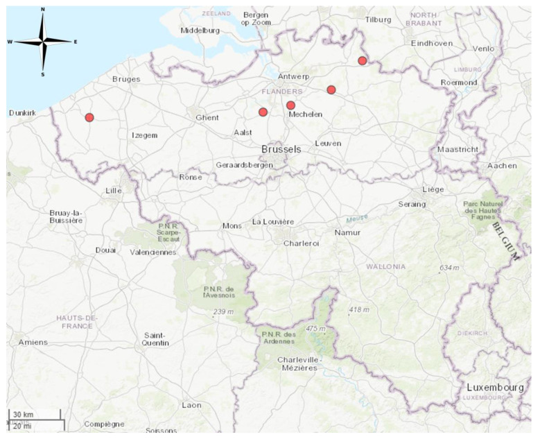
For the survey in live animals, five sampling sites that were never sampled before for lyssavirus surveillance were selected in Flanders. The sites were attended 6 h per night and were located at Arendonk (three nights), Diksmuide (one night), Duffel (four nights), Herentals (one night) and Liezele (one night) (Figure 1). Apart from a foraging location (Herentals), all other sites were near known hibernation sites for bats. Bats were captured at these sites for sample collection during the swarming season between mid-July and mid-October of 2018. During this period, different species of bats from different summer colonies visited hibernation sites to “swarm”, a behaviour linked to prospecting suitable hibernation sites and to mating [29,30].
Figure 1.
Collection sites in Flanders where bats were captured for saliva and blood collection and sampling. Sampling sites indicated on the map from left to right are Diksmuide, Liezele, Duffel, Herentals and Arendonk. Map created in R using the mapview package (https://cran.r-project.org/web/packages/mapview/index.html, accessed on 7 June 2024).
At all locations, bats were captured using bat mist nets (Ecotone, Poland) that were kept under constant surveillance. Bats were disentangled immediately after capture and then individually placed in lightweight cotton holding bags until inspection and sampling. Upon inspection, bat species were determined based on morphological characteristics by an expert in the field [1]. Captured bats were marked with a non-toxic marker to avoid repeated sampling if they were recaptured. Bats were released immediately after sampling. Recaptured bats were released immediately as well.
Both blood and saliva samples were collected for further research. Saliva samples were collected from the tongue using cotton swabs and stored at 4 °C in 200 µL of sterile phosphate-buffered saline (PBS). Blood samples from the interfemoral vein medial of the femur [31] were collected on filter paper as no more than one drop of blood could be obtained to ensure the safety of the bats. Saliva samples were used for real-time quantitative (RT-q) PCR to detect the presence of lyssaviruses. Blood samples were used to detect the presence of antibodies neutralising EBLV-1 using the method described below.
Passive surveillance consisted of bats being brought in from wildlife rehabilitation centres that received animals that were severely weakened and died or were found dead. Bats from all parts of Belgium were included if the date and place of discovery was available and the cadaver was in a good enough state for sample collection. Brain tissue was collected from the bats for RT-qPCR testing to determine the presence of different lyssaviruses, as specified below.
2.3. Detection of Lyssaviruses
Detection of lyssavirus RNA was conducted using a pan-lyssa RT-qPCR according to the method described by Fischer et al. [21]. Some samples were tested with the fluorescent antigen test (FAT) instead according to protocol of the WHO [32].
Prior to the RT-qPCR, total RNA was extracted using the Qiagen RNeasy kit (Qiagen, Hilden, Germany) according to the manufacturer’s instructions, using 50 µL of PBS in which cotton swabs were placed mixed with 300 µL of lysis buffer (Qiagen, Hilden, Germany).
The RT-qPCR was performed with primers JW12 and N145-165 [21], and extraction control was performed using primers VETINHR1 and VETINHF2 targeting 18S. The enzyme used was Q-script One Step RT (Quantabio, Beverly, MA, USA). This RT-qPCR can detect RABV, Lagos bat virus, BBLV, Mokola virus, Duvenhage virus, Australian bat lyssavirus, EBLV-1 and EBLV-2 [21]. Positive samples were sent to the European Reference Centre for Rabies (ANSES, Nancy, France) to be sequenced for genotyping.
2.4. Detection of Past EBLV-1 Infection through Modified Seroneutralisation Assay
Filter paper blood samples were incubated overnight in 200 µL of sterile PBS. The solution was tested for the presence of antibodies against EBLV-1 using the modified rapid fluorescent focus inhibition test (RFFIT) based on the Manual of Diagnostic Tests and Vaccines for Terrestrial Animals [33]. To increase specificity for EBLV-1-neutralising antibodies, an in-house modified neutralisation assay was designed using EBLV-1 virus instead of the standard CVS-11 RABV. The EBLV-1 strain used was called AF-2010, cultured from a positive bat from Spain [26]. Briefly, 1 in 3 serial dilutions of the sample were incubated at 37 °C for 90 min with 217.2 Median Cell Culture Infectious Dose (CCID50)/mL of EBLV-1. To control consistency between tests, the same viral dose of EBLV-1 had to neutralise the positive control to the same dilution for each test. For the positive control, OIE International Standard Serum for Rabies from dog origin (ANSES, LOT 2014-1) was diluted to 0.5 IU/mL for RABV neutralisation and used as EBLV-1 control as well. Antibodies bound the virus if any were present. Then, baby hamster kidney (BHK-21) cells (ATCC CCL-10, ordered from DSMZ, Leibniz, Germany, in 2013) were added to the mixture and incubated for 24 h. Infected BHK-21 cells were stained with fluorescent anti-nucleocapsid antibody and microscopically counted. The dilution that inhibited 50% of infection was determined. The cut-off value for positivity was chosen at a reciprocal titre of ≥27 to ensure no false positive reactions were read, as previously applied by Harris et al. [34]. The only goal was to detect the presence of antibodies that neutralise EBLV-1, not to determine exact titres. Cross-reaction of antibodies against other lyssaviruses that also neutralise EBLV-1 cannot be excluded.
3. Results
3.1. Bat Prevalence in Flanders
3.1.1. Life Animal Survey
During the survey in live animals in 2018, bats were captured at five sampling sites in Flanders, Belgium, none of which were sampled before (Figure 1). Five different species were identified. A total of 120 bats were captured. Of these, 79 were M. daubentonii, 12 were M. emarginatus, 3 were M. mystacinus, 3 were M. nattereri and 23 were Plecotus auritus. A full list of samples can be found in the Supplementary Materials (Table S1).
In total, 120 bats were captured of which 113 saliva samples and 87 blood samples were collected (Table 1). Multiple species were captured at 4 of the 5 sites, as indicated in Table 1.
Table 1.
Overview of samples taken during the survey in live animals. Samples were taken from five locations in Flanders, Belgium. Saliva and blood samples were taken where possible.
| Location/Bat Species | Number of Bats Captured | Number of Saliva Samples Taken | Number of Blood Samples Taken |
|---|---|---|---|
| Arendonk | 12 | 12 | 11 |
| M. emarginatus | 1 | 1 | 1 |
| M. nattereri | 1 | 1 | 1 |
| P. auritus | 10 | 10 | 9 |
| Diksmuide | 12 | 11 | 12 |
| M. mystacinus | 1 | 1 | 1 |
| M. daubentonii | 11 | 10 | 11 |
| Duffel | 83 | 77 | 54 |
| M. daubentonii | 64 | 63 | 40 |
| M. emarginatus | 3 | 3 | 3 |
| M. mystacinus | 1 | 1 | 1 |
| M. nattereri | 2 | 2 | 2 |
| P. auritus | 13 | 8 | 8 |
| Herentals | 7 | 7 | 7 |
| M. emarginatus | 7 | 7 | 7 |
| Liezele | 6 | 6 | 6 |
| M. daubentonii | 4 | 4 | 4 |
| M. emarginatus | 1 | 1 | 1 |
| M. mystacinus | 1 | 1 | 1 |
3.1.2. Passive Surveillance
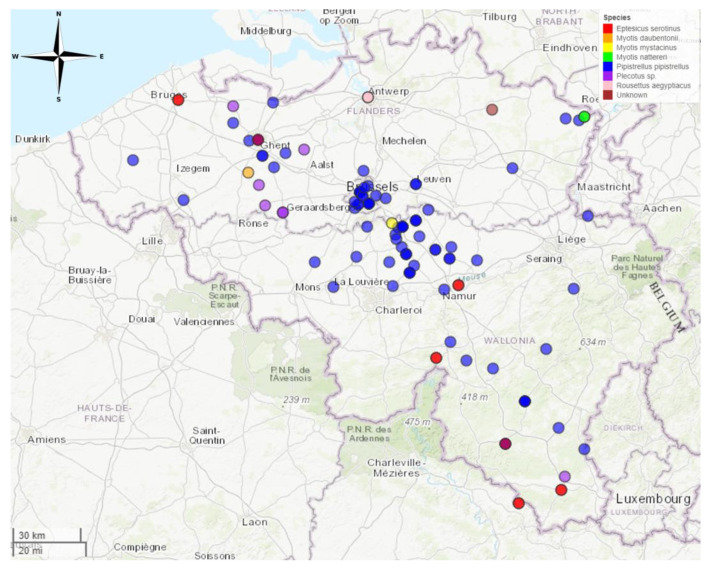
Passive surveillance was performed on bats brought in from wildlife rehabilitation centres between 2016 and 2018. In total, 133 bats were collected. A full list of samples can be found in the Supplementary Materials (Table S2). Nine samples did not have location information and/or could not be analysed due to the degraded state of the samples. A total of 124 animals were included in this study. Of these, 32 (25.8%) were from the Flemish region, 60 (48.4%) were from the Walloon region and 31 (25.0%) were from the Brussels region. One bat had an unknown origin (0.8%) (Figure 2).
Figure 2.
Locations in Belgium where bats were found during passive surveillance. Map created in R using the mapview package (https://cran.r-project.org/web/packages/mapview/index.html, accessed on 7 June 2024).
Out of 124 animals, 98 (79.0%) were Pipistrellus pipistrellus, 13 (10.5%) were E. serotinus, 2 (1.6%) were M. nattereri, 1 (0.8%) was M. daubentonii, 1 (0.8%) was M. mystacinus and 9 (7.3%) could not be identified. No saliva or blood samples could be obtained from these bats due to the condition in which they were collected, so only brain samples were tested.
Eptesicus serotinus was found at 7 locations. Myotis daubentonii was found at one location (Gavere). Myotis mystacinus was found at one location (Lasne). Myotis nattereri was found at one location (Kinrooi). Pipistrellus pipistrellus was found at 58 different locations. Plecotus sp. were found at 6 different locations. Rousettus aegyptiacus was found at one location (illegally imported through Antwerp airport) (Table 2).
Table 2.
Results of seroneutralisation assay for EBLV-1-neutralising antibodies per bat species. The causative agent can be any lyssavirus from phylogroup I as cross-protection cannot be excluded.
| Bat Species | Number of Bats Tested | Number of Samples Positive for Antibodies (%) |
|---|---|---|
| M. daubentonii | 55 | 16 (29%) |
| Diksmuide | 11 | 1 |
| Duffel | 44 | 13 |
| Liezele | 4 | 2 |
| M. emarginatus | 9 | 2 (22%) |
| Arendonk | 1 | 0 |
| Duffel | 3 | 0 |
| Herentals | 4 | 2 |
| Liezele | 1 | 0 |
| M. mystacinus | 3 | 2 (67%) |
| Diksmuide | 1 | 1 |
| Duffel | 1 | 0 |
| Liezele | 1 | 1 |
| M. nattereri | 3 | 0 (0%) |
| Arendonk | 1 | 0 |
| Duffel | 2 | 0 |
| P. auritus | 17 | 8 (47%) |
| Arendonk | 9 | 3 |
| Duffel | 8 | 5 |
| TOTAL | 87 | 28 (32%) |
In 2016, 48 bats were brought in of which 7 could not be analysed, so 41 were tested. In 2017, 43 bats were brought in and all were tested. In 2018, 43 bats were brought in of which 2 could not be tested, so 41 were included in this study.
3.2. Detection of Lyssavirus in Belgian Bats
Saliva samples taken during the survey in live animals were tested for the presence of lyssaviruses using RT-qPCR. In total, 113 samples were tested of which zero tested positive for any lyssavirus. The numbers of samples tested per location were 12, 11, 77, 7 and 6 for Arendonk, Diksmuide, Duffel, Herentals and Liezele, respectively.
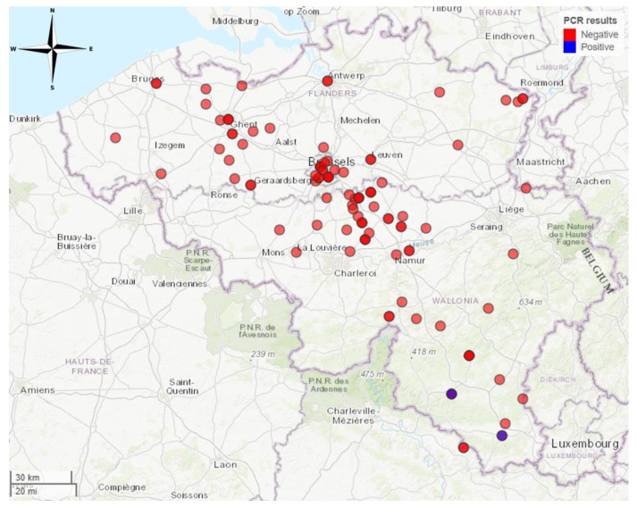
Samples of passive surveillance were tested using the same RT-qPCR and/or by FAT. In 2016, one bat tested positive for a lyssavirus by RT-qPCR methods out of 41 tested cadavers (2%). The severely weakened and immobile E. serotinus was brought to the Belgian National Reference Laboratory on 28 September 2016 after a biting incident with a hiker in Bertrix (Figure 3). The hiker received post-exposure prophylaxis afterwards and remained in good health.
Figure 3.
Locations in Belgium where bats were found during passive surveillance. Two E. serotinus bats tested positive for EBLV-1, one in 2016 in Bertrix and one in 2017 in Étalle, which is 30 km southeast of Bertrix. Map created in R using the mapview package (https://cran.r-project.org/web/packages/mapview/index.html, accessed on 7 June 2024).
In 2017, one other E. serotinus bat tested positive for lyssavirus by RT-qPCR out of 43 cadavers (2%). It was found on 16 October 2017 in Étalle (Figure 3), which is about 30 km southeast of Bertrix. Upon discovery of the weakened bat, a bat care centre in Bertrix was contacted and no human exposure was reported. Sequencing of the N gene by the European Reference Centre for Rabies (ANSES) confirmed EBLV-1 infection in both cases. Not enough sample was left to sequence the full genome.
In 2018, none of the 41 samples (0%) tested positive by RT-qPCR.
3.3. Modified Neutralisation Assay Shows Presence of EBLV-1-Neutralising Antibodies
Antibody assays can detect immune responses mounted against previous infections. Bats captured during the survey in 2018 were tested using this method. Since blood cannot be drawn from bat cadavers, it was not possible to test passive surveillance samples for antibody presence.
Out of 87 bats from which blood samples were collected, 32% tested positive for EBLV-1-neutralising antibodies with a range of 0% to 67% depending on the bat species (Table 2). Antibodies neutralising EBLV-1 might be mounted against EBLV-1 or against another lyssavirus and cross-protect against EBLV-1. The causative agent is therefore a lyssavirus but cannot be proven to be EBLV-1 specifically. A total of 16 specimens of M. daubentonii and 8 specimens of P. auritus tested positive for antibodies neutralising EBLV-1. Notably, there were also two specimens of M. emarginatus and two specimens of M. mystacinus that tested positive for antibodies neutralising EBLV-1.
In Arendonk, 27% (3/11) of bats showed antibodies neutralising EBLV-1. In Diksmuide, 18% (2/9) had antibodies. In Duffel, 33% (18/55) showed antibodies neutralising EBLV-1. In Herentals, two out of four were positive. In Liezele, three out of six showed presence of antibodies.
Overall, EBLV-1-neutralising antibodies could be detected in all bat species apart from M. nattereri. The number of positive samples ranged from 22% for M. emarginatus to 67% for M. mystacinus. The numbers were not large enough to infer conclusions about the presence of lyssaviruses per bat species (Table 2).
4. Discussion
The primary objective of this study centred upon the detection of European Bat Lyssavirus 1 (EBLV-1) as well as the detection of antibodies directed against this virus among bat populations in Belgium. EBLV-1 was successfully identified for the first time in Belgium during the years 2016 and 2017 in two E. serotinus specimens, and antibodies were detected at varying levels in different bat species. This goal was accomplished through the combined implementation of a survey in live animals and passive surveillance methodologies. It needs to be noted that antibodies neutralising EBLV-1 might be mounted against other lyssavirus and are detected due to cross-neutralisation.
Passive surveillance between 2016 and 2018 exclusively allowed for the testing of deceased bats. RT-qPCR and/or FAT testing was conducted on cerebral tissue extracted from bat cadavers. One sample did exhibit positive results for EBLV-1 through both RT-qPCR and FAT testing in the year 2016. This specific sample originated from an E. serotinus collected in Bertrix, situated within the southern part of Belgium. This particular bat specimen showed no wounds, and despite manifesting signs of despondency, it exhibited an aggressive response when subjected to handling. Subsequent to this initial case, a second E. serotinus yielded a positive outcome in RT-qPCR analysis one year later and was found in Étalle, situated approximately 30 km southeast of Bertrix. Although the possibility exists that these bats belonged to the same colony, this could not be verified. Serological and RNA evidence for EBLV-1 in this species has already been described by Robardet et al. for the northeast of France, which is not far from the Belgian border [10].
In the region of Flanders, five different bat species, namely M. daubentonii, M. emarginatus, M. mystacinus, M. nattereri and P. auritus, were captured during a survey in 2018. The investigations encompassed collection of fluid samples, including saliva and blood, from apparently healthy adult bats. Salivary specimens obtained from animals captured in live conditions yielded negative results across all samples for all tested lyssaviruses. This observation might be attributed to the intermittent nature of viral shedding, which ceases following seroconversion, as highlighted by Fooks et al. [35]. As such, the efficacy of RT-qPCR in detecting viral presence within saliva samples is inherently limited. These bats also showed a marked mycological diversity, including a new Pseudogymnoascus species, as described by Becker et al. [36].
Blood samples collected in 2018 were utilised for the purpose of detecting neutralizing antibodies specific to EBLV-1. EBLV-1-neutralising antibodies were detected in 32% of bats, ranging from 0% to 67% depending on bat species. Plecotus auritus samples have been shown before to be positive in neighbouring countries [10,13,37], and this study has confirmed the presence of antibodies against EBLV-1 in this species in Belgium.
This high percentage of antibodies in blood samples implies that infection with lyssaviruses is not uncommon in several of the captured bat species.
Remarkably, a notable proportion of blood samples from M. daubentonii tested positive as well, which is particularly significant due to the previous recognition of M. daubentonii as a host for EBLV-2 in other countries [28]. It is therefore most likely that these animals have antibodies against EBLV-2 yet are cross-neutralising against EBLV-1.
Myotis emarginatus and M. mystacinus also tested positive for EBLV-1-neutralising antibodies, which has not yet been shown before. It should be noted that the sample quantities were insufficient to also allow testing for antibodies against other lyssaviruses, but it is most likely that the antibodies mounted by these species are targeting other lyssaviruses and are cross-neutralising EBLV-1 rather than antibodies mounted against EBLV-1 specifically. Consequently, cross-reactivity of antibodies against other lyssaviruses from the same phylogroup remains likely, which is in line with the insights put forth by Weir et al. [37] and Inoue et al. [38]. Future research will focus on alternate blood sampling methods to allow for more tests and to be able to distinguish between antibodies targeting specific lyssaviruses. This will support further studies to yield more insight into the different lyssaviruses in Belgium.
In conclusion, this is the first study that shows that bats in Flanders have antibodies neutralising EBLV-1, suggesting the virus is circulating in Belgium, like it is in neighbouring countries. However, cross-neutralising antibodies against other lyssaviruses from the same phylogroup cannot be excluded. Furthermore, two cases of active EBLV-1 infection were detected in E. serotinus specimens in 2016 and 2017, confirming its presence in at least one bat species. Surveillance efforts should be strengthened to further monitor the current situation of lyssaviruses in Belgium, to be able to distinguish between them and to follow up on its effects on important and threatened bat populations. Belgium is already declared free of rabies in terrestrial mammals and therefore meets the objective of a rabies-free Europe. However, this research shows that rabies in free-flying mammals should be taken into account, in addition to it currently being a blind spot in the mapping of lyssaviruses in Europe.
Acknowledgments
The authors thank Agentschap voor Natuur en Bos and the Bats Working Group of Natuurpunt for their collaboration during sampling and identification of bat species, more specifically Conings, B., Boers, K., Lenaerts, A. and Hautekiet, D.
Supplementary Materials
The following supporting information can be downloaded at https://www.mdpi.com/article/10.3390/tropicalmed9070151/s1, Table S1: Detailed list of information on the bats captured during surveillance at 5 sites in Belgium.; Table S2: Detailed list of information on the bats tested during passive surveillance which were collected from 124 sites in Belgium.
Author Contributions
Conceptualization, C.V.d.E., S.T. and S.V.G.; methodology, C.V.d.E., S.T., W.W., D.D. and B.V.; formal analysis, I.N., C.V.d.E. and S.T.; data curation, I.N.; writing—original draft preparation, I.N.; writing—review and editing, I.N., S.T. and S.V.G.; visualization, I.N. All authors have read and agreed to the published version of the manuscript.
Institutional Review Board Statement
The animal study protocol was approved by the Ethics Committee of SCIENSANO (protocol code 20180413-01 on 31 May 2018).
Informed Consent Statement
Not applicable.
Data Availability Statement
Data can be requested by contacting the corresponding author.
Conflicts of Interest
The authors declare no conflicts of interest.
Funding Statement
This research received no external funding.
Footnotes
Disclaimer/Publisher’s Note: The statements, opinions and data contained in all publications are solely those of the individual author(s) and contributor(s) and not of MDPI and/or the editor(s). MDPI and/or the editor(s) disclaim responsibility for any injury to people or property resulting from any ideas, methods, instructions or products referred to in the content.
References
- 1.Dietz C., Kiefer A. Bats of Britain and Europe. Bloomsbury Publishing; London, UK: 2016. [Google Scholar]
- 2.Boyles J.G., Cryan P.M., McCracken G.F., Kunz T.H. Economic importance of bats in agriculture. Science. 2011;332:41–42. doi: 10.1126/science.1201366. [DOI] [PubMed] [Google Scholar]
- 3.Genus: Lyssavirus|ICTV. [(accessed on 7 June 2024)]. Available online: https://ictv.global/report/chapter/rhabdoviridae/rhabdoviridae/lyssavirus.
- 4.Van Gucht S., Le Roux I. Rabies control in Belgium: From Eradication in Foxes to Import of a Contaminated Dog. [(accessed on 11 August 2023)];Vlaams Diergeneeskundig Tijdschrift. 2008 Volume 77 Available online: https://openjournals.ugent.be/vdt/article/id/87230/ [Google Scholar]
- 5.Freuling C. Novel Lyssavirus in Natterer’s Bat, Germany. Emerg. Infect. Dis. 2011;17:1519–1522. doi: 10.3201/eid1708.110201. [DOI] [PMC free article] [PubMed] [Google Scholar]
- 6.Arechiga Ceballos N., Vazquez Moron S., Berciano J.M., Nicolas O., Aznar Lopez C., Juste J., Rodriguez Nevado C., Aguilar Setien A., Echevarria J.E. Novel Lyssavirus in Bat, Spain. Emerg. Infect. Dis. 2013;19:793–795. doi: 10.3201/eid1905.121071. [DOI] [PMC free article] [PubMed] [Google Scholar]
- 7.Botvinkin A.D., Poleschuk E.M., Kuzmin I.V., Borisova T.I., Gazaryan S.V., Yager P., Rupprecht C.E. Novel Lyssaviruses Isolated from Bats in Russia. Emerg. Infect. Dis. 2003;9:1623–1625. doi: 10.3201/eid0912.030374. [DOI] [PMC free article] [PubMed] [Google Scholar]
- 8.Nokireki T., Tammiranta N., Kokkonen U.-M., Kantala T., Gadd T. Tentative novel lyssavirus in a bat in Finland. Transbound Emerg. Dis. 2018;65:593–596. doi: 10.1111/tbed.12833. [DOI] [PubMed] [Google Scholar]
- 9.Černe D., Hostnik P., Toplak I., Presetnik P., Maurer-Wernig J., Kuhar U. Discovery of a novel bat lyssavirus in a Long-fingered bat (Myotis capaccinii) from Slovenia. PLoS Neglected Trop. Dis. 2023;17:e0011420. doi: 10.1371/journal.pntd.0011420. [DOI] [PMC free article] [PubMed] [Google Scholar]
- 10.Robardet E., Borel C., Moinet M., Jouan D., Wasniewski M., Barrat J., Boué F., Montchâtre-Leroy E., Servat A., Gimenez O., et al. Longitudinal survey of two serotine bat (Eptesicus serotinus) maternity colonies exposed to EBLV-1 (European Bat Lyssavirus type 1): Assessment of survival and serological status variations using capture-recapture models. PLoS Negl. Trop. Dis. 2017;11:e0006048. doi: 10.1371/journal.pntd.0006048. [DOI] [PMC free article] [PubMed] [Google Scholar]
- 11.Picard-Meyer E., Servat A., Wasniewski M., Gaillard M., Borel C., Cliquet F. Bat rabies surveillance in France: First report of unusual mortality among serotine bats. BMC Vet. Res. 2017;13:387. doi: 10.1186/s12917-017-1303-1. [DOI] [PMC free article] [PubMed] [Google Scholar]
- 12.Muller T., Cox J., Peter W., Schafer R., Johnson N., McElhinney L.M., Geue J.L., Tjornehoj K., Fooks A.R. Spill-over of European Bat Lyssavirus Type 1 into a Stone Marten (Martes foina) in Germany. J. Vet. Med. Ser. B. 2004;51:49–54. doi: 10.1111/j.1439-0450.2003.00725.x. [DOI] [PubMed] [Google Scholar]
- 13.Schatz J., Freuling C.M., Auer E., Goharriz H., Harbusch C., Johnson N., Kaipf I., Mettenleiter T.C., Mühldorfer K., Mühle R.-U., et al. Enhanced Passive Bat Rabies Surveillance in Indigenous Bat Species from Germany—A Retrospective Study. PLoS Neglected Trop. Dis. 2014;8:e2835. doi: 10.1371/journal.pntd.0002835. [DOI] [PMC free article] [PubMed] [Google Scholar]
- 14.Folly A.J., Marston D.A., Golding M., Shukla S., Wilkie R., Lean F.Z.X., Núñez A., Worledge L., Aegerter J., Banyard A.C., et al. Incursion of European Bat Lyssavirus 1 (EBLV-1) in Serotine Bats in the United Kingdom. Viruses. 2021;13:1979. doi: 10.3390/v13101979. [DOI] [PMC free article] [PubMed] [Google Scholar]
- 15.Van der Poel W.H., Van der Heide R., Verstraten E.R., Takumi K., Lina P.H.C., Kramps J.A. European bat lyssaviruses, the Netherlands. Emerg. Infect. Dis. 2005;11:1854. doi: 10.3201/eid1112.041200. [DOI] [PMC free article] [PubMed] [Google Scholar]
- 16.Takumi K., Lina P.H.C., Van Der Poel W.H.M., Kramps J.A., Van Der Giessen J.W.B. Public health risk analysis of European bat lyssavirus infection in The Netherlands. Epidemiol. Infect. 2009;137:803–809. doi: 10.1017/S0950268807000167. [DOI] [PubMed] [Google Scholar]
- 17.Serra-Cobo J., López-Roig M., Seguí M., Sánchez L.P., Nadal J., Borrás M., Lavenir R., Bourhy H. Ecological Factors Associated with European Bat Lyssavirus Seroprevalence in Spanish Bats. PLoS ONE. 2013;8:e64467. doi: 10.1371/journal.pone.0064467. [DOI] [PMC free article] [PubMed] [Google Scholar]
- 18.Seidlova V., Zukal J., Brichta J., Anisimov N., Apoznański G., Bandouchova H., Bartonička T., Berková H., Botvinkin A.D., Heger T., et al. Active surveillance for antibodies confirms circulation of lyssaviruses in Palearctic bats. BMC Vet. Res. 2020;16:482. doi: 10.1186/s12917-020-02702-y. [DOI] [PMC free article] [PubMed] [Google Scholar]
- 19.Rabies-Bulletin-Europe Classification. [(accessed on 11 August 2023)]. Available online: https://www.who-rabies-bulletin.org/site-page/classification.
- 20.Banyard A.C., Hayman D., Johnson N., McElhinney L., Fooks A.R. Advances in Virus Research. Elsevier; Philadelphia, PA, USA: 2011. [(accessed on 11 August 2023)]. Bats and Lyssaviruses; pp. 239–289. Available online: https://linkinghub.elsevier.com/retrieve/pii/B9780123870407000123. [DOI] [PubMed] [Google Scholar]
- 21.Fischer M., Freuling C.M., Müller T., Wegelt A., Kooi E.A., Rasmussen T.B., Voller K., Marston D.A., Fooks A.R., Beer M., et al. Molecular double-check strategy for the identification and characterization of European Lyssaviruses. J. Virol. Methods. 2014;203:23–32. doi: 10.1016/j.jviromet.2014.03.014. [DOI] [PubMed] [Google Scholar]
- 22.Johnson N., Aréchiga-Ceballos N., Aguilar-Setien A. Vampire Bat Rabies: Ecology, Epidemiology and Control. Viruses. 2014;6:1911–1928. doi: 10.3390/v6051911. [DOI] [PMC free article] [PubMed] [Google Scholar]
- 23.Amengual B., Bourhy H., López-Roig M., Serra-Cobo J. Temporal Dynamics of European Bat Lyssavirus Type 1 and Survival of Myotis myotis Bats in Natural Colonies. PLoS ONE. 2007;2:e566. doi: 10.1371/journal.pone.0000566. [DOI] [PMC free article] [PubMed] [Google Scholar]
- 24.Regnault B., Evrard B., Plu I., Dacheux L., Troadec E., Cozette P., Chrétien D., Duchesne M., Vallat J.-M., Jamet A., et al. First Case of Lethal Encephalitis in Western Europe Due to European Bat Lyssavirus Type 1. Clin. Infect. Dis. 2022;74:461–466. doi: 10.1093/cid/ciab443. [DOI] [PubMed] [Google Scholar]
- 25.Fooks A.R., McElhinney L.M., Pounder D.J., Finnegan C.J., Mansfield K., Johnson N., Brookes S.M., Parsons G., White K., McIntyre P.G., et al. Case report: Isolation of a European bat lyssavirus type 2a from a fatal human case of rabies encephalitis. J. Med. Virol. 2003;71:281–289. doi: 10.1002/jmv.10481. [DOI] [PubMed] [Google Scholar]
- 26.Van Gucht S., Verlinde R., Colyn J., Vanderpas J., Vanhoof R., Roels S., Francart A., Brochier B., Suin V. Favourable Outcome in a Patient Bitten by a Rabid Bat Infected with the European Bat Lyssavirus-1. Acta Clin. Belg. 2013;68:54–58. doi: 10.2143/ACB.68.1.2062721. [DOI] [PubMed] [Google Scholar]
- 27.McElhinney L.M., Marston D.A., Wise E.L., Freuling C.M., Bourhy H., Zanoni R., Moldal T., Kooi E.A., Neubauer-Juric A., Nokireki T., et al. Molecular Epidemiology and Evolution of European Bat Lyssavirus 2. Int. J. Mol. Sci. 2018;19:156. doi: 10.3390/ijms19010156. [DOI] [PMC free article] [PubMed] [Google Scholar]
- 28.Shipley R., Wright E., Selden D., Wu G., Aegerter J., Fooks A.R., Banyard A.C. Bats and Viruses: Emergence of Novel Lyssaviruses and Association of Bats with Viral Zoonoses in the EU. TropicalMed. 2019;4:31. doi: 10.3390/tropicalmed4010031. [DOI] [PMC free article] [PubMed] [Google Scholar]
- 29.Van Schaik J., Janssen R., Bosch T., Haarsma A.-J., Dekker J.J.A., Kranstauber B. Bats Swarm Where They Hibernate: Compositional Similarity between Autumn Swarming and Winter Hibernation Assemblages at Five Underground Sites. PLoS ONE. 2015;10:e0130850. doi: 10.1371/journal.pone.0130850. [DOI] [PMC free article] [PubMed] [Google Scholar]
- 30.Dekeukeleire D., Janssen R., Haarsma A.J., Bosch T., Van Schaik J. Swarming Behaviour, Catchment Area and Seasonal Movement Patterns of the Bechstein’s Bats: Implications for Conservation. Acta Chiropterolog. 2016;18:349–358. doi: 10.3161/15081109ACC2016.18.2.004. [DOI] [Google Scholar]
- 31.Hooper S.E., Amelon S.K. Handling and blood collection in the little brown bat (Myotis lucifugus) Lab Anim. 2014;43:197–199. doi: 10.1038/laban.543. [DOI] [PubMed] [Google Scholar]
- 32.World Health Organization. Rupprecht C.E., Fooks A.R., Abela-Ridder B. Laboratory Techniques in Rabies. 5th ed. Volume 1. World Health Organization; Geneva, Switzerland: 2018. [(accessed on 15 September 2023)]. Available online: https://apps.who.int/iris/handle/10665/310836. [Google Scholar]
- 33.World Organization for Animal Health. Stear M.J. OIE Manual of Diagnostic Tests and Vaccines for Terrestrial Animals (Mammals, Birds and Bees) 5th ed. Volume 130. Cambridge University Press; Cambridge, MA, USA: 2005. p. 727. [Google Scholar]
- 34.Harris S.L., Aegerter J.N., Brookes S.M., McElhinney L.M., Jones G., Smith G.C., Fooks A.R. Targeted Surveillance for European Bat Lyssaviruses in English Bats (2003–06) J. Wildl. Dis. 2009;45:1030–1041. doi: 10.7589/0090-3558-45.4.1030. [DOI] [PubMed] [Google Scholar]
- 35.Fooks A.R., Banyard A.C., Horton D.L., Johnson N., McElhinney L.M., Jackson A.C. Current status of rabies and prospects for elimination. Lancet. 2014;384:1389–1399. doi: 10.1016/S0140-6736(13)62707-5. [DOI] [PMC free article] [PubMed] [Google Scholar]
- 36.Becker P., Eynde C.v.D., Baert F., D’hooge E., De Pauw R., Normand A.-C., Piarroux R., Stubbe D. Remarkable fungal biodiversity on northern Belgium bats and hibernacula. Mycologia. 2023;115:484–498. doi: 10.1080/00275514.2023.2213138. [DOI] [PubMed] [Google Scholar]
- 37.Weir D.L., Coggins S.A., Vu B.K., Coertse J., Yan L., Smith I.L., Laing E.D., Markotter W., Broder C.C., Schaefer B.C. Isolation and Characterization of Cross-Reactive Human Monoclonal Antibodies That Potently Neutralize Australian Bat Lyssavirus Variants and Other Phylogroup 1 Lyssaviruses. Viruses. 2021;13:391. doi: 10.3390/v13030391. [DOI] [PMC free article] [PubMed] [Google Scholar]
- 38.Inoue Y., Kaku Y., Harada M., Ishijima K., Kuroda Y., Tatemoto K., Virhuez-Mendoza M., Nishino A., Yamamoto T., Inoue S., et al. Cross-neutralization activities of antibodies against 18 lyssavirus glycoproteins. Jpn. J. Infect. Dis. 2023;77:169–173. doi: 10.7883/yoken.JJID.2023.400. [DOI] [PubMed] [Google Scholar]
Associated Data
This section collects any data citations, data availability statements, or supplementary materials included in this article.
Supplementary Materials
Data Availability Statement
Data can be requested by contacting the corresponding author.


