Abstract
The influenza virus neuraminidase (NA) is a tetrameric, virus surface glycoprotein possessing receptor-destroying activity. This enzyme facilitates viral release and is a target of anti-influenza virus drugs. The NA structure has been extensively studied, and the locations of disulfide bonds within the NA monomers have been identified. Because mutation of cysteine residues in other systems has resulted in temperature-sensitive (ts) proteins, we asked whether mutation of cysteine residues in the influenza virus NA would yield ts mutants. The ability to rationally design tight and stable ts mutations could facilitate the creation of efficient helper viruses for influenza virus reverse genetics experiments. We generated a series of cysteine-to-glycine mutants in the influenza A/WSN/33 virus NA. These were assayed for neuraminidase activity in a transient expression system, and active mutants were rescued into infectious virus by using established reverse genetics techniques. Mutation of two cysteines not involved in intrasubunit disulfide bonds, C49 and C146, had modest effects on enzymatic activity and on viral replication. Mutation of two cysteines, C303 and C320, which participate in a single disulfide bond located in the β5L0,1 loop, produced ts enzymes. Additionally, the C303G and C320G transfectant viruses were found to be attenuated and ts. Because both the C303G and C320G viruses exhibited stable ts phenotypes, they were tested as helper viruses in reverse genetics experiments. Efficiently rescued were an N1 neuraminidase from an avian H5N1 virus, an N2 neuraminidase from a human H3N2 virus, and an N7 neuraminidase from an H7N7 equine virus. Thus, these cysteine-to-glycine NA mutants allow the rescue of a variety of wild-type and mutant NAs into influenza virus.
The neuraminidase (NA) of influenza A and B viruses is an enzymatic, tetrameric, type II integral membrane glycoprotein found on the surface of virions. The NA possesses receptor-destroying neuraminidase (acylneuraminyl hydrolase; EC 3.2.1.18) activity and cleaves the α-ketosidic linkage connecting terminal sialic acid residues to adjacent d-galactose or d-galactosamine residues (17). This activity facilitates viral release from infected cells by removing sialic acid residues from glycoproteins and glycolipids at the cell surface, which are potential ligands for the viral hemagglutinin (HA). The presence of these residues during viral budding otherwise results in virus-virus aggregation and retention of viral particles at the cell surface. Thus, when temperature-sensitive (ts) NA mutant viruses are grown at nonpermissive temperature, or when influenza viruses are grown in the presence of NA inhibitors, viral particles form aggregates at the cell surface (40, 41).
Because NA activity is required for efficient viral release, drugs which inhibit NA activity can be effective anti-influenza virus agents. This was first demonstrated in the mid-1970s when the sialic acid analogues DANA and FANA were shown to inhibit influenza virus replication in vitro (36, 41). Recently, using the X-ray crystal structure of NA, additional NA inhibitors have been designed (39, 53). Two such compounds, zanamivir (GG167 or 4-guanidino-Neu5Ac2en) and GS4104, are in clinical use in humans (20, 21).
Other functions have also been attributed to NA. For example, the NA of influenza A/WSN/33 (H1N1) (WSN) virus is required for the neuropathogenicity of this strain in mice (29) and has been postulated to be a virulence factor (16, 34). Also, compatibility of new HA-NA combinations generated by reassortment may influence the emergence of pandemic strains (46). Because NA is a structural protein, it may contribute to the assembly of viral particles (24, 55). NA may also contribute to virus-induced apoptosis (38, 47). Additionally, avian influenza viruses possess NAs with hemagglutinating activity of unknown significance (25, 26, 52).
The influenza virus NA proteins have been extensively studied. Mutant proteins have been generated and assayed in transient expression systems; viruses possessing mutant NAs have been isolated and characterized; and detailed X-ray crystal structures of NA head structures have been solved for two of the nine influenza A NA types, N2 and N9, as well as for the influenza B virus NA. Reverse genetics technology has allowed the introduction of specific NA mutants into influenza viruses (9, 12). Most of these reverse genetics experiments have used the WSN virus NA, because of its relatively efficient rescue system (10). However, other rescue systems based on either a host range selection or antibody selection have been described (25, 29, 30).
Structurally, all of the NA types have a common head structure. Each monomer head consists of a propeller-like structure with six blades each composed of a four-stranded, anti-parallel β-sheet. The strands of each β-sheet are connected via short loops, and separate sheets are also connected by intervening loops. Among the various NA types, particular residues are absolutely conserved. These conserved residues include amino acids in or near the active site, residues at interfaces between β-sheets and at reverse turns, and cysteine residues involved in disulfide bonds (6).
A number of the conserved NA cysteines participate in intrasubunit disulfide bonds, and most of these bonds link cysteines located within strands of β-sheets. Additionally, a disulfide bond forms between two cysteines (C303 and C320 [WSN numbering]), located in an extended loop, β5L0,1 (51). In addition, some of the nonconserved cysteines participate in intersubunit disulfides which join monomers in the mature tetrameric enzyme.
Disulfide bonds are thought to be important for maintaining protein stability but may not be essential for proper protein folding (8). Previous analyses of several viral proteins, including the influenza virus HA, the herpes simplex virus type 1 (HSV-1) gD, and the HSV-1 αTIF (α trans-inducing factor; VP16) proteins, indicated that mutation of cysteine residues, including those known to participate in disulfide bonds, can alter protein stability and/or function (3, 32, 48, 54). Additionally, studies with the HSV-1 gD-1 and αTIF proteins indicated that mutation of cysteine residues may result in temperature-sensitive proteins (31, 44).
We were interested in determining whether ts influenza viruses could be generated by mutation of NA cysteine residues. Such mutants might prove useful as helper viruses in reverse genetics experiments. Successful generation of ts NA mutants would also recommend this approach for other viral proteins, either for the generation of novel helper viruses or for the introduction of attenuating mutations into live vaccine strains. Therefore, using the available influenza NA X-ray crystal structures as a guide to the location of disulfide bonds (51), we generated a series of cysteine-to-glycine mutations within the WSN NA. These mutants were constructed such that each disulfide bond would be disrupted. The resulting mutants were screened for NA activity after transient expression at different temperatures. We identified four individual cysteines which, when mutated, yielded active enzymes. Two C-to-G mutants (C303G and C320G) were attenuated and ts, while two C-to-G mutants (C49G and C146G) were similar to the wild type. These four mutant NAs were introduced into transfectant viruses via the established WSN NA rescue system. The C303G and C320G transfectant viruses displayed tight ts growth properties and failed to produce properly folded, stable NAs at the nonpermissive temperature. Additionally, these ts viruses have been used in an efficient reverse genetics system to rescue an N1 from an avian H5N1 virus, an N2 from a human H3N2 virus, and an N7 from an equine H7N7 virus.
MATERIALS AND METHODS
Cell lines and viruses.
COS-1 cells were maintained in Dulbecco modified Eagle medium (DMEM)–10% fetal bovine serum (FBS). MDBK cells were maintained in REM–10% FBS, and viruses were plaqued on MDBK cells in REM. MDCK cells were maintained in minimal essential medium–10% FBS, and viruses were plaqued on MDCK cells in DMEM–F-12, supplemented with 5 μg of trypsin (Trypsin 1:250; Difco) per ml. Vero cells were maintained in serum-free AIM-V medium. Stocks of WSN virus and the transfectant WSN NA mutants C49G, C146G, C303G, and C320G were grown on MDBK cells. Stocks of WSN-HK virus and WSN-N1(HK/97), WSN-N2(LA/87), and WSN-N7(Cor/74) (see below for explanation of designations) transfectant viruses were grown in 10-day-old embryonated chicken eggs.
Generation and expression of mutant WSN NA proteins.
The NA sequence from pT3NAm1, which encodes wild-type WSN NA protein, was subcloned between the EcoRI and HindIII sites of pGEM-11zf(−) such that NA expression could be driven by the T7 promoter. The resulting plasmid, pGEM-NAm1, was used as template for site-directed mutagenesis. Mutagenesis reactions were performed by using a Stratagene QuikChange site-directed mutagenesis kit. Primers used in these reactions are listed in Table 1.
TABLE 1.
Site-directed mutagenesis primers
| Residue | Primera | Restriction siteb |
|---|---|---|
| C49 | 152-CATACTGGAATAGGTAACCAAGGCAGC-178 | BstEII |
| C76 | 236-AATTCATCTCTTGGGCCCATCCGTGGG-262 | ApaI |
| C109 | 331-GCCTTTTATTTCAGGATCCCACTTGGAATGC-361 | BamHI |
| C146 | 443-GCCTTAATGAGCGGGCCCGTCGGTGAAGCT-472 | ApaI |
| C169 | 507-CTTGGTCAGCTAGCGCAGGACATGGATGGAG-536 | NheI |
| C218 | 656-GAGTCTGAATGCACCGGAGTAAATGGTTC-684 | BsmI |
| C264 | 797-CACTACGAGGAAGGATCCTGTTACCC-822 | BamHI |
| C266 | 805-GGAATGTTCCGGATATCCGGATACCGGCAAAG-836 | BspEI |
| C303 | 917-GGATACATCGGATCCGGGGTTTTCG-941 | BamHI |
| C320 | 965-GGAACAGGCAGCGGGGGCCCAGTGTCT-991 | ApaI |
| C406 | 1222-CTGTATGAGGCCCGGGTTCTGGGTTG-1247 | SmaI |
The indicated deoxyoligonucleotide and a complementary deoxyoligonucleotide were used for mutagenesis reactions. Boldface characters indicate differences between the primer and the template, pGEM-NAm1; underlines indicate codons mutated from cysteine to glycine.
To facilitate the identification of mutant clones, the restriction sites were introduced without changing the amino acid sequence.
NAs were expressed via the vaccinia virus-T7 expression system (11) previously used to express other NA mutants (14). Confluent 35-mm-diameter dishes of COS-1 cells were infected with the T7 RNA polymerase-expressing vaccinia virus, vTF7-3, at 1 PFU/cell; 45 min after addition of virus, cells were transfected with 5 μg of NA expression plasmid by using DOTAP liposomal transfection reagent (Boehringer Mannheim) and then grown at 33, 37, or 39.5°C for 15 h.
NA assays.
COS-1 cells transfected with NA expression plasmids were washed once in phosphate-buffered saline, scraped from the dish, pelleted at 4°C, resuspended in lysis buffer (10 mM Tris-HCl [pH 7.4], 1 M NaCl, 10 mM CaCl2, 2% Triton X-100, 1 mM Pefabloc [Boehringer Mannheim]), and incubated on ice 30 min. The lysate was then microcentrifuged at 15,000 rpm for 30 min at 4°C. The pellets were removed, and the supernatants were assayed for NA activity.
NA assays were performed at 30°C for 1 h. Reaction mixtures contained 0.1 M potassium phosphate buffer (pH 6.0), 1 mM CaCl2, and 0.5 mM 2′-(4-methylumbelliferyl)-α-d-N-acetylneuraminic acid (Mu-NANA). Reactions were terminated by addition of 2 ml of 0.5 M glycine-NaOH (pH 10.4). Fluorescence, resulting from cleavage of the ketosidic bond, was measured using a Turner model 112 fluorometer. Tenfold dilutions of each lysate were prepared and assayed in order to compare samples in a linear range.
Cloning of N1, N2, and N7 NA genes.
The N1 from influenza A/chicken/Hong Kong/27402/97 (H5N1) virus [N1(HK/97)], the N2 from influenza A/Los Angeles/2/87 (H3N2) virus [N2(LA/87)], and the N7 from influenza A/equine/Cornell/16/74 (H7N7) virus [N7(Cor/74)] were amplified by reverse transcription-PCR (RT-PCR) from viral RNA (vRNA). The H5N1 vRNA was provided by Michael Perdue, and the H7N7 virus was provided by Gary Whittaker. The primers (influenza virus sequences in uppercase; other sequences, [restriction enzyme sites for cloning or plasmid linearization; bacteriophage promoters] in lowercase) used to amplify the NA genes were as follows: for N1, 5′gcgcaagcttgaagacgcagcaaaAGCAGGAGTTTAAAATG-3′ and 5′-gcgctctagaattaaccctcactaaaAGTAGAAACAAGGAGTTTTTTG-3′; for N7, 5′gcgatcatcgatctcttcgAGCAAAAGCAGGGTAATTTTGAAATGAATCCTAATCAAAAACTC-3′ and 5′gcgcctcgagaattaaccctcactaaaAGTAGAAACAAGGGTTTTTTTCGTTTTACG-3′; for N2, 5′-tctagaggacgcgggggAGCAAAAGCAGGAGTGAAGATG-3′ and 5′-gcgcaagctttaatacgactcactataAGCGAAAGCAGGAGT-3′ (these primers possess noncoding sequences corresponding to those of influenza A/Bangkok/1/79 (H3N2) virus).
RNP transfections.
Rescue of the WSN NA mutants was performed as described elsewhere (10) except that transfectant viruses were grown at 33°C. Briefly, MDBK cells were infected with WSN-HK virus (multiplicity of infection of 1). One hour after addition of virus, cells were transfected with in vitro-synthesized NA vRNAs transcribed in the presence of influenza A virus polymerase. Following transfection, the cells were incubated at 33°C for 18 h. Supernatants were then harvested and plaqued at 33°C on MDBK cells. Resulting transfectant viruses were plaque purified three times on MDBK cells and subsequently amplified on MDBK cells.
Rescue experiments for N1(HK/97), the N2(LA/97), and N7(Cor/74) viruses were performed as follows. Vero cells, maintained in serum-free medium, were infected with C320G virus at 1 PFU/cell. One hour after addition of virus, the cells were transfected with NA vRNAs synthesized in vitro from linearized plasmids. These vRNAs were transcribed in the presence of influenza A virus polymerase which, to inactivate endogenous vRNAs, was previously UV irradiated for 90 s at a distance of 10 cm, using an 8W G3T5 GL-8 germicidal UV bulb. Following transfection, cells were grown in MEM–0.2% bovine serum albumin–3 μg of trypsin per ml at 33°C for 24 h. Supernatants were then harvested and plaqued on MDCK cells at 39.5°C. Additionally, 10-day-old embryonated chicken eggs were infected with 0.1 ml of supernatant from transfected cells and incubated at 39.5°C for 2 days. Rescued transfectant viruses were plaque purified three times on MDCK cells at 39.5°C.
Analysis of transfectant vRNAs.
Wild-type WSN, WSN-N1(HK/97), WSN-N2(LA/87), and WSN-N7(Cor/74) viruses were grown in 10-day-old embryonated eggs for 2 days at 37°C. Allantoic fluid containing virus was harvested from the eggs and centrifuged at 3,000 × g for 15 min at 4°C in a tabletop centrifuge. Subsequently, the virus-containing supernatants were centrifuged at 10,000 rpm in an SW28 rotor for 30 min at 4°C, virus from the supernatants was pelleted through a 30% sucrose cushion by centrifugation at 25,000 rpm in an SW28 rotor for 90 min at 4°C, the viral pellet was resuspended in Trizol Reagent (Gibco BRL), and vRNA was extracted by following the manufacturer’s protocol.
RT-PCR amplification of the vRNAs was performed with the Titan one-tube RT-PCR system (Boehringer Mannheim). Reactions were performed as follows: 30 min at 60°C for one cycle; 2 min at 94°C for one cycle; 30 s at 94°C, 30 s at 60°C, and 1 min 10 s at 68°C for 35 cycles; 7 min at 68°C for one cycle. The primers used to amplify the WSN N1 and N1(HK/97) were the same as those used to clone the N1(HK/97) segment (see above). The primers used to amplify a portion of N2(LA/87) were 5′-AATAGAAAGAAAATAACAGAGATAGTGTA-3′ and 5′-TGGTTCCCTGCCCAAGTGCAAATTGATAAC-3′ and correspond to nucleotides 171 to 200 and the complement of nucleotides 386 to 415 of the influenza A/Shanghai/11/87 (H3N2) virus N2 sequence (GenEMBL accession no. U42633). The primers used to amplify N7(Cor/74) were the same as those used to clone this segment (see above).
Polyacrylamide gel analysis of vRNAs was performed with a 2.8% polyacrylamide gel (acrylamide:bisacrylamide, 30:0.8) containing 7.7 M urea and 1× Tris-borate-EDTA. Gels were run at 150 V for 1 h 50 min and silver stained (10).
RESULTS
Generation and assay of NA mutants.
A series of single cysteine-to-glycine mutations were generated in the WSN NA ectodomain such that two nucleotide changes would be required for the glycine to revert to a cysteine (Table 2). A schematic diagram of locations of the WSN NA cysteines is shown in Fig. 1. Both cysteines not involved in intramolecular disulfide bonds (C49 and C146) were individually mutated. Additionally, each disulfide bond was individually disrupted. To accomplish this, the amino-terminal member of each disulfide-bonded cysteine pair was mutated. Additionally, two double cysteine-to-glycine mutants (C49G,C146G and C303G,C320G) were generated.
TABLE 2.
Relative activities of mutant NAs in the vaccinia virus-T7 system
| Mutation | Codon change(s) | % Activitya at:
|
||
|---|---|---|---|---|
| 33°C | 37°C | 39.5°C | ||
| C49G | TGC→GGT | 130 | 130 | 124 |
| C76G | TGT→GGG | <0.1 | <0.1 | <0.1 |
| C109G | TGT→GGA | <0.1 | <0.1 | <0.1 |
| C146G | TGT→GGG | 27.6 | 30.4 | 17.4 |
| C169G | TGT→GGA | <0.1 | <0.1 | <0.1 |
| C218G | TGT→GGA | <0.1 | <0.1 | <0.1 |
| C264G | TGT→GGA | <0.1 | <0.1 | <0.1 |
| C266G | TGT→GGA | <0.1 | <0.1 | <0.1 |
| C303G | TGC→GGA | 2.3 | 2.7 | 0.3 |
| C320G | TGT→GGG | 2.4 | 1.0 | 0.2 |
| C406G | TGC→GGA | <0.1 | <0.1 | <0.1 |
| C49G,C146G | TGT→GGA, TGT→GGG | 21.6 | 16.7 | 10.2 |
| C303G,C320G | TGC→GGA, TGT→GGG | <0.1 | <0.1 | <0.1 |
100% activity corresponds to the activity of wild-type WSN NA transfected in parallel at the indicated temperature. The activities of wild-type WSN NA were 3.64 ± 0.72 at 33°C, 4.41 ± 0.44 at 37°C, and 1.06 ± 0.14 pmol of Mu-NANA cleaved/s at 39.5°C.
FIG. 1.
Locations of cysteine residues of the NA of WSN virus. In the schematic diagram of the WSN NA primary structure, locations of all 19 cysteine residues are indicated (numbering according to Hiti and Nayak [22]), and thin lines connecting cysteines represent intrasubunit disulfide bonds. Domains of the protein are indicated as follows: tail, the six-amino-acid cytoplasmic tail; T.M., the transmembrane domain; stalk, indicates the long, slender stalk which extends from the virion surface; head, the large, globular domain possessing enzymatic activity. Equivalent cysteines in the heads of the WSN N1 and the influenza A/RI/5-/57 virus N2 (6) are as follows (WSN/N2): 76/92, 109/124, 113/129, 169/183, 218/232, 223/237, 264/278, 266/280, 275/289, 303/318, 320/337, 277/291, 216/230, 402/417, 406/421, and 431/447. Equivalent cysteines form equivalent disulfide bonds in the two proteins. However, the N2 contains an additional intramolecular disulfide bond (C175-C193 [N2 numbering]) not found in N1 NAs.
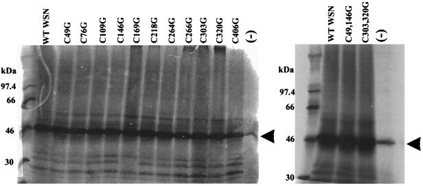
Each mutant was expressed in vitro via a coupled in vitro transcription-translation system to confirm that each clone could be expressed from the T7 RNA promoter (Fig. 2).
FIG. 2.
In vitro translation of WSN NA cysteine-to-glycine mutants. Wild-type (WT) WSN NA and the indicated mutant WSN NAs were transcribed and translated in vitro in the presence of [35S]methionine. (−), no-DNA negative control reaction. The products were run on a sodium dodecyl sulfate–10% polyacrylamide gel, which was then exposed to film. The arrowheads indicate positions of full-length, unmodified NA.
The resulting mutant NA molecules were then expressed at 33, 37, or 39.5°C, using the vaccinia virus-T7 expression system. The mutants were assayed for activity at 30°C, using as substrate the low-molecular-weight sialic acid analog Mu-NANA; the results are summarized in Table 2. The relative activities reported in Table 2 do not necessarily reflect the specific activities of the mutants, since the amount produced of each mutant was not precisely quantified.
Mutation of either cysteine not involved in intramolecular disulfide bonds, C49 or C146, had relatively little effect on neuraminidase activity following expression at any temperature. C49G yielded activity levels similar to those of wild-type NA when expressed at 33, 37, or 39.5°C. C146G was somewhat attenuated and slightly ts. When expressed at 33 or 37°C, C146G was approximately one-third as active as wild-type NA; however, when expressed at 39.5°C, C146G displayed only 17.4% of the activity of wild-type NA. The C49G,C146G double mutant displayed defects only slightly more severe than those of the single C146G mutant.
Mutation of any of the cysteines involved in intramolecular disulfide bonds severely affected the production of functional enzyme, regardless of expression temperature. The only mutations affecting cysteine residues involved in intrasubunit disulfide bonds which retained activity targeted the same disulfide bond. When either C303 or C320 was mutated to glycine, NA activity was significantly impaired after expression at 33, 37, or 39.5°C. Interestingly, both mutant enzymes were ts, displaying 2% of wild-type activity at 33°C and approximately 10-fold-lower activity (0.2%) at 39.5°C. The C303G,C320G double mutant, however, exhibited no NA activity. Of note, both C303 and C320 lie within a single loop, β5L0,1, of the NA, whereas the remainder of the disulfide bonds found in the NA head involve cysteines within β-sheets (51).
Effects of cysteine-to-glycine mutations on viral growth.
To study the role of NA cysteines in the context of an infectious virus, the four active NA mutants were rescued into a WSN virus background by using the established WSN NA rescue system. Rescue of the C49G and C146G mutants was as efficient as rescue of wild-type WSN NA. Rescue of the attenuated C303G and C320G mutants was approximately 10-fold less efficient than rescue of wild-type NA. Upon rescue, both C303G and C320G were found to form small plaques on MDBK cells at 33°C, while C49G and C146G plaques were similar in size to wild-type WSN plaques (data not shown).
The plaque-forming phenotype of the mutant viruses was further analyzed on MDCK cells in the presence of trypsin, at 33 or 39.5°C. The plaques formed by C49G and C146G at either 33 or 39.5°C were similar in size to those formed by wild-type WSN (data not shown). However, both C303G and C320G were found to form small plaques at 33°C and no plaques, even after 4 days, at 39.5°C. In these experiments, the efficiency of plaque formation of both C303G and C320G viruses was more than 3 logs lower at 39.5°C than at 33°C.
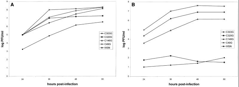
To further assess the impact of the cysteine-to-glycine mutations on viral replication, multicycle growth of the transfectant viruses was analyzed on MDCK cells at either 33 or 39.5°C (Fig. 3). At 33°C, the C49G and C146G viruses grew similarly to wild-type WSN virus, achieving titers of 108 PFU/ml. However, at 33°C both C303G and C320G were attenuated 1 and 2 logs, respectively, compared with wild-type WSN virus (Fig. 3A). At 39.5°C, C49G again grew as well as or better than wild-type WSN. C146G grew to a slightly lower titer, perhaps reflecting the slightly ts phenotype seen in the transient expression experiments. At 39.5°C, the ts phenotype of mutants C303G and C320G was apparent. These viruses grew to titers 5 to 6 logs lower than wild-type WSN titers (Fig. 3B). These results demonstrate that there is a good correlation between the relative amount of NA activity obtained in the transient expression assay (Table 2) and the extent of viral growth in tissue culture (Fig. 3). These experiments also indicate that the mutant viruses C303G and C320G exhibit tight ts phenotypes.
FIG. 3.
Growth curves of transfectant viruses C49G, C146G, C303G, and C320G. (A) Multicycle growth of the transfectant viruses at 33°C. (B) Multicycle growth of the transfectant viruses at 39.5°C. MDCK cells were infected with wild-type WSN, C49G, C146G, C303G, or C320G virus at a multiplicity of infection of 0.001. Plaque assays were performed on MDCK cells at 33°C to determine the titer of the collected samples.
Clearly, C49 and C146 are not required for efficient function of the WSN NA in the context of viral infection. However, disruption of the disulfide bond between C303 and C320 significantly impairs NA function, rendering the molecule ts. In the context of an infectious virus, C303G appears to be a more severe mutation despite the similar levels of activity seen in the NA proteins expressed from plasmids. This may reflect the use of the low-molecular-weight Mu-NANA as substrate in the in vitro NA assays. There may be differences in the ability of the two NAs to cleave sialic acid residues from large glycoconjugates, but such differences may not be seen with a low-molecular-weight substrate.
C303G and C320G fail to produce stable, properly folded NA at the nonpermissive temperature.
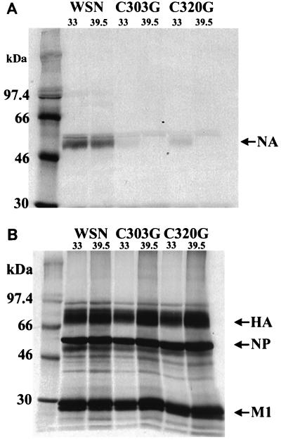
To better understand the specific defect in the C303G and C320G viruses, we examined viral protein expression levels at 33 and 39.5°C. MDCK cells were infected with WSN, C303G, or C320G virus, grown at 33 or 39.5°C for 12 h, and then labeled with [35S]methionine and [35S]cysteine for 1 h. After the label was chased for 1 h with unlabeled medium to allow maturation of the labeled proteins, lysates were prepared and analyzed by immunoprecipitation with a mixture of two monoclonal antibodies that do not recognize denatured WSN NA protein or with polyclonal antiserum raised against whole WSN virus.
At 33°C, the C303G virus produced 6.9% and the C320G virus produced 9.6% as much immunoprecipitable NA as did wild-type WSN virus. At 39.5°C, neither mutant virus produced detectable levels of immunoprecipitated NA (Fig. 4A). In contrast, slightly more HA, NP, and M1 were produced at 39.5°C than at 33°C. Additionally, mutants C303G and C320G produced HA, NP, and M1 at levels similar to those for wild-type WSN at both 33 and 39.5°C (Fig. 4B).
FIG. 4.
Immunoprecipitation assays indicating that C303G and C320G NAs do not fold normally at 39.5°C. (A) Immunoprecipitation of NA proteins. (B) Immunoprecipitation of viral structural proteins HA, NP, and M1. Immunoprecipitations were performed on lysates of MDCK cells infected with the indicated viruses and incubated at the indicated temperatures. The cultures were labeled with [35S]methionine-[35S]cysteine for 1 h and chased with unlabeled medium for 1 h, beginning 12 h postinfection. Immunoprecipitation of NA was performed with a mixture of two monoclonal antibodies, 3C8 and 10C9 (43). Immunoprecipitations of HA, NP, and M1 were performed with a polyclonal anti-WSN virus antiserum, 8236.
These results suggest that the C303G and C320G NA molecules are not simply defective in enzymatic activity. Rather these NAs likely possess gross structural defects, perhaps due to the loss of the C303-C320 disulfide bond. Thus, both C303G and C320G virus-infected cells have decreased amounts of stable, properly folded NA compared with wild-type virus, even at 33°C. At 39.5°C, stable, properly folded NAs are essentially absent from mutant-virus infected cells.
A general reverse genetics system for the rescue of influenza A virus NAs using C303G and C320G as helper viruses.
The tight ts phenotype exhibited by the C303G and C320G viruses suggested that these viruses might serve as efficient helper viruses in reverse genetics experiments. Following RNP transfection of wild-type WSN NA, helper virus-infected MDBK cells were incubated for 24 h at 33°C. Supernatants for the RNP-transfected/infected cells were then passaged on MDBK cells at 39.5°C to select against the helper virus. More than 104 transfectant viruses could be rescued by this method, an efficiency that compares favorably with that of the established WSN NA rescue system using WSN-HK as the helper virus (10). The resulting viruses appeared to be transfectants and not revertants, because no viruses were recovered from C303G and C320G virus-infected, mock-transfected controls (data not shown). These results suggested that C303G and C320G can function as efficient helper viruses. In addition, the rescue of the ts phenotype by introduction of new NA genes provides genetic confirmation that the ts lesions encoded by the C303G and C320G viruses reside in the NA segment.
Rescue of N1, N2, and N7 NA genes into virus.
The better-growing mutant virus, C320G, was then used in reverse genetics experiments designed to rescue non-WSN NAs. Vero cells were transfected in these experiments because of their relatively high transfection efficiency and because they withstand the presence of trypsin in the growth medium. These cells were infected with 1 PFU of C320G virus per cell and RNP-transfected with in vitro-synthesized vRNAs corresponding to the N1(HK/97), N2(LA/87), or N7(Cor/74) virus. Following incubation of the RNP-transfected cells at 33°C for 24 h, the supernatants were plaqued on MDCK cells at 39.5°C or passaged in 10-day-old, embryonated chicken eggs at 39.5°C. Transfectant viruses were obtained for the N1, N2, and N7 NAs [WSN-N1(HK/97), WSN-N2(LA/87), and WSN-N7(Cor/74), respectively]; 60 WSN-N1(HK/97) transfectants, <15 WSN-N2(LA/87) transfectants, and 30 WSN-N7(Cor/74) transfectants were obtained in a transfection. After passage at 39.5°C, no viruses were obtained from the mock-transfected controls.
The transfectant viruses were plaque purified three times at 39.5°C and amplified at 39.5°C in 10-day-old embryonated chicken eggs. vRNA was then extracted from these viruses and analyzed by RT-PCR and by polyacrylamide gel analysis.
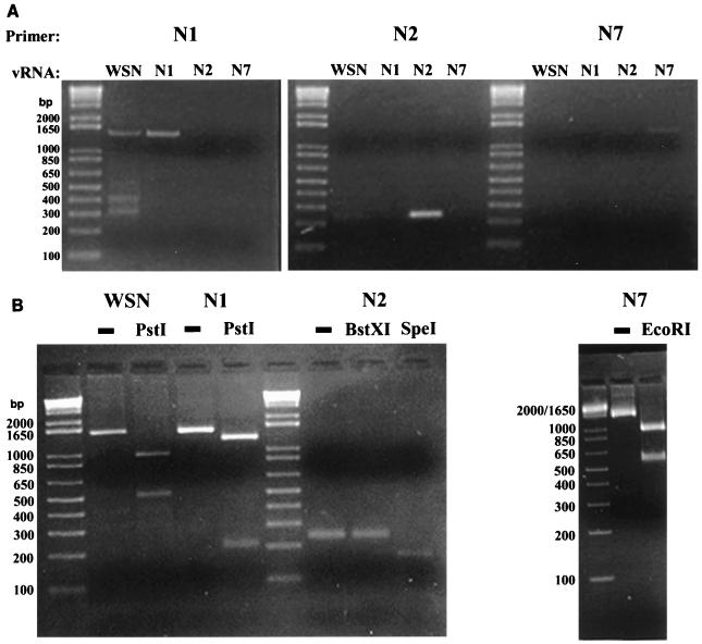
RT-PCR was performed with primers designed to amplify full-length N1 [WSN or N1(HK/97)] vRNA, a fragment of N2 vRNA, or full-length N7 vRNA. The N1 primers yielded products of the expected sizes of 1,453 and 1,445 bp (longer than the NA vRNA due to sequences at the 5′ and 3′ ends of the primers), for the WSN control and the WSN-N1(HK/97) transfectant virus, respectively. The N1 primers did not yield a product for the WSN-N2(LA/87) or WSN-N7(Cor/74) virus (Fig. 5A). These data indicate that the WSN-N2(LA/87) and WSN-N7(Cor/74) viruses do not possess N1 NAs and therefore are not C320G revertants. Additionally, the WSN-N1(HK/97) virus possessed an N1 vRNA as expected. To confirm that the WSN-N1(HK/97) virus was not a revertant, the WSN and WSN-N1(HK/97) RT-PCR products were digested with PstI. The WSN NA and HK/97 N1 have different PstI restriction patterns, with WSN NA expected to yield bands of 1,224 and 221 bp and the HK/97 N1 expected to yield bands of 944 and 509 bp. As seen in Fig. 5B, the WSN-N1(HK/97) virus possesses an NA with the PstI restriction pattern characteristic of the HK/97 N1. The NA of the WSN-N1 virus was therefore derived from the transfected N1 segment.
FIG. 5.
RT-PCR analysis of transfectant viruses WSN-N1(HK/97), WSN-N2(LA/87), and WSN-N7(Cor/74). (A) RT-PCR was performed on vRNAs obtained from the indicated viruses, using primers specific for the N1, N2, or N7 vRNA. (B) Restriction enzyme analysis of the N1, N2, or N7 RT-PCR products. Products were left undigested (−) or were digested with the indicated restriction endonuclease(s).
RT-PCR of the WSN-N2(LA/87) NA yielded a product of the expected 244 bp (Fig. 5A). It was necessary to confirm that the rescued N2 segment corresponded to the transfected N2(LA/87) and not the N2 of the X-31 virus from which the viral polymerase preparation (used during creation of the in vitro-synthesized N2 vRNA) was generated. The WSN-N2(LA/87) RT-PCR product should possess an SpeI site 70 bp from one end. The X-31 N2 RT-PCR product should possess a BstXI site 73 bp from one end but no SpeI site. The WSN-N2(LA/87) RT-PCR product could be cleaved 70 bp from its end by SpeI but was not cleaved by BstXI, as expected for the N2(LA/87) (Fig. 5B). The WSN-N2(LA/87) NA was therefore derived from the transfected N2 segment.
RT-PCR of the WSN-N7(Cor/74) NA yielded a product of the expected 1,504 bp (longer than the NA due to sequences at the 5′ and 3′ ends of the primers) (Fig. 5A). The N7(Cor/74) RT-PCR product was expected to contain an EcoRI site, while the WSN, N1(HK/97), N2(LA/87), and N2(X-31) vRNAs do not possess EcoRI sites. The N7(Cor/74) was indeed cleaved by EcoRI, yielding the expected 927- and 531-bp products, confirming that this virus possessed the transfected N7 segment (Fig. 5B).
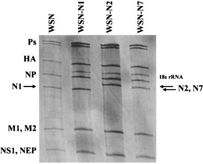
As a final confirmation that the transfectant viruses possessed the transfected NA vRNAs in a stable form, the transfectants were amplified once again in 10-day-old embryonated chicken eggs, and vRNAs were prepared. These vRNAs were then analyzed by polyacrylamide gel electrophoresis (Fig. 6). The WSN-N1(HK/97) virus possessed an NA of the expected 1,401 nucleotides, slightly smaller than the 1,409-bp WSN NA. The WSN-N2(LA/87) and the WSN-N7(Cor/74) vRNAs migrated slower than the WSN NA vRNA, as expected. These results confirm that the ts C303G and C320G viruses serve as efficient helper viruses for the rescue of a variety of NA types.
FIG. 6.
RNA gel analysis of transfectant vRNAs. vRNAs are labeled according to the gene products which they encode, and the location of the 18S rRNA is indicated. Positions of NA vRNAs are indicated by arrows. RNA samples were run on a 2.8% 7.7 M urea-containing polyacrylamide gel and silver stained. Ps, polymerase (PA, PB1, and PB2) segment; NEP, nuclear export protein.
DISCUSSION
The cysteine-to-glycine mutants analyzed in this study can be classified into three groups. The first group consists of enzymatically inactive mutants in which intramolecular disulfides formed between β-sheets were disrupted (51). These mutants may be defective due to loss of the disulfide bond, due to the loss of the side chain of a single cysteine residue, or due to the presence of a single, unpaired cysteine. By analogy with the N2 structure, the disulfides C218-C223 and C264-C275 will link β3S1 to β3S2 and β4S1 and β4S2, respectively. These disulfide bonds are located near the loops β3L0,1 and β4L0,1 which contain charged residues which may be involved in catalysis or in maintaining the structure of the active site (51). These disulfide bonds may therefore be necessary to maintain the structure or function of the enzymatic active site. Alternatively, loss of a cysteine side chain or the presence of an unpaired cysteine in this region may perturb the structure of the active site. It is also possible that the mutation of cysteines in β-sheets might cause defects in NA processing or stability. Precedents do exist for single amino acid changes yielding both stable, inactive NA mutants and degraded NA mutants (27, 28).
In contrast to the first group of NA mutants, mutation of the residues which form the C303-C320 disulfide bond resulted in ts proteins. Neuraminidase activity detected following expression at 33°C of either of these mutants was reduced significantly (about 50-fold) compared to wild-type NA, while both mutant proteins were almost inactive at 39.5°C. When these mutant genes were rescued into virus, both mutant viruses formed small plaques at 33°C and grew to titers 1 to 2 logs lower than those of wild-type WSN virus. At 39.5°C, no plaques could be detected for either mutant virus. In liquid culture, a 5- to 6-log decrease in titer was observed at 39.5°C compared with 33°C. These results demonstrated that an NA which has only 2% of wild-type activity (as measured with a low-molecular-weight substrate) is able to support replication of an influenza virus. They also demonstrate that mutation of the residues which form the C303-C320 disulfide bond yields a virus with a very tight ts phenotype.
Temperature-sensitive cysteine mutations have been seen in other systems as well. These may appear because disulfide bonds are important for stabilization of protein structure but are not usually required for a protein to assume its proper configuration (8). However, the ts phenotypes may also arise due to the properties of the side chain of the substitute amino acid. When several cysteines of the HSV-1 gD protein were individually changed to serine, the resulting mutant proteins were ts in their transport and/or processing. Additionally, while the mutants Cys-2 and Cys-4 were able to complement a gD-minus virus at 31.5°C, these two mutants were unable to complement the gD-minus virus at 37°C (32). Likewise, when any of the three cysteines of the HSV-1 αTIF protein (VP16) were individually changed to glycine, the resulting mutants were ts for binding to host cell proteins and for binding to the αTIF response element. Double cysteine-to-glycine αTIF mutations were also found to be ts in the context of viral infection. These mutant viruses grew to a titer 3 to 5 logs lower than that of a wild-type virus at nonpermissive temperature (44).
The third group of mutants were those affecting the two ectodomain cysteines which do not participate in intrasubunit disulfide bonds (C49 and C146 [WSN numbering]). Mutation of C49 resulted in an enzyme which functioned as well as wild-type NA at any temperature tested, both in the transient expression system and in the context of an infectious virus. This result was not surprising given that viruses possessing deletions encompassing C49 grew as well as wild-type WSN (33). Despite its nonessential nature, even at 39.5°C, C49 was found to participate in intersubunit disulfide bonding in the fowl plague virus N1 (19). Similarly, intersubunit disulfide bonds of the influenza A virus M2 protein are nonessential for protein function (5, 23). Mutation of C146 resulted in an enzyme which was about one-third as active as wild-type NA at 33°C and about one-fifth as active as wild-type NA at 39.5°C. However, this modest decrease in NA activity had little impact on replication of viruses possessing this mutation. The slightly ts nature of this enzyme likely reflects some disturbance in NA structure which is enhanced at higher temperatures. Although it was suggested that this residue might participate in intersubunit disulfide bonds in N1 molecules (51), no biochemical evidence for the participation of this cysteine in a subunit-subunit link was found (19). When a C49G,C146G double mutant was tested, it behaved similarly to the C146G mutant. Given these data, it is clear that intersubunit disulfide bonds are not required for the formation of stable, functioning influenza virus NAs, even at elevated temperatures.
The C303G and C320G mutants described here display interesting and useful properties (42, 49). In particular, both mutants are extremely tight. At the nonpermissive temperature, they displayed a greater than 3-log decrease in efficiency of plaque formation and a 5- to 6-log decrease in titer after multicycle growth in liquid culture. The frequency at which revertant or suppressor mutations of C303G or C320G arise must be explored further, however. Mutations in HA, which result in decreased affinity for sialic acid, can confer resistance to NA inhibitors (1, 18, 35). It remains possible that similar HA mutations which suppress the ts phenotype of C303G and C320G will arise.
The C303G and C320G mutants have also been shown to be useful as helper viruses in reverse genetics experiments, permitting the rescue of WSN NA, an avian virus N1, a human virus N2, and an equine virus N7. The ability to efficiently rescue non-WSN NAs should facilitate future studies of NA structure and function. Possible examples include the rescue of wild-type and mutant forms of NAs from clinically important viruses. The avian N1 that we rescued is 99% identical to the N1 of an H5N1 human isolate which caused a fatal infection in a child (50), and the N2 that we rescued is from a human H3N2 virus. Additionally, since we can also rescue an N7, it should now be possible to rescue other NA types which have interesting properties, such as hemagglutination. Also, cytotoxic T-cell epitopes have been expressed from viruses possessing the WSN NA segment (4, 13, 43), and these constructs have been useful in generating effective cytotoxic T-lymphocyte responses against other pathogens and against tumor cells (15, 37, 45).
In summary, we have demonstrated that ts influenza viruses can be generated by mutation of specific cysteine residues within the viral NA. These ts viruses displayed tight and stable ts phenotypes which permitted their use as helper viruses in reverse genetics experiments.
ACKNOWLEDGMENTS
This work was partially supported by grants from the National Institutes of Health to A.G.-S. and P.P. and an NIH NRSA postdoctoral fellowship to C.F.B.
We thank Louis Nguyenvu for excellent technical assistance and Hongyong Zheng for helpful discussions.
REFERENCES
- 1.Blick T J, Sahasrabudhe A, McDonald M, Ownes I J, Morley P J, Fenton R J, McKimm-Breschkin J L. The interaction of neuraminidase and hemagglutinin mutations in influenza virus in resistance to 4-guanidino-Neu5Ac2en. Virology. 1998;246:95–103. doi: 10.1006/viro.1998.9194. [DOI] [PubMed] [Google Scholar]
- 2.Bos T J, Nayak D. Identification of defects in the neuraminidase gene of four temperature-sensitive mutants of A/WSN/33 influenza virus. Virology. 1986;154:85–96. doi: 10.1016/0042-6822(86)90432-0. [DOI] [PubMed] [Google Scholar]
- 3.Braakman I, Hoover-Litty H, Wagner K R, Helenius A. Folding of influenza virus hemagglutinin in the endoplasmic reticulum. J Cell Biol. 1991;114:401–411. doi: 10.1083/jcb.114.3.401. [DOI] [PMC free article] [PubMed] [Google Scholar]
- 4.Castrucci M R, Bilsel P, Kawaoka Y. Attenuation of influenza A virus by insertion of a foreign epitope into the neuraminidase. J Virol. 1992;66:4647–4653. doi: 10.1128/jvi.66.8.4647-4653.1992. [DOI] [PMC free article] [PubMed] [Google Scholar]
- 5.Castrucci M R, Hughes M, Donatelli I, Wells K, Takada A, Kawaoka Y. The cysteine residues of the M2 protein are not required for influenza A virus replication. Virology. 1997;238:128–134. doi: 10.1006/viro.1997.8809. [DOI] [PubMed] [Google Scholar]
- 6.Colman P M. Neuraminidase: enzyme and antigen. In: Krug R M, editor. The influenza viruses. New York, N.Y: Plenum Press; 1989. pp. 175–218. [Google Scholar]
- 7.Colman P M, Varghese J N, Laver W G. Structure of the catalytic and antigenic sites in influenza virus neuraminidase. Nature. 1983;303:41–44. doi: 10.1038/303041a0. [DOI] [PubMed] [Google Scholar]
- 8.Creighton T E. Protein folding coupled to disulfide bond formation. Biol Chem. 1997;378:731–744. doi: 10.1515/bchm.1997.378.8.731. [DOI] [PubMed] [Google Scholar]
- 9.Enami M, Luytjes W, Krystal M, Palese P. Introduction of site-specific mutations into the genome of influenza virus. Proc Natl Acad Sci USA. 1990;87:3802–3805. doi: 10.1073/pnas.87.10.3802. [DOI] [PMC free article] [PubMed] [Google Scholar]
- 10.Enami M, Palese P. High-efficiency formation of influenza virus transfectants. J Virol. 1991;65:2711–2713. doi: 10.1128/jvi.65.5.2711-2713.1991. [DOI] [PMC free article] [PubMed] [Google Scholar]
- 11.Fuerst T R, Earl P L, Moss B. Use of a hybrid vaccinia virus-T7 RNA polymerase system for expression of target genes. Mol Cell Biol. 1987;7:2538–2544. doi: 10.1128/mcb.7.7.2538. [DOI] [PMC free article] [PubMed] [Google Scholar]
- 12.García-Sastre A, Palese P. The cytoplasmic tail of the neuraminidase protein of influenza A virus does not play an important role in the packaging of this protein into viral envelopes. Virus Res. 1995;37:37–47. doi: 10.1016/0168-1702(95)00017-k. [DOI] [PubMed] [Google Scholar]
- 13.García-Sastre A, Muster T, Barclay W S, Percy N, Palese P. Use of a mammalian internal ribosomal entry site element for expression of a foreign protein by a transfectant influenza virus. J Virol. 1994;68:6254–6261. doi: 10.1128/jvi.68.10.6254-6261.1994. [DOI] [PMC free article] [PubMed] [Google Scholar]
- 14.Ghate A A, Air G M. Site-directed mutagenesis of catalytic residues of influenza virus neuraminidase as an aid to drug design. Eur J Biochem. 1998;258:320–331. doi: 10.1046/j.1432-1327.1998.2580320.x. [DOI] [PubMed] [Google Scholar]
- 15.Gonzalo R M, Rodriguez D, García-Sastre A, Rodriguez J R, Palese P, Esteban M. Enhanced CD8+ T cell response to HIV-1 env by combined immunization with influenza and vaccinia virus recombinants. Vaccine. 1999;17:887–892. doi: 10.1016/s0264-410x(98)00274-6. [DOI] [PubMed] [Google Scholar]
- 16.Goto H, Kawaoka Y. A novel mechanism for the acquisition of virulence by a human influenza A virus. Proc Natl Acad Sci USA. 1998;95:10224–10228. doi: 10.1073/pnas.95.17.10224. [DOI] [PMC free article] [PubMed] [Google Scholar]
- 17.Gottschalk A. The specific enzyme of influenza virus and Vibrio cholerae. Biochim Biophys Acta. 1957;23:645–646. doi: 10.1016/0006-3002(57)90389-x. [DOI] [PubMed] [Google Scholar]
- 18.Gubareva L V, Bethell R, Hart G J, Murti K G, Penn C R, Webster R G. Characterization of mutants of influenza A virus selected with the neuraminidase inhibitor 4-guanidino-Neu5Ac2en. J Virol. 1996;70:1818–1827. doi: 10.1128/jvi.70.3.1818-1827.1996. [DOI] [PMC free article] [PubMed] [Google Scholar]
- 19.Hausmann J, Kretzchmar E, Garten W, Klenk H-D. Biosynthesis, intracellular transport and enzymatic activity of an avian influenza A virus neuraminidase: role of unpaired cysteines and individual oligosaccharides. J Gen Virol. 1997;78:3233–3245. doi: 10.1099/0022-1317-78-12-3233. [DOI] [PubMed] [Google Scholar]
- 20.Hayden F G, Treanor J J, Betts B F, Lobo M, Esinhart J D, Hussey E K. Saftey and efficacy of the neuraminidase inhibitor GG167 in experimental human influenza. JAMA. 1996;275:295–299. [PubMed] [Google Scholar]
- 21.Hayden F G, Osterhaus A D, Treanor J J, Fleming D M, Aoki F Y, Nicholson K G, Bohnen A M, Hirst H M, Keene O, Wightman K. Efficacy and safety of the neuraminidase inhibitor zanamivir in the treatment of influenza virus infections. GG167 Influenza Study Group. N Engl J Med. 1997;337:874–880. doi: 10.1056/NEJM199709253371302. [DOI] [PubMed] [Google Scholar]
- 22.Hiti A L, Nayak D P. Complete nucleotide sequence of the neuraminidase gene of human influenza virus A/WSN/33. J Virol. 1982;41:730–734. doi: 10.1128/jvi.41.2.730-734.1982. [DOI] [PMC free article] [PubMed] [Google Scholar]
- 23.Holsinger L J, Lamb R A. Influenza virus M2 integral membrane protein is a homotetramer stabilized by formation of disulfide bonds. Virology. 1991;183:32–43. doi: 10.1016/0042-6822(91)90115-r. [DOI] [PubMed] [Google Scholar]
- 24.Jin H, Leser G P, Zhang J, Lamb R A. Influenza virus hemagglutinin and neuraminidase cytoplasmic tails control particle shape. EMBO J. 1997;16:1236–1247. doi: 10.1093/emboj/16.6.1236. [DOI] [PMC free article] [PubMed] [Google Scholar]
- 25.Kobasa D, Rodgers M E, Wells K, Kawaoka Y. Neuraminidase hemadosorption activity, conserved in avian influenza viruses, does not influence viral replication in ducks. J Virol. 1997;71:6706–6713. doi: 10.1128/jvi.71.9.6706-6713.1997. [DOI] [PMC free article] [PubMed] [Google Scholar]
- 26.Laver W G, Colman P M, Webster R G, Hinshaw V S, Air G M. Influenza virus neuraminidase with hemagglutinin activity. Virology. 1984;137:314–323. doi: 10.1016/0042-6822(84)90223-x. [DOI] [PubMed] [Google Scholar]
- 27.Lentz M R, Air G M. Loss of enzyme activity in a site-directed mutant of influenza neuraminidase compared to expressed wild-type protein. Virology. 1986;148:74–83. doi: 10.1016/0042-6822(86)90404-6. [DOI] [PubMed] [Google Scholar]
- 28.Lentz M R, Webster R G, Air G M. Site-directed mutation of the active site of influenza neuraminidase and implications for the catalytic mechanism. Biochemistry. 1987;26:5351–5358. doi: 10.1021/bi00391a020. [DOI] [PubMed] [Google Scholar]
- 29.Li S, Schulman J, Itamura S, Palese P. Glycosylation of neuraminidase determines the neurovirulence of influenza A/WSN/33 virus. J Virol. 1993;67:6667–6673. doi: 10.1128/jvi.67.11.6667-6673.1993. [DOI] [PMC free article] [PubMed] [Google Scholar]
- 30.Liu C, Air G M. Selection and characterization of a neuraminidase-minus mutant of influenza virus and its rescue by cloned neuraminidase genes. Virology. 1993;194:403–407. doi: 10.1006/viro.1993.1276. [DOI] [PubMed] [Google Scholar]
- 31.Long D, Cohen G H, Muggeridge M I, Eisenberg R J. Cysteine mutants of herpes simplex virus type 1 glycoprotein D exhibit temperature-sensitive properties in structure and function. J Virol. 1990;64:5542–5552. doi: 10.1128/jvi.64.11.5542-5552.1990. [DOI] [PMC free article] [PubMed] [Google Scholar]
- 32.Long D, Wilcox W C, Abrams W R, Cohen G H, Eisenberg R J. Disulfide bond structure of glycoprotein D of herpes simplex virus types 1 and 2. J Virol. 1992;66:6668–6685. doi: 10.1128/jvi.66.11.6668-6685.1992. [DOI] [PMC free article] [PubMed] [Google Scholar]
- 33.Luo G, Chung J, Palese P. Alterations of the stalk of influenza virus neuraminidase: deletions and insertions. Virus Res. 1993;29:141–153. doi: 10.1016/0168-1702(93)90055-r. [DOI] [PubMed] [Google Scholar]
- 34.Mastrovich M, Zhou N, Kawaoka Y, Webster R G. The surface glycoproteins of H5 influenza viruses isolated from humans, chickens, and wild aquatic birds have distinguishable properties. J Virol. 1999;73:1146–1155. doi: 10.1128/jvi.73.2.1146-1155.1999. [DOI] [PMC free article] [PubMed] [Google Scholar]
- 35.McKimm-Breschkin J L, Blick T J, Sahastabudhe A, Tiong T, Marshall D, Hart G J, Bethell R C, Penn C R. Generation and characterization of variants of NWS/G70C influenza virus after in vitro passage in 4-amino-Neu5Ac2en and 4-guanido-Neu5Ac2en. Antimicrob Agents Chemother. 1996;40:40–46. doi: 10.1128/aac.40.1.40. [DOI] [PMC free article] [PubMed] [Google Scholar]
- 36.Meindl P, Bodo G, Palese P, Schulman J, Tuppy H. Inhibition of neuraminidase activity by derivatives of 2-deoxy-2,3-dehydro-N-acetylneuraminic acid. Virology. 1974;58:457–463. doi: 10.1016/0042-6822(74)90080-4. [DOI] [PubMed] [Google Scholar]
- 37.Miyahara Y, García-Sastre A, Rodriguez D, Rodriguez J R, Murata K, Tsuji M, Palese P, Esteban M, Zavala F, Nussenzweig R S. Recombinant viruses expressing a human malaria antigen can elicit potentially protective immune CD8+ responses in mice. Proc Natl Acad Sci USA. 1998;95:3954–3959. doi: 10.1073/pnas.95.7.3954. [DOI] [PMC free article] [PubMed] [Google Scholar]
- 38.Morris S J, Price G E, Barnett J M, Hiscox S A, Smith H, Sweet C. Role of neuraminidase in influenza virus-induced apoptosis. J Gen Virol. 1999;80:137–146. doi: 10.1099/0022-1317-80-1-137. [DOI] [PubMed] [Google Scholar]
- 39.Ogawa, S., M. Yoshikawa, and T. Taki. 1992. Synthesis of a carbocyclic analogue of N-acetylneuraminic acid (pseudo-N-acetylneuraminic acid). J. Chem. Soc. Chem. Commun. 406–408.
- 40.Palese P, Tobita K, Ueda M, Compans R W. Characterization of temperature-sensitive influenza virus mutants defective in neuraminidase. Virology. 1974;61:397–410. doi: 10.1016/0042-6822(74)90276-1. [DOI] [PubMed] [Google Scholar]
- 41.Palese P, Compans R W. Inhibition of influenza virus replication in tissue culture by 2-deoxy-2,3-dehydro-N-fluoroacetylneuraminic acid (FANA): mechanism of action. J Gen Virol. 1976;33:2142–2146. doi: 10.1099/0022-1317-33-1-159. [DOI] [PubMed] [Google Scholar]
- 42.Parkin N T, Chiu P, Coelingh K L. Temperature sensitive mutants of influenza A virus generated by reverse genetics and clustered charged to alanine mutagenesis. Virus Res. 1996;46:31–44. doi: 10.1016/s0168-1702(96)01373-1. [DOI] [PubMed] [Google Scholar]
- 43.Percy N, Barclay W S, García-Sastre A, Palese P. Expression of a foreign protein by influenza A virus. J Virol. 1994;68:4486–4492. doi: 10.1128/jvi.68.7.4486-4492.1994. [DOI] [PMC free article] [PubMed] [Google Scholar]
- 44.Poon A P W, Roizman B. The phenotype in vitro and in infected cells of herpes simplex virus 1 α trans-inducing factor (VP16) carrying temperature-sensitive mutations introduced by substitution of cysteines. J Virol. 1995;69:7658–7667. doi: 10.1128/jvi.69.12.7658-7667.1995. [DOI] [PMC free article] [PubMed] [Google Scholar]
- 45.Restifo N P, Surman D R, Zheng H, Palese P, Rosenberg S A, García-Sastre A. Transfectant influenza A viruses are effective recombinant immunogens in the treatment of experimental cancer. Virology. 1998;249:89–97. doi: 10.1006/viro.1998.9330. [DOI] [PMC free article] [PubMed] [Google Scholar]
- 46.Rudneva I E, Sklyanskaya E I, Barulina O S, Yamnikova S S, Kovaleva V P, Tsvetkova I V, Kaverin N V. Phenotypic expression of HA-NA combinations in human-avian influenza A virus reassortants. Arch Virol. 1996;141:1091–1099. doi: 10.1007/BF01718612. [DOI] [PubMed] [Google Scholar]
- 47.Schultz-Cherry S, Hinshaw V S. Influenza virus neuraminidase activates latent transforming growth factor β. J Virol. 1996;70:8624–8629. doi: 10.1128/jvi.70.12.8624-8629.1996. [DOI] [PMC free article] [PubMed] [Google Scholar]
- 48.Segal M S, Bye J M, Sambrook J F, Gething M-J H. Disulfide bond formation during the folding of influenza virus hemagglutinin. J Cell Biol. 1992;118:227–244. doi: 10.1083/jcb.118.2.227. [DOI] [PMC free article] [PubMed] [Google Scholar]
- 49.Subbarao E K, Park E J, Lawson C M, Chen A Y, Murphy B R. Sequential addition of temperature-sensitive missense mutations into the PB2 gene of influenza A transfectant viruses can effect an increase in temperature sensitivity and attenuation and permits the rational design of a genetically engineered live influenza A virus vaccine. J Virol. 1995;69:5969–5977. doi: 10.1128/jvi.69.10.5969-5977.1995. [DOI] [PMC free article] [PubMed] [Google Scholar]
- 50.Subbarao K, Klimov A, Katz J, Regenery H, Lim W, Hall H, Perdue M, Swayne D, Bender C, Huang J, Hemphill M, Rowe T, Shaw M, Xu X, Fukada K, Cox N. Characterization of an avian influenza A (H5N1) virus isolated from a child with a fatal respiratory illness. Science. 1998;279:393–396. doi: 10.1126/science.279.5349.393. [DOI] [PubMed] [Google Scholar]
- 51.Varghese J N, Laver W G, Colman P M. Structure of the influenza virus glycoprotein antigen neuraminidase at 2.9 Å resolution. Nature. 1983;303:35–40. doi: 10.1038/303035a0. [DOI] [PubMed] [Google Scholar]
- 52.Varghese J N, Colman P M, van Donkelaar A, Blick T J, Sahasrabudhe A, McKimm-Breschkin J L. Structural evidence for a second sialic acid binding site in avian influenza virus neuraminidase. Proc Natl Acad Sci USA. 1997;94:11808–11812. doi: 10.1073/pnas.94.22.11808. [DOI] [PMC free article] [PubMed] [Google Scholar]
- 53.von Itzstein M, Wu W Y, Kok G B, Pegg M S, Dyason J C, Jin B, Van Phan T, Smythe M L, White H F, Oliver S W, et al. Rational design of potent sialidase-based inhibitors of influenza virus replication. Nature. 1993;363:418–423. doi: 10.1038/363418a0. [DOI] [PubMed] [Google Scholar]
- 54.Wilcox W C, Long D, Sodora D L, Eisenberg R J, Cohen G H. The contribution of cysteine residues to antigenicity and extent of processing of herpes simplex virus type 1 glycoprotein D. J Virol. 1988;62:1941–1947. doi: 10.1128/jvi.62.6.1941-1947.1988. [DOI] [PMC free article] [PubMed] [Google Scholar]
- 55.Yamamoto-Goshima F, Maeno K, Morishits T, Ueda M, Fujita Y, Nakajima K, Yoshii S. Role of neuraminidase in the morphogenesis of influenza B virus. J Virol. 1994;68:1250–1254. doi: 10.1128/jvi.68.2.1250-1254.1994. [DOI] [PMC free article] [PubMed] [Google Scholar]


