Abstract
Background
155Tb represents a potentially useful radionuclide for diagnostic medical applications, but its production remains a challenging problem, in spite of the fact that many production routes have been already investigated and tested. A recent experimental campaign, conducted with low-energy proton beams impinging on a 155Gd target with 91.9% enrichment, demonstrated a significant co-production of 156gTb, a contaminant of great concern since its half-life is comparable to that of 155Tb and its high-energy γ emissions severely impact on the dose released and on the quality of the SPECT images. In the present investigation, the isotopic purity of the enriched 155Gd target necessary to minimize the co-production of contaminant radioisotopes, in particular 156gTb, was explored using various computational simulations.
Results
Starting from the recent experimental data obtained with a 91.9% 155Gd-enriched target, the co-production of other Tb radioisotopes besides 155Tb has been theoretically evaluated using the Talys code. It was found that 156Gd, with an isotopic content of 5.87%, was the principal contributor to the co-production of 156gTb. The analysis also demonstrated that the maximum amount of 156Gd admissible for 155Tb production with a radionuclidic purity higher than 99% was 1%. A less stringent condition was obtained through computational dosimetry analysis, suggesting that a 2% content of 156Gd in the target can be tolerated to limit the dose increase to the patient below the 10% limit. Moreover, it has been demonstrated that the imaging properties of the produced 155Tb are not severely affected by this level of impurity in the target.
Conclusions
155Tb can be produced with a quality suitable for medical applications using low-energy proton beams and 155Gd-enriched targets, if the 156Gd impurity content does not exceed 2%. Under these conditions, the dose increase due to the presence of contaminant radioisotopes remains below the 10% limit and good quality images, comparable to those of 111In, are guaranteed.
Keywords: Terbium radioisotopes, 155Tb production, Theranostics, SPECT imaging, Gadolinium targets, Proton-induced nuclear-reaction calculations, Radioisotopic contaminants effects, Dosimetric calculations, Compton noise in SPECT imaging
Background
Terbium is one of the few rare earth radiometals that could be used in nuclear medicine for tumor diagnosis and treatment, due to the favorable physical decay properties such as half-lives, type and energy of emissions. The four Tb radionuclides with higher clinical interest are 152Tb and 155Tb, positron and γ emitters respectively, relevant for diagnostic purposes, and 149Tb and 161Tb, α and β− emitters respectively, suitable for therapeutic applications [1]. The matched pairs therapeutic/diagnostic radioisotopes of this element could be ideal candidates for theranostic applications, with no differences in chemical and pharmacokinetics behaviours [2]. Indeed, significant differences could be observed when using pairs of diverse elements for diagnostics and therapy, because it is well known that each metal ion has specific chemical demands arising from its fundamental characteristics, giving rise to different coordination and geometry numbers [3], which influence the pharmacological properties of the labelled molecules.
Positron emission tomography (PET) imaging is the clinical nuclear imaging technique with higher sensitivity and resolution. Although, single photon emission computed tomography (SPECT) is still the predominant technology worldwide, due to the easy availability of γ-emitter radionuclides and the low cost of SPECT camera compared to PET scanner [4]. Therefore, the production of new γ-emitting radionuclides that could be detected with the large number of available SPECT scanners is encouraged. Among Tb isotopes, 155Tb is the only suitable for SPECT imaging, thanks to its two γ emissions at 87 keV (32%) and 105 keV (25%). Preclinical studies in nude mice bearing tumor xenograft demonstrated that 155Tb-labelled biomolecules were able to visualize tumor sites using a small-animal SPECT/CT scanner even 7 days after radiocomplexes administration [5]. 155Tb has been produced by different methods, although most of them generated a large number of isotopes. First investigations on the production of 155Tb via proton-induced reactions on Gd targets date back to 1989 with the work by Dmitriev et al. [6]. In this case, no cross-section measurements were performed, but rather irradiation experiments of natGd thick targets for yields and activities, with incident proton energies between 11 and 22 MeV. Using natGd targets, cross-section measurements were performed much later by Vermeulen et al. [7]. Besides the detailed experimental work that included the measurement of cross sections for the formation of a variety of terbium radionuclides, theoretical calculations were also performed for all contributing reactions. Since natGd has seven stable isotopes, it is not suited as target material for radioisotope production with high radionuclidic purity (RNP). Therefore, comparisons were made with accurate theoretical simulations in order to identify the most promising reactions that could be effectively employed in studies with enriched targets [7]. More recently Formento-Cavaier et al. [8] extended the measurements with natGd targets up to 70 MeV. Experimental investigations with enriched targets of 155Gd and 156Gd have been undertaken by Favaretto et al. [9], with the result that 156Gd(p,2n)155Tb provides high production yields, but implies the use of higher-energy cyclotrons and a significant contamination by the co-production of 156Tb. The 155Gd(p,n)155Tb reaction, on the other hand, can be performed with medical cyclotrons, with incident protons up to around 18 or 20 MeV, and has the potential to provide significant yields with high purity. In a subsequent publication, Dellepiane et al. [10] used the same enriched gadolinium oxide (155Gd 91.9% enrichment or 156Gd 93.3% enrichment) as target materials, measuring the 155Tb production cross sections as well as a variety of contaminants produced for such highly enriched targets. In the most favorable case of the 155Gd target, using an input energy of 10.5 MeV, contamination from 156Tb was still significant, leading to a maximum RNP not greater than 93% after a decay time of about 96 h. Thus, it was suggested, as possible solution, to purify the final product through an off-line mass separation technique at the expense of a lower production yield, due the low efficiency of the current mass separation approaches. The production of 155Tb and other Tb-isotopes (149Tb, 152Tb and 161Tb) has been explored also at CERN-ISOLDE using spallation of high-energy proton beams on Tantalum targets, followed by ionization and mass separation [1, 11]. The drawback stands in the limited quantity of 155Tb that can be produced with this method, due to the observed low efficiency of the accumulation procedure [12, 13]. It has been suggested also to irradiate a 159Tb target by intermediate energy (60, 70 MeV) proton beams [14, 15]. This would open the possibility to have a 155Dy/155Tb generator system, similarly to the renowned 99Mo/99mTc one. However, the double separation chemistry among lanthanides represents a crucial step that still needs to be solved and the possible co-production of very long-lived terbium contaminants could imply a too low specific activity.
In this work Talys calculations of 155Tb cross section have been benchmarked with data measured by Dellepiane et al. [10] with an enriched 155Gd target. The contribution of each isotopic component has been disentangled to expose the effects deriving from the impurities of the target. Once the modeling has been tested on the specific isotopic 155Gd abundance of the enriched target used by Dellepiane et al., the level of enrichment of the 155Gd target necessary to produce 155Tb with high purity has been investigated. However, the assessment of RNP is not enough to evaluate the suitability of the produced 155Tb for clinical purposes. In fact, different produced contaminants can have a different impact on the dose delivered to the patient and on the quality of the SPECT images. Detailed biodistribution data of the DOTA-folate conjugate 161Tb-cm09 in IGROV-1 tumour-bearing mice have been reported [11]. As the biodistribution is independent of the radioisotope used to label the molecule, these data can be used to estimate the dosimetric properties of the same radiopharmaceutical when labeled with different Tb radioisotopes. In this way, it was possible to assess the dose increase (DI) [16] to the patient following 155Tb-cm09 injection, due to the presence of contaminants in the 155Tb produced supposing different enrichment of the 155Gd target. Moreover, the effect of the contaminants with high-energy γ emissions on the SPECT images quality was evaluated.
Methods
Cross sections and thick target irradiation
The study of the nuclear reaction routes implies the adoption of different models to consider both the compound nucleus formation/decay and pre-equilibrium dynamics. To this purpose the Talys code [17] has been used, specifically version 1.95 which includes the geometry dependent hybrid (GDH) model [18, 19]. To describe the nuclear reaction mechanisms this Talys version provides 5 pre-equilibrium (PE) and 6 level-density (LD) models, for a total of 30 possible combinations of models. The description of the PE processes is mainly based on the exciton model. Transition matrices are the basic building blocks of the exciton formalism and they are described (1) analytically; (2) numerically; or (3) derived from the imaginary component of the optical potential. A fully quantum–mechanical approach, based upon the Feshbach-Kerman-Koonin theory and alternative to the exciton model, is included and labelled as (4). The already mentioned GDH model, which considers nuclear surface effects in the exciton model, is denoted as option (5). Another important ingredient for the compound nucleus formation is the nuclear LD, which affects the cross section through the Hauser-Feshbach formalism. Talys considers three phenomenological LD models, namely (1) the Fermi gas with constant temperature; (2) the back-shifted Fermi gas; (3) the generalized superfluid model; and three microscopic models, that is (4) the Goriely’s tabulated Hartree–Fock densities; (5) the Hilaire’s tabulation based upon the combinatorial model, and (6) the Hartree–Fock–Bogoliubov temperature-dependent formalism. The variability of these models can be described statistically introducing an interquartile band, measuring the model dispersion between the lower Q1 and the upper Q3 quartile, as has been discussed in a recent paper [20]. In addition, the modeling has been tested on the specific isotopic abundances of the enriched 155Gd target used by Dellepiane et al.
The modeled cross sections have been employed to evaluate the radionuclides produced under specific irradiation conditions, including the level of enrichment of the target. The computational approach discussed by Canton et al. [21] has been used for the evaluation of rates, activities, yields, and purities.
The rate R of production of a radionuclide from a beam colliding on a thick target can be derived from the expression
| 1 |
where I0 is the beam current, zproj is the projectile charge (1 for a proton beam), e the electron charge, Na the Avogadro number, A the atomic mass of the target element, Ein and Eout the energy of the beam hitting the target and the one leaving the target after traveling through its thickness, respectively. The production cross section for the nuclide is σ(E), while the target density ρt and the stopping power of the projectile in the target dE/dx, described by the Bethe-Bloch formula (22).
Once the rates given in Eq. (1) for all the Tb radionuclides of interest are determined, the time evolution of the number of produced radionuclides, and the evolution of the activities during and after an irradiation can be calculated from the Bateman equations. The time dependence of the number of Tb radionuclides produced and their corresponding activities are used to evaluate the isotopic and radionuclidic purities for the production of 155Tb. The main decay data of interest for the present analysis are reported in Table 1.
Table 1.
Main decay data of xxxTb radionuclides.
| Radionuclide | Decay mode (%) | T1/2 | Emitted energy (MeV/nt) | ||
|---|---|---|---|---|---|
| Electron | Photon | Total | |||
| 154gTb | EC, β+ (100%) | 21.5 h | 0.0681 | 2.2831 | 2.3512 |
| 154m1Tb | EC (78.20%), IT (21.80%), β−(< 0.10%) | 9.4 h | – | – | – |
| 154m2Tb | EC (98.20%), IT (1.80%) | 22.7 h | – | – | – |
| 155 Tb | EC (100%) | 5.32 d | 0.0434 | 0.1777 | 0.2211 |
| 156gTb | EC (100%) | 5.35 d | 0.0835 | 1.9371 | 2.0206 |
| 156m1Tb | IT (100%) | 24.4 h | 0.0171 | 0.0370 | 0.0540 |
| 156m2Tb | IT (100%) | 5.3 h | 0.0874 | 0.0048 | 0.0922 |
Assessment of organ absorbed doses and effective dose due to xxxTb-cm09 injection
Dosimetric assessment were carried out using biodistribution data of the DOTA-folate conjugate 161Tb-cm09 in IGROV-1 tumour-bearing mice [11]. The cm09 is composed by a targeting vector (which selectively binds to the folate receptor expressed on a variety of tumour types) conjugated to small-molecular-weight albumin (which improves the blood circulation time and tissue distribution profile of folate conjugates) and to the DOTA chelating agent. Dosimetric evaluation has been performed supposing the cm09 labelled with 155Tb and also with other Tb-radioisotopes expected to be produced by proton irradiation of 155Gd-enriched targets.
Biodistribution data of 161Tb-cm09, acquired in a time window of 7 days post injection in IGROV-1 tumour-bearing female nude mice [11], were used to estimate absorbed doses in humans due to the various Tb radioisotopes produced during the irradiation of all the considered enriched 155Gd target. This evaluation was done through the relative mass scaling method, which takes into account the differences in human (H) and animal (A) organ masses compared to the total body masses [25]. The activity concentrations in the different animal source organs (blood, lung, spleen, kidneys, stomach, intestines, liver, salivary glands, muscle and bone), reported as per cent of injected activity per gram of tissue ([%IA/g]A), were scaled from mice to adult male humans to obtain the decay-corrected per cent of injected activity for each human source organ ([%IA/organ]H) through the following formula:
| 2 |
with OWH the weight of human organ, TBWA and TBWH the total body weight for animal and human, respectively. OWH and TBWH values were obtained from the adult male phantom implemented in the Organ Level Internal Dose Assessment (OLINDA) software code [26]. Biodistribution data were then plotted as a function of post injection time and fitted with CoKiMo software [26] by a tri-exponential equation, representing the phase of accumulation and the possibility of both a fast and a slow elimination of the radiopharmaceutical [27]. At last, the number of disintegrations per unit of administered activity in the source organs was obtained by integration of organ activity curves for each xxxTb-radioisotope, considering its physical half-life. These data were then used as input values in a human adult male phantom to perform dosimetric calculations with the OLINDA software code (version 2.2.3), as reported before [28], obtaining the organ absorbed doses per unit of administered activity. For each xxxTb radioisotope, the specific effective dose, xxxTbED corresponds to the sum of the product of the organ equivalent dose per unit of administered activity, xxxTbDorg, and the respective tissue-weighting factor, worg, recommended by ICRP 103 [29],
| 3 |
The irradiation simulations with different target enrichment leads to a distinct distribution of xxxTb activities, described by the fraction of total activity:
| 4 |
The total effective dose (EDtot) of Tb-cm09 was calculated at different times after the end of bombardment (EoB) by summing all the contributions of the produced Tb radioisotopes:
| 5 |
Finally, the evaluation of the DI generated by the co-produced Tb-impurities is determined according to the following equation:
| 6 |
and represents the ratio between the total effective dose and the effective dose due to an ideal injection of pure 155Tb compound. The doses discussed above refer to the total doses of an injection delivered at time t post-production.
Assessment of the imaging properties of 155Tb in the presence of other contaminant radioisotopes
The main problem in the quality of SPECT images when 155Tb contains γ-emitting contaminants is the noise produced by the Compton scattering of high energy γ rays. Even using energy windows to acquire only the γ rays of interest, higher energy γ rays can be Compton scattered and registered by the acquisition system. The effect of such signals results in blurry and bad contrast images. Therefore, a study of the noise contribution of high-energy γ rays emitted by contaminant terbium radioisotopes has been carried out using a preclinical PET/SPECT/CT system (VECTor 5, Milabs) as a model of the acquisition system.
To simulate the γ-ray spectrum of each Tb radionuclide in VECTor 5, a homemade menu-driven spreadsheet software Visual Gamma was used. It makes use of specific initial system parameters such as efficiency and peak shape and simulates the spectrum of the radionuclides, using as input data the energy and the intensity of the γ ray, taken from the NUDAT3 database [24]. Parameters such as γ-ray efficiency, the peak width and the shape of the Compton area as a function of the γ-rays energies have been determined using standard calibration point-like sources (57Co, 22Na, 60Co and 137Cs) in air. The process of assessment of the image quality, obtained using a particular γ-ray peak, was based on two main factors: the intensity of the γ-ray emission and the presence of Compton-scattered γ rays in the energy window of the peak selected for imaging. An energy window of 10% interval around the peak barycenter was chosen. To evaluate the influence of Compton-scattered γ rays on the final quality of the image, the ratio δ = Nextr/Nintr was used, where Nextr is the number of all higher-energy γ rays that underwent Compton scattering and fell into the selected energy window, and Nintr is the number of γ rays from the selected photo-peak that fell into the energy window, i.e. not including Compton scattering contribution. It is convenient to use such Compton-to-peak ratio since in the ideal case (lack of other noises) its value directly corresponds to the amount of the noises in the image.
Imaging qualities were evaluated for 155Tb produced from the proton irradiation of 155Gd targets with enrichment of 100, 99 and 98%, at the EoB and 96 h later.
Results
Cross sections
The same composition of the enriched 155Gd targets employed by Dellepiane [10] for cross-section measurements (154Gd 0.5%, 155Gd 91.90%, 156Gd 5.87%, 157Gd 0.81%, 158Gd 0.65%, 160Gd 0.27%) was used in the irradiation simulations In Fig. 1 the experimental cross sections for 155Tb production are compared with theoretical calculations. The gray band represents the models variability between Q1 and Q3, the dashed lines are the minimum/maximum values of the cross sections obtained with all models, and the blue solid line is the Talys default option (PE2-LD1), commonly referred to as the standard simulation in the literature. For the 155Tb case all models reproduce equally the cross section up to 10 MeV, and for higher energy the band is relatively thin and this corresponds to a limited model variability, as expected from a typical (p,n) reaction. On account of this, and because the Talys default reproduces the very recent data measured by Dellepiane [10] quite satisfactorily, the analysis will be presented considering the Talys default as the benchmark calculation.
Fig. 1.
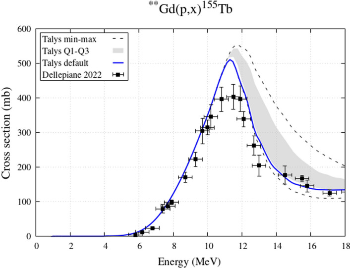
Experimental 155Tb cross sections from enriched 155Gd target and theoretical curves expressing the variability of nuclear reaction models
Figure 2 shows the contribution of each isotopic component of the target to the 155Tb production. The main contribution to the 155Tb cross section comes, as should be expected, from the 155Gd component of the target. For energies higher than 10 MeV, also the contribution from the 156Gd component becomes significant, and this explains the increase of the experimental data with respect to an ideal target with 100% 155Gd enrichment. The contribution from 157Gd is quite small and that from other Gadolinium components of the targets, such as 154Gd, is negligible. This figure includes cross-section data for 100%-enriched targets assessed by Dellepiane et al., 2023 [30]; they were published after the submission of this study and added in the final revision of the text.
Fig. 2.
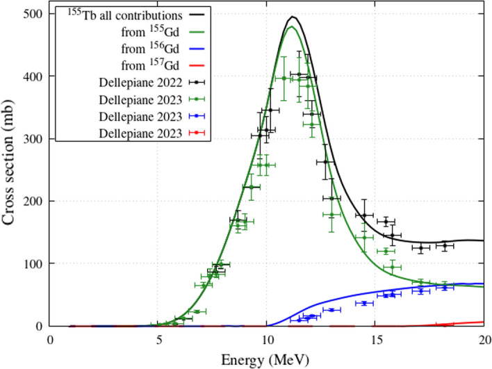
155Tb cross sections from enriched 155Gd target: Talys calculations and contributions from different Gd isotopes of the target
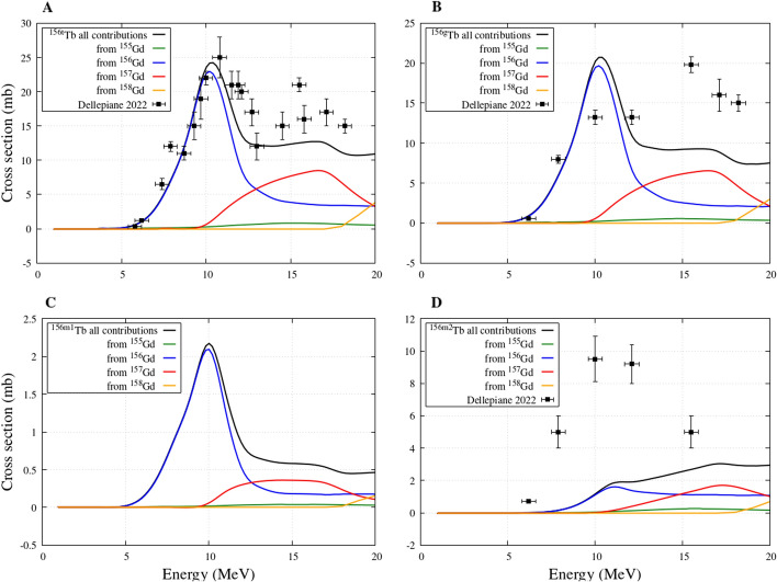
Next, the 156tTb total cross section, which refers to the cumulative cross sections of 156g,156m1,156m2Tb (ground and the first two metastable states), is presented in Fig. 3A. The curve including all contributions is in agreement with the data measured by Dellepiane et al. It is evident that the main contribution belongs to the 156Gd component of the target, followed by the 157Gd component at slightly higher energies. At even higher energies, about 19 MeV, the contribution from 158Gd also becomes significant. Instead, the contribution to the 156Tb cross section from the main 155Gd component of the target remains quite small in the entire range of considered energies.
Fig. 3.
156g, 156m1, 156m2, 156tTb (where t in panel A is the cumulative cross section of 156g, 156m1, 156m2Tb) cross sections from enriched 155Gd target obtained from Talys calculations: contributions from different Gd isotopes of the target
Figure 3B, C and D describe the 156Tb production cross sections for ground and the two metastable states, respectively. For the ground state, the model calculations are in fair agreement with the measurements, although at higher energies there is a tendency to underestimate the data. For the first metastable state there are no measured production data, while for the second one the curves underestimate the measurements. In all cases, by separating the individual isotopic components in the Gd target, it is evident that the main contributions to the production of the contaminants derive from the Gd isotopes heavier than 155.
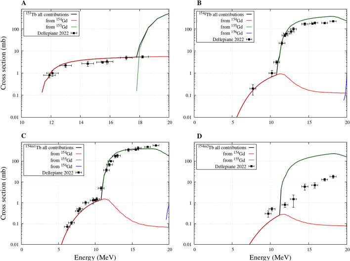
The remaining contaminants that may have an impact on the quality of the produced 155Tb are 153Tb and 154Tb (separated in ground and the first two metastable states) and their cross sections are given in Fig. 4. The theoretical curves are in clear agreement with the measurements, with the exception of 154m2Tb where slight discrepancies can be observed. In all figures it is evident that the contamination at energies lower that 10 MeV can be entirely ascribed to the small presence of 154Gd in the target, responsible of a characteristic bump seen in the lower energy data. At higher energies, 155Gd becomes the principal contributor to the production of these contaminants.
Fig. 4.
153, 154g, 154m1,154m2Tb cross sections from enriched 155Gd target obtained from Talys calculations: contributions from different Gd isotopes of the target
Yields, isotopic, and radionuclidic purity
Starting from the cross section analysis of the effects of the isotopic components of the target used for 155Tb production, yields, isotopic purity and RNP were assessed for different target compositions. Four different targets were considered in the simulations to evaluate the minimum enrichment required for a 155Tb production with the purity needed for medical applications: the first target with the exact isotopic composition considered in measurements by Favaretto et al. [9] and Dellepiane et al. [10] (in particular with 91.90% of 155Gd and 5.87% of 156Gd), two highly-enriched targets (99% and 98% of 155Gd and 156Gd the only contaminant), and the ideal target with 100% of 155Gd. The irradiation conditions, for all cases, were set to 1 µA current, 1 h irradiation time and 10.5–8 MeV energy interval, corresponding to the optimal energy selection defined by Dellepiane.
Table 2 reports the yields of the main Tb radionuclides involved in the production. The 156Tb contamination grows proportionally with the fraction of 156Gd in the target. At the energies considered, the production of 153Tb is negligible in all cases. The 154g, 154m1, 154m2Tb contamination remains small and stable when varying the 156Gd component in the target. It increases only if the target contains a fraction of 154Gd, as in the case of the target employed by Dellepiane, with a 0.5% contribution. Table 2 also exhibits the yields 72 h and 96 h after EoB, when the activities of 154gTb and all metastable states are considerably reduced, since their half-lives are about 1 d or less. Figure 5 shows the time evolution of the 155Tb RNP considering the four different target enrichment. The solid green line (156Gd contamination 5.87%) levels at 93.5%, in agreement with the RNP measured [10] after 96 h from EoB. Significantly higher values are reached in the other three cases. In particular, 97.8, 98.8, and 99.8% RNP is obtained, after 96 h, with a target enrichment of 98, 99, and 100%, respectively. It is evident that the contamination of the target with 156Gd directly affects the 155Tb RNP, so it is crucial to limit it as much as possible.
Table 2.
xxxTb radioisotopes yields (MBq/µA·h) for different 155Gd-enriched targets at the EoB, 72, and 96 h after
| Target enrichment | 155Tb | 156gTb | 156m1Tb | 156m2Tb | 154gTb | 154m1Tb | 154m2Tb |
|---|---|---|---|---|---|---|---|
| 155Gd-100% [EoB] | 4.383 | 0.0027 | 0.0013 | 0.0121 | 0.0293 | 0.121 | 0.0 |
| 155Gd-99% [EoB] | 4.3424 | 0.03743 | 0.0217 | 0.05467 | 0.0290 | 0.120 | 0.0 |
| 155Gd-98% [EoB] | 4.3021 | 0.07209 | 0.04214 | 0.0972 | 0.0287 | 0.119 | 0.0 |
| 155Gd-91.90% [EoB] | 4.04829 | 0.2123 | 0.12422 | 0.2682 | 0.07016 | 0.274 | 0.008 |
| 155Gd-100% [72 h] | 2.9648 | 0.00239 | 0.00017 | 9.85E-07 | 0.00479 | 0.0006 | 0.0 |
| 155Gd-99% [72 h] | 2.9375 | 0.02977 | 0.00281 | 4.4496E-06 | 0.00475 | 0.0006 | 0.0 |
| 155Gd-98% [72 h] | 2.910 | 0.0571 | 0.0054 | 7.914E-06 | 0.004698 | 0.0006 | 0.0 |
| 155Gd-91.90% [72 h] | 2.73853 | 0.16776 | 0.01144 | 2.183E-05 | 0.01125 | 0.0013 | 0.0009 |
| 155Gd-100% [96 h] | 2.6026 | 0.002118 | 8.63E-05 | 4.26905E-08 | 0.00224 | 0.0001 | 0.0 |
| 155Gd-99% [96 h] | 2.57866 | 0.02639 | 0.0014 | 1.928E-07 | 0.00222 | 0.0001 | 0.0 |
| 155Gd-98% [96 h] | 2.5547 | 0.05067 | 0.00276 | 3.4296E-07 | 0.002196 | 0.0001 | 0.0 |
| 155Gd-91.90% [96 h] | 2.40399 | 0.14878 | 0.00812 | 9.4601E-07 | 0.00526 | 0.0002 | 0.0004 |
Fig. 5.
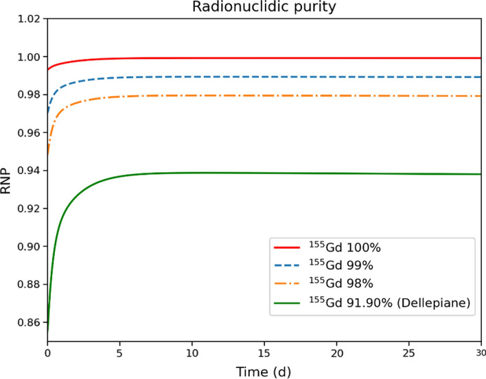
Time-evolution of 155Tb radionuclidic purity for the different target enrichments. The curves start 1 min after EoB to eliminate the transient effects of the rapidly decaying products
Figure 6 shows the fraction of total activity of 154gTb, 156gTb, 156m1Tb, and 156m2Tb. Clearly, the amount of 156Gd in the target proportionally influences the activity of all three states of 156Tb. However, the main problem is represented by 156gTb because its long half-life is comparable to the one of 155Tb. Conversely, the production of 154gTb represents a minor issue, because of its shorter half-life. Moreover, in the selected energy region, any residual amount of 156Gd does not produce 154gTb. This contaminant is mainly produced from 154Gd, which appears, with a residue of 0.5%, only in the less enriched target.
Fig. 6.
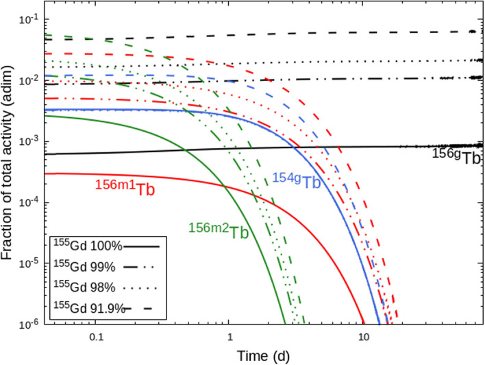
Fraction of total activity for the main contaminants 154gTb (blue curves), 156gTb (black curves), 156m1Tb (red curves), 156m2Tb (green curves), for different enrichment of the 155Gd targets
In addition, we have evaluated the isotopic purities and they correspond to 93.9, 97.9, 98.9, and 99.9% for the target enrichments of 91.90, 98, 99, and 100%, respectively, 96 h after the EoB.. This implies that the production route is essentially carrier-free, without the presence of contaminants, including stable or long-lived ones.
Organ absorbed doses and effective dose due to XXXTb-cm09 injection
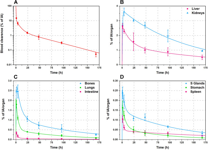
Biokinetics curves were obtained plotting the radiopharmaceutical concentration corrected by the radioactive decay vs. time for each source organ of the ICRP 89 male phantom of 73 kg [31]. The total volume of the blood (5110 ml) was obtained using the specific volume value of 70 ml/kg. Figure 7 shows a fast blood clearance followed by a quick radiopharmaceutical uptake by the main organs, with a slow wash-out. Liver and kidneys were the organs with slower clearance.
Fig. 7.
Time-activity curves of Tb-cm09 in the main source organs: symbols show experimental data obtained from biodistribution studies; the lines depict fitted time-activity curves
The number of disintegrations in the source organs, calculated by assuming that the injected radiopharmaceutical was labelled with only one of the radioisotopes 154gTb, 155Tb, 156gTb, 156m1Tb and 156m2Tb, are reported in Table 3. These are the main radionuclides expected to be produced by proton irradiation of 155Gd-enriched targets. The dosimetric properties of 154m1Tb and 154m2Tb were not assessed because these two metastable states are not included in the OLINDA software. However, at an irradiation energy of 10.5 MeV, 154m1Tb and 154m2Tb production is essentially due to the presence of 154Gd in the target, not considered in the case of 155Gd target enrichment ≥ 98%. Besides, since the energy of these two metastable states is very close to that of the ground state, their contribution to the absorbed dose occurs mainly through the decay of the ground state, properly taken into account through the application of the Bateman equations.
Table 3.
Number of nuclear transitions (MBq × h/MBq) in source organs per unit administered activity of xxxTb-cm09 for male ICRP 89 human phantom
| Organ/tissue | 154gTb-cm09 | 155Tb-cm09 | 156gTb-cm09 | 156m1Tb-cm09 | 156m2Tb-cm09 |
|---|---|---|---|---|---|
| Heart contents | 0.118 | 0.168 | 0.168 | 0.122 | 0.075 |
| Lung | 0.177 | 0.391 | 0.392 | 0.190 | 0.079 |
| Spleen | 0.014 | 0.048 | 0.048 | 0.016 | 0.005 |
| Kidneys | 0.912 | 2.885 | 2.891 | 1.012 | 0.244 |
| Stomach | 0.020 | 0.043 | 0.044 | 0.022 | 0.008 |
| Left colon | 0.0031 | 0.0072 | 0.0072 | 0.0033 | 0.0014 |
| Small intestine | 0.0146 | 0.0335 | 0.0335 | 0.0156 | 0.0066 |
| Right colon | 0.0062 | 0.0144 | 0.0144 | 0.0067 | 0.0028 |
| Rectum | 0.0031 | 0.0072 | 0.0072 | 0.0033 | 0.0014 |
| Liver | 0.498 | 1.443 | 1.446 | 0.545 | 0.166 |
| Cortical bone | 0.405 | 0.991 | 0.993 | 0.438 | 0.152 |
| Salivary glands | 0.041 | 0.112 | 0.113 | 0.044 | 0.014 |
| Remaining | 3.985 | 8.819 | 8.833 | 4.274 | 1.719 |
For the dosimetric assessment, the total activity in the intestines was distributed in left colon, small intestine, right colon and rectum, according to their mass ratio with respect to the total intestine mass. The mean maximum volume of blood that can be contained in the four chambers of the heart, two atria and two ventricles, of an adult man is about 505 ml [32], therefore, just the 10% of the blood activity was assigned to the “heart contents” and the rest to “Remaining”. The activity in mouse muscle was extrapolated to humans considering that the human muscle is 40% of total body weight. Muscle activity was also assigned to the”Remaining” organs when performing calculations with OLINDA 2.2.3, because this tissue is not included in the source organs of the ICRP 89 phantom model.
For each xxxTb-radiopharmaceutical, the organs with the highest number of disintegrations were the kidneys, followed by the liver and the cortical bone. Comparing the different radioisotopes, it was found that the radionuclides with the highest number of disintegrations were 155Tb and 156gTb, due to their long half-life. 156m2Tb showed the lowest number of disintegrations because of its relatively short half-life.
The absorbed doses per unit of administered activity in the main human male organs, calculated for different xxxTb-cm09, are reported in Table 4. For all the radioisotopes, the organs receiving the highest absorbed dose are the kidneys, followed by the osteogenic cells for 155Tb, 156m1Tb and 156m2Tb, and by the adrenals for 154gTb and 156gTb. 156gTb is the radioisotope giving the highest values of absorbed doses, due to its long half-life and the large amount of energy emitted per decay (see Table 1). The absorbed doses due to 154gTb administration are also higher than those of 155Tb, because, even if the 154gTb half-life is much shorter than that of 155Tb, the energy emitted per decay is about ten times higher. The absorbed doses due to 156m1Tb and 156m2Tb are about one order of magnitude lower than those of 155Tb. The ED after administration of 156m1Tb-cm09 and 156m2Tb-cm09 are lower than the one of 155Tb-cm09, consequently their presence as contaminants in the produced 155Tb will not increase the dose to the patient. In contrast, when the radiopharmaceutical is labelled with 156gTb or 154gTb, the ED is 5.9 and 2.4 times the ED values of 155Tb-cm09. Therefore, to guarantee a dose increment per unit of activity administered lower than 10%, the presence of 156gTb or 154gTb as contaminants must be lower than about 2% or 7%, respectively.
Table 4.
Organ absorbed doses (mGy/MBq) and ED values (mSv/MBq) per unit administered activity calculated for xxxTb-cm09 for male ICRP 89 phantoms
| Target organ | 154gTb-cm09 | 155Tb-cm09 | 156gTb-cm09 | 156m1Tb-cm09 | 156m2Tb-cm09 |
|---|---|---|---|---|---|
| Adreanals | 1.68E-01 | 6.56E-02 | 4.69E-01 | 6.78E-03 | 1.44E-03 |
| Brain | 2.61E-02 | 9.25E-03 | 5.68E-02 | 1.38E-03 | 1.22E-03 |
| Esophagus | 4.93E-02 | 1.52E-02 | 1.10E-01 | 1.98E-03 | 1.24E-03 |
| Eyes | 2.58E-02 | 9.09E-3 | 5.57E-02 | 1.36E-03 | 1.22E-03 |
| Gallbladder wall | 7.75E-02 | 2.66E-02 | 1.97E-01 | 3.05E-03 | 1.27E-03 |
| Left colon | 5.91E-02 | 2.04E-02 | 1.44E-01 | 2.50E-03 | 1.71E-03 |
| Small intestine | 5.10E-02 | 1.67E-02 | 1.19E-01 | 2.11E-03 | 1.71E-03 |
| Stomach wall | 5.60E-02 | 1.87E-02 | 1.29E-01 | 2.49E-03 | 2.05E-03 |
| Right colon | 5.59E-02 | 1.87E-02 | 1.34E-01 | 2.31E-03 | 1.71E-03 |
| Rectum | 4.13E-02 | 1.33E-02 | 9.11E-02 | 1.80E-03 | 1.70E-03 |
| Heart wall | 6.75E-02 | 2.14E-02 | 1.35E-01 | 3.64E-03 | 4.98E-03 |
| Kidneys | 3.92E-01 | 3.48E-01 | 1.29E00 | 4.27E-02 | 3.99E-02 |
| Liver | 1.01E-01 | 5.06E-02 | 2.77E-01 | 6.10E-03 | 4.74E-03 |
| Lungs | 4.78E-02 | 1.99E-02 | 1.10E-01 | 3.00E-03 | 3.36E-03 |
| Pancreas | 6.14E-02 | 1.97E-02 | 1.46E-01 | 2.37E-03 | 1.22E-03 |
| Prostate | 4.15E-02 | 1.23E-02 | 9.27E-02 | 1.55E-03 | 1.22E-03 |
| Salivary Glands | 6.42E-02 | 4.67E-02 | 1.74E-01 | 6.57E-03 | 8.32E-03 |
| Red Marrow | 4.11E-02 | 1.15E-02 | 9.39E-02 | 1.41E-03 | 1.03E-03 |
| Osteogenic cells | 6.00E-02 | 8.61E-02 | 1.75E-01 | 1.78E-02 | 1.04E-02 |
| Spleen | 7.72E-02 | 3.27E-02 | 2.12E-01 | 3.53E-03 | 1.75E-03 |
| Testes | 2.92E-02 | 8.58E-03 | 6.09E-02 | 1.20E-03 | 1.21E-03 |
| Thymus | 4.31E-02 | 1.22E-02 | 8.86E-02 | 1.75E-03 | 1.24E-03 |
| Thyroid | 3.55E-02 | 1.10E-02 | 7.60E-02 | 1.54E-03 | 1.22E-03 |
| Urinary bladder wall | 3.82E-02 | 1.12E-02 | 8.24E-02 | 1.48E-03 | 1.22E-03 |
| Total body | 3.57E-02 | 1.36E-02 | 8.13E-02 | 1.91E-03 | 1.74E-03 |
| Effective dose | 4.44E-02 | 1.86E-02 | 1.09E-01 | 2.47E-03 | 2.06E-03 |
The dose increase due to 155Tb contaminants obtained with different levels of target enrichment
The DI due to 155Tb-cm09 obtained with different levels of target enrichment is plotted in Fig. 8 vs. time t post-production. The DI is well above 25% for the 91.9% target enrichment and this confirms that the isotopic contamination of that target is inadequate for medical purposes. The ideal case of a 155Gd target with 100% enrichment is described by the solid red line. Here, the RNP is very close to 100% and the DI is negligible. Quite interesting are the dashed and dot-dashed curves obtained with 99% and 98% enrichment. The former reaches a 5% DI, while the latter remains within the 10% limit.
Fig. 8.
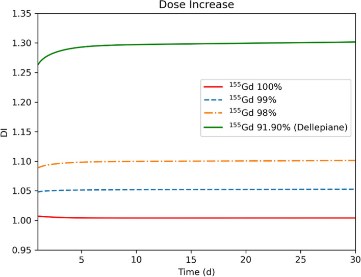
Dose increase for 155Tb-cm09 radiopharmaceutical labelled at different times after the end of irradiation of different 155Gd-enriched targets
Impact on the image quality
The imaging quality of 155Tb produced with the different 155Gd-enriched targets was assessed at the EoB and 96 h later by considering the 156gTb and 154gTb yields reported in Table 2. The metastable states 156m1Tb, 156m2Tb and 154m1Tb were disregarded in the simulations because of their lower energy γ-ray emission.
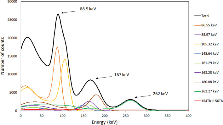
Using the data in Table 2, a γ-ray spectrum produced at the EoB from the irradiation of 100% 155Gd-enriched target was simulated, as shown in Fig. 9. The γ rays with the energies of 86.55 keV and 105.318 keV emitted from 155Tb and the low intensity 88.97 keV γ ray emitted by 156gTb are situated close with respect to the energy resolution of the imaging system and form a compound peak with a barycenter of 88.5 keV. Other four peaks from 155Tb with energies of 148.64 keV, 161.29 keV, 163.28 keV and 180.08 keV form a less intense compound peak with a barycenter of 167 keV. A smaller 155Tb peak at 262 keV (5.3%) is exposed to the higher energy γ rays from the isotopes of 154gTb and 156gTb only. For imaging purposes, the compound peak with the energy of 88.5 keV is most convenient, for its higher intensity, although the other two peaks at 167 keV and 262 keV can also be used for imaging.
Fig. 9.
Simulated spectrum for 155Tb, obtained immediately after the proton bombardment of 100% 155Gd-enriched target. The predicted observable spectrum is presented with a thicker black line
Table 5 shows the calculated Compton-to-peak ratio for the three principal peaks of 155Tb with respect to the γ-ray background, generated by the contaminant isotopes 156gTb and 154gTb. The analysis of the data reveals that in all cases the level of noise in the γ-ray images reconstructed using a 10% energy window around the barycenter of the peaks does not exceed 27%. For the intense peak at 88.5 keV the Compton-to-peak ratio remains within the interval of 19%– 23% for all enriched targets immediately after the EoB and no significant improvement could be achieved 96 h later. Lower Compton-to-peak values, slightly below 20%, are obtained for the compound peak at 167 keV. This suggests its use as a single peak for image reconstruction in some particular cases. The lower intensity of the peak at 262 keV makes the ratio more affected by the presence of the contaminant nuclides 156gTb and 154gTb, although its maximum value still remains below 27%. However, it is worth noting that the present image quality estimation is made for point-like γ-ray sources in air, hence the values quoted refer to the imaging system only and do not take into account Compton scattering inside the tissues. To estimate this contribution for the case of small animals imaging, a water phantom was used in our laboratory, obtaining a noise increase level smaller than 5%.
Table 5.
Compton-to-peak ratio calculated from the simulation of 155Tb γ-ray spectra, obtained for 100%, 99% and 98% enrichment of 155Gd targets, at the EoB and 96 h later
| Peak energy (keV) | Compton-to-peak ratio EoB | Compton-to-peak ratio 96 h | ||||
|---|---|---|---|---|---|---|
| 100% 155Gd | 99% 155Gd | 98% 155Gd | 100% 155Gd | 99% 155Gd | 98% 155Gd | |
| 88.5 | 19.44% | 20.99% | 22.59% | 18.89% | 21.52% | 22.61% |
| 167 | 10.24% | 14.54% | 19.06% | 8.91% | 15.95% | 19.32% |
| 262 | 5.84% | 16.12% | 26.9% | 1.71% | 19.8% | 26.55% |
Discussion
The use of low-energy proton beams on 155Gd-enriched target represents a promising possibility for the production of 155Tb, however the isotopic purity of the target strongly influences the amount of co-produced radioisotopes. The most problematic contaminant is 156gTb, because its half-life is comparable to that of 155Tb, and its γ emissions have severe impact on the dose released and on the quality of the SPECT images. For these reasons, its production should be kept as low as possible. The co-production of 156gTb can be minimized by limiting the 156Gd content in the target. In particular, a RNP higher than 98.5% can be obtained starting from about 30 h after the EoB with a 156Gd content lower than 1%. For longer times, the RNP levels at 99%. However, the 99% value is not an established limit and a smaller one can be tolerated if the DI due to the presence of contaminant radioisotopes is low. By assuming a 10% limit in DI as an acceptable condition for the contamination of a production route [33], the maximum content of 156Gd in the target could be increased to 2%. It is worth to note that in this case the DI does not exceed the 10% value in the entire time range shown in Fig. 8, namely from 0 to 30 days. Therefore the available activity can be utilized much earlier with respect to the timing when the RNP is close to 98%, and specifically, the product can be available right after the time needed to perform a radiochemical purification [9].
The main limits of these results is that extreme care should always be taken when using biodistribution data from animals to predict absorbed doses to humans and that the DI due to contaminant radioisotopes must be determined for each specific radiopharmaceutical, because it directly depends on its biodistribution and kinetics characteristics [34]. Taking into account these limitations, the dosimetric results obtained in this study should be interpreted as useful to have an idea of the contribution of Tb impurities to the absorbed doses imparted after injection of 155Tb-cm09 and to select the 155Gd target requirements for 155Tb production, however a more precise estimation is possible only using biodistribution data collected on humans.
Enriched 155Gd is currently commercially available from various companies (ISOFLEX, CIL, AMT, etc.) with 155Gd purity greater than 90%, but with a 156Gd component larger than 2%, not yet sufficient for the production of 155Tb with the necessary purity for medical application. Gd-isotopes separation is performed commercially using the "Calutron method", namely using a very large mass spectrometer for electro-magnetic separation. Since this technique is very energy inefficient, the prices for the enriched Gd material are high. However, it is possible to tune the Calutron to produce highly enriched 155Gd with isotopic enrichment > 98.0%, or with 156Gd content < 2%. To make a producer interested in changing the operational plan implies an economical return and profitability, which presently is not yet expected by the isotope-producer companies (Allan Pashkovski, Managing Director, Isoflex, Private communications, 6th July, 2023). A scenario of a diffuse worldwide production of 155Tb by hospital cyclotrons may be much more favorable of economical return for the producer companies.
The targeted therapeutic agents currently used in nuclear medicine are based on biological structures such as peptides, monoclonal antibodies and their fragments, coupled to α and β- emitters radionuclides. These agents, due to their nature, exhibit maximum absorption time range from several hours to a few days after injection. To perform SPECT imaging of these radiocomplexes, a radiopharmaceutical labeled with a radionuclide having half-life comparable to this maximum absorption time is required [35]. For this reason, 111In is the only radionuclide currently used in the clinic to perform SPECT imaging of these radiocomplexes, since its half-life (2.8 d) is long enough for image acquisition even several days after the radiocomplex administration. Therefore, to evaluate the 155Tb potential as matched pair for targeted theranostic agents labelled with 149Tb and 161Tb, it is interesting to compare its imaging and dosimetric properties with those of 111In.
Imaging simulations were performed for pure 111In using the main photon emissions at energies of 171 keV (91%) and 245 keV (94%) to compare its Compton-to-peak ratio with those of 155 Tb. The Compton-to-peak ratio of the peak at 171 keV, the closest in energy to the 88.5 keV and 167 keV peaks of 155Tb, turned out to be 22.6%, which is slightly larger than the values obtained for 155Tb, although they are comparable within 10% of uncertainty interval. These results are supported by the excellent imaging properties of 155Tb reported by Favaretto [9], where a spatial resolution up to 1 mm was obtained from a Derenzo phantom. Moreover, Müller et al. [5] published a comparison between SPECT images of Derenzo phantoms filled with solutions of pure 155Tb (2.6 MBq) and 111In (4 MBq), demonstrating comparable spatial resolution and the ability to produce equal images with lower activity of 155Tb.
To evaluate the influence of the physical properties of the labeling radionuclide, the dosimetric properties of 111In-labelled-cm09 were estimated with OLINDA by assuming the same biodistribution of Tb-cm09 compound. The calculated ED of 111In-cm09 was 0.0217 mSv/MBq, very similar to that of 155Tb-cm09 (0.0186 mSv/MBq). These results are a consequence of the fact that, despite the half-life of 155Tb is almost 2 times longer than that of 111In, its total energy emission per decay is about 2 times lower compared to 111In (0.4409 MeV/nt). It should therefore be expected that the imaging and dosimetric properties of 155Tb-radiocomplexes are comparable and even better than those of 111In ones.
Conclusions
155Tb can be produced with a quality suitable for medical applications using low-energy proton beams and 155Gd-enriched targets, if the 156Gd impurity content does not exceed 2%. Under these conditions, the dose increase due to the presence of contaminant radioisotopes remains below the 10% limit and good quality images, comparable to those of 111In, are guaranteed.
Acknowledgements
The authors thank Allan Pashkovski for private communications. One of the authors (N. Uzunov) would like to acknowledge the support received from the Abdus Salam International Center for Theoretical Physics Trieste, Italy, and in particular the Program for Training and Research in Italian Laboratories (TRIL). F. Barbaro thanks for a fellowship from the Department of Physics and Astronomy, University of Padua, funded by "Bando PARD-2023" (CUP C93C23004310005). This work was carried out as part of the activities of the APHRODITE-155 (P2022TTCAZ) project, funded by the national call PRIN PNRR 2022.
Abbreviations
- DI
Dose increase
- ED
Effective dose
- EoB
End of bombardment
- GDH
Geometry dependent hybrid
- LD
Level-density
- OLINDA
Organ Level Internal Dose Assessment
- PE
Pre-equilibrium
- PET
Positron emission tomography
- RNP
Radionucludic purity
- SPECT
Single photon emission computed tomography
Author contributions
LC, LDN, and LMA contributed to the study conception and design. FB and LC performed cross-section calculation and assessed yields, isotopic and radionuclidic purity for targets with different compositions. LDN, LMA, and FB obtained biokinetic curves and performed dosimetric studies. NU evaluated the impact of impurities on image quality. The manuscript was written, read and approved by all authors.
Funding
This work was supported by the REMIX-CSN5 research program (2021–2023), funded by Italian National Institute of Nuclear Physics (INFN) as part of the activities of the LARAMED project of the INFN-Legnaro National Laboratories. The work of LC and FB was also funded by NUCSYS-CSN4 INFN research program.
Availability of data and materials
The datasets used and/or analysed during the current study are available from the corresponding author on reasonable request.
Declarations
Ethics approval and consent to participate
Not applicable.
Consent for publication
Not applicable.
Competing interests
The authors declare that they have no competing interests.
Footnotes
Publisher's Note
Springer Nature remains neutral with regard to jurisdictional claims in published maps and institutional affiliations.
Co-senior authors: Laura De Nardo and Laura Melendez-Alafort.
References
- 1.Müller C, Zhernosekov K, Köster U, Johnston K, Dorrer H, Hohn A, et al. A unique matched quadruplet of terbium radioisotopes for PET and SPECT and for α- and β–radionuclide therapy: an in vivo proof-of-concept study with a new receptor-targeted folate derivative. J Nucl Med. 2018;53(12):1951–9. 10.2967/jnumed.112.107540 [DOI] [PubMed] [Google Scholar]
- 2.Sadler AWE, Hogan L, Fraser B, Rendina LM. Cutting edge rare earth radiometals: prospects for cancer theranostics. EJNMMI Radiopharm Chem. 2022. 10.1186/s41181-022-00173-0. 10.1186/s41181-022-00173-0 [DOI] [PMC free article] [PubMed] [Google Scholar]
- 3.Price EW, Orvig C. Matching chelators to radiometals for radiopharmaceuticals. Chem Soc Rev Chem Soc Rev. 2014;260(43):260–90. 10.1039/C3CS60304K [DOI] [PubMed] [Google Scholar]
- 4.Israel O, Pellet O, Biassoni L, De Palma D, Estrada-Lobato E, Gnanasegaran G, et al. Two decades of SPECT/CT – the coming of age of a technology: an updated review of literature evidence. Eur J Nucl Med Mol Imaging. 2019;46(10):1990–2012. 10.1007/s00259-019-04404-6 [DOI] [PMC free article] [PubMed] [Google Scholar]
- 5.Müller C, Fischer E, Behe M, Köster U, Dorrer H, Reber J, et al. Future prospects for SPECT imaging using the radiolanthanide terbium-155 — production and preclinical evaluation in tumor-bearing mice. Nucl Med Biol. 2014;41:e58-65. 10.1016/j.nucmedbio.2013.11.002 [DOI] [PubMed] [Google Scholar]
- 6.Dmitriev PP, Molin GA, Dmitrieva ZP. Production of155Tb for nuclear medicine in the reactions 155Gd(p, n),156Gd(p,2n), and 155Gd(d,2n). Sov At Energy. 1989;66(6):470–2. 10.1007/BF01123521 [DOI] [Google Scholar]
- 7.Vermeulen C, Steyn GF, Szelecsényi F, Kovács Z, Suzuki K, Nagatsu K, et al. Cross sections of proton-induced reactions on natGd with special emphasis on the production possibilities of 152Tb and 155Tb. Nucl Instru Methods Phys Res Sect B Beam Interact with Mater Atoms. 2012;275:24–32. 10.1016/j.nimb.2011.12.064. 10.1016/j.nimb.2011.12.064 [DOI] [Google Scholar]
- 8.Formento-Cavaier R, Haddad F, Alliot C, Sounalet T, Zahi I. New excitation functions for proton induced reactions on natural gadolinium up to 70 MeV with focus on 149Tb production. Nucl Instruments Methods Phys Res Sect B Beam Interact with Mater Atoms. 2020;478(May):174–81. 10.1016/j.nimb.2020.06.029 [DOI] [Google Scholar]
- 9.Favaretto C, Talip Z, Borgna F, Grundler PV, Dellepiane G, Sommerhalder A, et al. Cyclotron production and radiochemical purification of terbium-155 for SPECT imaging. EJNMMI Radiopharm Chem. 2021;6(1):37. 10.1186/s41181-021-00153-w. 10.1186/s41181-021-00153-w [DOI] [PMC free article] [PubMed] [Google Scholar]
- 10.Dellepiane G, Casolaro P, Favaretto C, Grundler PV, Mateu I, Scampoli P, et al. Cross section measurement of terbium radioisotopes for an optimized 155Tb production with an 18 MeV medical PET cyclotron. Appl Radiat Isot. 2022;184(January):110175. 10.1016/j.apradiso.2022.110175. 10.1016/j.apradiso.2022.110175 [DOI] [PubMed] [Google Scholar]
- 11.Müller C, Reber J, Haller S, Dorrer H, Bernhardt P, Zhernosekov K, et al. Direct in vitro and in vivo comparison of 161Tb and 177Lu using a tumour-targeting folate conjugate. Eur J Nucl Med Mol Imaging. 2014;41(3):476–85. 10.1007/s00259-013-2563-z. 10.1007/s00259-013-2563-z [DOI] [PubMed] [Google Scholar]
- 12.Webster B, Ivanov P, Russell B, Collins S, Stora T, Ramos JP, et al. Chemical purification of terbium-155 from pseudo-isobaric impurities in a mass separated source produced at CERN. 2019;(April):1–9. [DOI] [PMC free article] [PubMed]
- 13.Fiaccabrino DE, Kunz P, Radchenko V. Potential for production of medical radionuclides with on-line isotope separation at the ISAC facility at TRIUMF and particular discussion of the examples of 165Er and 155Tb. Nucl Med Biol. 2021;94–95:81–91. 10.1016/j.nucmedbio.2021.01.003. 10.1016/j.nucmedbio.2021.01.003 [DOI] [PubMed] [Google Scholar]
- 14.Steyn GF, Vermeulen C, Szelecsényi F, Kovács Z, Hohn A, Van Der Meulen NP, et al. Cross sections of proton-induced reactions on 152Gd, 155Gd and 159Tb with emphasis on the production of selected Tb radionuclides. Nucl Instruments Methods Phys Res Sect B Beam Interact with Mater Atoms. 2014;319:128–40. 10.1016/j.nimb.2013.11.013. 10.1016/j.nimb.2013.11.013 [DOI] [Google Scholar]
- 15.Tárkányi F, Hermanne A, Ditrói F, Takács S, Ignatyuk AV. Activation cross-sections of longer lived radioisotopes of proton induced nuclear reactions on terbium up to 65 MeV. Appl Radiat Isot. 2017;127(May):7–15. 10.1016/j.apradiso.2017.04.030. 10.1016/j.apradiso.2017.04.030 [DOI] [PubMed] [Google Scholar]
- 16.Barbaro F, Canton L, Carante MP, Colombi A, DeNardo L, Fontana A, et al. The innovative 52gMn for positron emission tomography (PET) imaging: Production cross section modeling and dosimetric evaluation. Med Phys. 2023;50(3):1843–54. 10.1002/mp.16130 [DOI] [PubMed] [Google Scholar]
- 17.Goriely S, Hilaire S, Koning AJ. Improved reaction rates for astrophysics applications with the TALYS reaction code. AIP Conf Proc. 2009;1090:629–30. 10.1063/1.3087118 [DOI] [Google Scholar]
- 18.Konobeyev AY, Fischer U, Pereslavtsev PE, Koning A, Blann M. Implementation of GDH model in TALYS-1 . 7 code.
- 19.Blann M. Importance of the nuclear density distribution on pre-equilibrium decay. Phys Rev Lett. 1972;28(12):757–9. 10.1103/PhysRevLett.28.757 [DOI] [Google Scholar]
- 20.Colombi A, Carante MP, Barbaro F, Canton L, Fontana A. Production of high-purity 52gMn from natV targets with alpha beams at cyclotrons. Nucl Technol. 2021. 10.1080/00295450.2021.1947122. 10.1080/00295450.2021.1947122 [DOI] [Google Scholar]
- 21.Canton L, Fontana A. Nuclear physics applied to the production of innovative radiopharmaceuticals. Eur Phys J Plus. 2020;135(9):1–21. 10.1140/epjp/s13360-020-00730-z. 10.1140/epjp/s13360-020-00730-z [DOI] [Google Scholar]
- 22.Leo WR. Techniques for Nuclear and Particle Physics Experiments. Berlin: Springer; 1994. [Google Scholar]
- 23.ICRP 2008. Nuclear Decay Data for Dosimetric Calculations. ICRP Publication 107. Ann. ICRP 38 (3). 2008. [DOI] [PubMed]
- 24.NuDat 3 - National Nuclear Data. https://www.nndc.bnl.gov/nudat3/.
- 25.Sparks R, Aydogan B. Comparison of the effectiveness of some common animal data scaling techniques in estimating human radiation dose. In: Sixth international radiopharmaceutical dosimetry symposium [Internet]. 1999. p. 705–16. Available from: https://www.osti.gov/servlets/purl/684479.
- 26.Stabin MG, Sparks RB, Crowe E. OLINDA/EXM: the second-generation personal computer software for internal dose assessment in nuclear medicine. J Nucl Med. 2005;46:1023–7. [PubMed] [Google Scholar]
- 27.Meléndez-Alafort L, Rosato A, Ferro-Flores G, Penev I, Uzunov N. Development of a five-compartmental model and software for pharmacokinetic studies. Comptes Rendus L’Academie Bulg des Sci. 2017;70(12):1649–54. [Google Scholar]
- 28.De Nardo L, Pupillo G, Mou L, Esposito J, Rosato A, Meléndez-Alafort L. A feasibility study of the therapeutic application of a mixture of 67/64 Cu radioisotopes produced by cyclotrons with proton irradiation. Med Phys. 2022;49(4):2709–24. 10.1002/mp.15524. 10.1002/mp.15524 [DOI] [PMC free article] [PubMed] [Google Scholar]
- 29.ICRP 2007. The 2007 Recommendations of the International Commission on Radiological Protection. ICRP Publication 103. Ann. ICRP 37 (2–4) [Internet]. 2007. Available from: http://www.icrp.org/publication.asp?id=ICRP Publication 103 [DOI] [PubMed]
- 30.Dellepiane G, Casolaro P, Gottstein A, Mateu I, Scampoli P, Braccini S. Experimental assessment of nuclear cross sections for the production of Tb radioisotopes with a medical cyclotron. Appl Radiat Isot. 2023;200(July):110969. 10.1016/j.apradiso.2023.110969. 10.1016/j.apradiso.2023.110969 [DOI] [PubMed] [Google Scholar]
- 31.ICRP 2002. Basic anatomical and physiological data for use in radiological protection reference values. ICRP Publication 89. Ann. ICRP 32 (3–4). [PubMed]
- 32.Kawel-Boehm N, Hetzel SJ, Ambale-Venkatesh B, Captur G, Francois CJ, Jerosch-Herold M, et al. Reference ranges (“normal values”) for cardiovascular magnetic resonance (CMR) in adults and children: 2020 update. J Cardiovasc Magn Resonance BioMed Central. 2020. 10.1186/s12968-020-00683-3. 10.1186/s12968-020-00683-3 [DOI] [PMC free article] [PubMed] [Google Scholar]
- 33.DeNardo L, Pupillo G, Mou L, Furlanetto D, Rosato A, Esposito J, et al. Preliminary dosimetric analysis of DOTA-folate radiopharmaceutical radiolabelled with 47Sc produced through natV(p, x)47Sc cyclotron irradiation. Phys Med Biol. 2021;66(2):025003. 10.1088/1361-6560/abc811. 10.1088/1361-6560/abc811 [DOI] [PubMed] [Google Scholar]
- 34.Meléndez-Alafort L, Ferro-Flores G, De Nardo L, Bello M, Paiusco M, Negri A, et al. Internal radiation dose assessment of radiopharmaceuticals prepared with cyclotron-produced 99m Tc. Med Phys. 2019;46(3):1437–46. 10.1002/mp.13393 [DOI] [PubMed] [Google Scholar]
- 35.Lo WL, Lo SW, Chen SJ, Chen MW, Huang YR, Chen LC, et al. Molecular imaging and preclinical studies of radiolabeled long-term rgd peptides in u-87 mg tumor-bearing mice. Int J Mol Sci. 2021;22(11). [DOI] [PMC free article] [PubMed]
Associated Data
This section collects any data citations, data availability statements, or supplementary materials included in this article.
Data Availability Statement
The datasets used and/or analysed during the current study are available from the corresponding author on reasonable request.


