

ERRATUM
This corrects the article published in the European Journal of Histochemistry 2014;58:2262, where the micrographs of panels C, D and F of Figure 1 partially overlap, as they where taken from the same microscope slide; the same did occur for the micrographs of panel CON and NC in Figure 6A. This was due to an unintentionally made material mistake.
The corresponding quantitative data were presented in Table 1 and Figure 3B of the same article, based on the morphological and morphometric evaluation of tens of images: this confirms the validity in the data presented and make the paper’s discussion and conclusions fully reliable.
The correct Figure 1 and Figure 6 and relevant legends are shown below.
We apologize for any inconvenience caused and appreciate the opportunity to correct and clarify.
Figure 1.
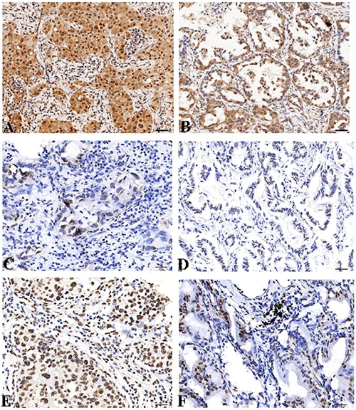
The protein expression of MMP-7 and COX-2 in LAC tissues (magnification ×200). LAC tissues were immunohistochemically stained with the anti-MMP-7 and COX-2 antibodies and classified as positive expression (A) and negative expression (C). Adjacent non-cancer tissues were immunohistochemically stained with an anti-MMP-7 antibody and classified as positive expression (B) and negative expression (D). COX-2 was highly expressed in LAC tissues (E) and lowly expressed in ANCT (F). Positive immunostaining of MMP-7 and COX-2 was mainly localized in the cytoplasm of tumor and tissue cells. Scale bars: 75 µm.
Figure 6.
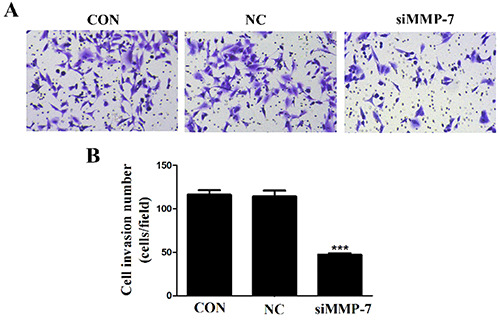
The effect of MMP-7 knockdown on cell invasion. (A) Transwell assay was performed to determine cell invasion; scale bars: 75 µm. (B) Cell invasive potential was markedly weakened in siMMP-7 group compared with the CON and NC groups (***p<0.001).