Abstract
Juvenile myelomonocytic leukemia (JMML) is a rare, generally aggressive myeloproliferative neoplasm affecting young children. It is characterized by granulomonocytic expansion, with monocytosis infiltrating peripheral tissues. JMML is initiated by mutations upregulating RAS signaling. Approximately 10% of cases remain without an identified driver event. Exome sequencing of two unrelated cases of familial JMML of unknown genetics and analysis of the French JMML cohort identified 11 patients with variants in SH2B3, encoding LNK, a negative regulator of the JAK-STAT pathway. All variants were absent from healthy population databases, and the mutation spectrum was consistent with a loss of function of the LNK protein. A stoploss variant was shown to affect both protein synthesis and stability. The other variants were either truncating or missense, the latter affecting the SH2 domain that interacts with activated JAK. Of the 11 patients, eight from five families inherited pathogenic bi-allelic SH2B3 germline variants from their unaffected heterozygous parents. These children represent half of the cases with no identified causal mutation in the French cohort. They displayed typical clinical and hematologic features of JMML with neonatal onset and marked thrombocytopenia. They had a hypomethylated DNA profile with fetal characteristics and did not have additional genetic alterations. All patients showed partial or complete spontaneous clinical resolution. However, progression to thrombocythemia and immunity-related pathologies may be of concern later in life. Bi-allelic SH2B3 germline mutations thus define a new condition predisposing to a JMML-like disorder, suggesting that JAK pathway deregulation is capable of initiating JMML, and opening new therapeutic options.
Introduction
Juvenile myelomonocytic leukemia (JMML) is a rare, aggressive myeloproliferative neoplasm affecting infants and young children. It is characterized by excessive granulomonocytic proliferation in bone marrow (BM) and peripheral blood (PB) leading to splenomegaly, leukocytosis with precursors in PB, monocytosis, infiltration of peripheral tissues with histiocytes and normal or moderately increased blast count.1,2
The natural course of JMML is generally rapidly fatal. The only potentially curative treatment is BM transplantation. However, the disease is characterized by a highly heterogeneous course, with a third of patients progressing to acute leukemia, while about 10% have indolent forms or even spontaneous resolutions.3
JMML arises from hematopoietic stem or myeloid progenitor cells.4-6 It is initiated by abnormal activation of RAS signaling, leading to in vitro hypersensitivity of myeloid progenitors to granulocyte-macrophage colony-stimulating factor.7 RAS pathway hyperactivation is due to mutations in genes encoding RAS proteins (NRAS, KRAS) or regulators (PTPN11, NF1 or CBL),8,9 which define genetic and clinical subgroups. More rarely, activating mutations affecting other small G proteins (RRAS, RRAS2, RIT1)10 have also been described, as well as fusions causing activation of genes coding for transducers upstream of the RAS pathway.11,12
A particular feature of JMML is its frequent occurrence in the context of a predisposing genetic syndrome, including Noonan syndrome, neurofibromatosis type 1 and CBL syndrome, which are due to a constitutional upregulation of the RAS-MAPK pathway (the so-called RASopathies).13-16 The identification of an activating mutation affecting the RAS pathway confirms the diagnosis of JMML in over 90% of cases.17 Nevertheless, there are still a small number of patients in whom no mutation has been identified (e.g., 5% in the French JMML cohort).
SH2B3 (OMIM 605093) encodes LNK (lymphocyte adaptor protein), an SH2-domain adaptor protein that acts as a negative regulator of intracellular signaling promoted by cytokines and growth factors activating the JAK-STAT pathway.18 Earlier work suggested that Lnk negatively regulates normal hematopoietic stem and progenitor cell expansion and self-renewal.19,20 Somatic SH2B3 variants have been reported in some JMML cases21 and Sh2b3-/-mice develop a myeloproliferative neoplasm.22,23 Germline mono-allelic,24-27 or more rarely bi-allelic28,29 LNK loss-of-function mutations have been previously identified in various hematologic diseases.
Here we provide evidence that bi-allelic germline inactivating variants in SH2B3 underlie a disease predisposing to a JMML-like disorder. We report 11 patients with SH2B3-associated JMML, including eight from five families who inherited bi-allelic pathogenic germline SH2B3 variants. SH2B3-associated JMML represents a new specific entity characterized by extremely early onset, probable persistence of fetal features, and partial or complete spontaneous clinical resolution. JMML in this group of patients is clearly distinct from other forms of JMML as a whole, but also from the disease associated with germ-line mono-allelic or somatic mutations of SH2B3 that have previously been reported.
Methods
Patients
The study included 234 patients with JMML referred to the French reference laboratory between 1995 and 2023, and one family with two probands from the Bambino-Gesù Children’s Hospital (OPBG) in Rome, Italy.
The diagnosis of JMML was based on clinical and hematologic findings, centrally reviewed cytomorphological examination of PB and BM smears, and genetic screening of genes known to initiate JMML or differential diagnoses (Wiskott-Aldrich syndrome, GATA2, osteopetrosis). Absence of BCR::ABL1 and KMT2A rearrangements was checked. In patients with no RAS-related variant, the presence of rare fusions involving FLT3, ALK, and PDGFRA/B was assessed by RNA-sequencing analysis. Karyotyping was performed using standard procedures. All patients fulfilled the World Health Organization’s criteria for JMML.
Ethical approval
The study was approved by the institutional review board of “Hôpitaux Universitaires Paris Nord Val-de-Seine” in Paris (Ref: 00006477) and the OPBG in Rome (Ref: 1702_ OPBG_2018), in accordance with the Helsinki declaration.
Samples
BM or PB samples were obtained from the patients at the time of diagnosis of JMML and from the patients’ parents. Mononuclear cells were isolated on a Ficoll gradient (Eurobio). The constitutional origin of the variant(s) was assessed using skin-derived fibroblasts. Healthy controls used for methylation and RNA-sequencing assays were males of 9, 10 and 12 years of age. The fetal BM sample was obtained from the product of a spontaneous abortion at 15 weeks of gestation. Online Supplementary Figure S1 shows the analyses performed in patients.
Genome-wide DNA array analysis
Genomic DNA samples were analyzed by single-nucleotide polymorphism array technologies using the Genome-Wide GeneChip Human SNP Array 6.0 (Affymetrix), and/or high-density array comparative genomic hybridization.12
Whole-exome sequencing and whole-genome sequencing
Target enrichment was performed using the SureSelect Human All Exon V4+UTRs or SureSelect AllExon V5 (Agilent Technologies, Santa Clara, CA, USA), and captured regions were sequenced with a HiSeq2000 or NextSeq500 instrument (Illumina, San Diego, CA, USA), as previously described.12,33 Details are provided in the Online Supplementary Material.
Sequencing the mRNA of mononucleated cells
Libraries were prepared with NEBNext Ultra II Directional RNA Library Prep Kit, according to the supplier’s recommendations. Paired-end 100-bp read sequencing was performed on a NovaSeq platform. Image analysis and base calling were performed using Illumina Real Time Analysis (3.4.4) with default parameters. The bioinformatic pipeline is described in the Online Supplementary Information. Unsupervised analyses of gene expression and differential expression were conducted using the Galileo tool (IntegraGen), as described in the Online Supplementary Information.
DNA methylation (capture enzymatic methyl sequencing)
Libraries were prepared using the Twist Targeted Methylation sequencing protocol system (Twist Bioscience) according to the guidelines and subjected to paired-end sequencing with 100-bp reads on a Novaseq 6000 platform (Online Supplementary Information). An unsupervised classification based on the 1,000 most variant 100 bp tiles was generated using hierarchical clustering (cosine distance, Ward method) in R software.
Further information
More details on the methods are provided in the Online Supplementary Material.
Results
Exome sequencing in two families segregating JMML identifies homozygous mutations in SH2B3
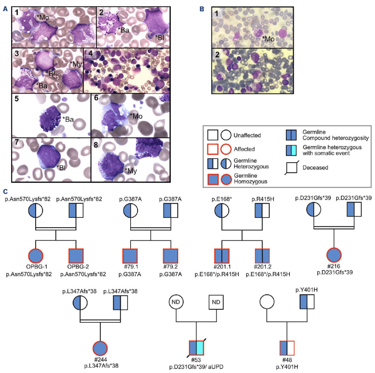
Trio exome sequencing was undertaken in proband #79.1 from the French JMML cohort, who suffered a syndromic neonatal JMML with no identified mutation. The analysis revealed the presence of a homozygous variant (c.1160G>C, p.Gly387Ala) in SH2B3 in the affected child. A review of the child’s records confirmed that he met the consensus clinical and hematologic criteria for the diagnosis of JMML, with hepatosplenomegaly, massive monocytosis (14×109/L), blasts, circulating myeloid precursors and thrombocytopenia (platelet count, 21×109/L) (Figure 1A; Online Supplementary Table S1). The BM smear showed hypercellularity associated with a decreased number of megakaryocytes. Spontaneous in vitro growth of myeloid progenitors was positive. The child’s brother (#79.2), born a few years later, also presented with neonatal JMML (Figure 1). Genetic analysis showed that he had inherited the familial SH2B3 mutation in a bi-allelic pattern.
Independently, whole-exome sequencing was performed in a family having two siblings diagnosed with an unclassified neonatal-onset syndromic JMML at the OPBG (Rome, Italy). The proband, #OPBG-2, was diagnosed at the age of 1 month, with elevated leukocyte counts, monocytosis (4.2×10⁹/L), thrombocytopenia (platelet count, 41×109/L) and 3% myeloblasts (Figure 1B; Online Supplementary Table S1). Immature monocytes and mild dysgranulopoiesis were observed in PB. Hypercellular BM, with granulocytic expansion, and 7% blasts, were consistent with a diagnosis of JMML. Similarly, his 5-year-old sister, #OPBG-1, had been diagnosed with JMML in the first days of life. Following delivery, she had splenomegaly and respiratory difficulties, which necessitated 24 h intubation. Her blood count showed leukocytosis (white blood cell count, 56×109/L), anemia (hemoglobin, 11.8 g/dL), and thrombocytopenia (platelet count, 35×109/L) (Online Supplementary Table S1). She experienced spontaneous resolution under active surveillance. Hypersensitivity of myeloid progenitor cells to granulocyte-macrophage colony-stimulating factor was observed in both children. Whole-exome analysis revealed the presence of a homozygous frameshift variant (c.1709dupA, p.Asn570LysfsTer82) in SH2B3 in both siblings.
Targeted sequencing of SH2B3 in the whole French JMML cohort
SH2B3 sequencing was then extended to the 234 patients of the French JMML cohort. Filtering out variants with a frequency >10-4 in the general population (GnomAD Non cancer v.2.1.1 database), at least one variant was identified in nine of the 234 (3.8%) patients, including six of the 12 (50%) mutation-negative cases (Online Supplementary Figure S2). Searching for mutations in patients’ fibroblasts and/or in their parents revealed that, in eight of the nine patients, the SH2B3 variants identified were germline. Of these eight patients, six (from 4 families) carried bi-allelic SH2B3 variants (Figure 1C). In the remaining two patients, the germline SH2B3 variant was mono-allelic. Whole-genome sequencing was performed and ruled out an alteration of the second SH2B3 allele.
Variant segregation in six families showed that germline variants were always inherited from unaffected heterozygous parents (Figure 1C). In four families, all affected children inherited bi-allelic SH2B3 variants, consistent with an autosomal recessive transmission of JMML predisposition. Three of these families were consanguineous. In family #48, the mono-allelic germline SH2B3 variant in the affected child was inherited from the unaffected father. Only one of the 234 patients (#209) had an exclusively somatic SH2B3 variant.
Assessing variant pathogenicity
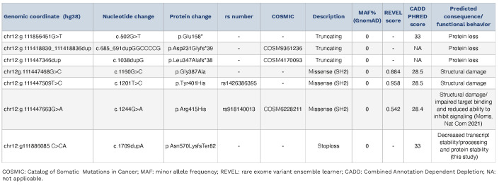
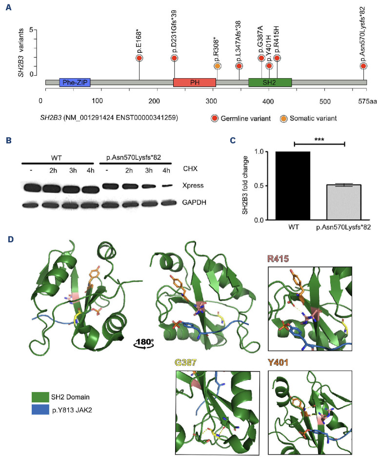
Including the Italian family, a total of seven different SH2B3 variants were identified. Among them, three were truncating (frameshift indels or stop codon), three were missense, and one caused protein elongation with a divergent C-terminus (Table 1; Figure 2A). Each variant was found as a single occurrence except p.Asp231Glyfs*39, which was found in two unrelated patients. All changes were predicted to be deleterious; none was referenced in the general population (Table 1) and, importantly, no homozygous truncating variant was referenced in gnomAD.
Based on the bi-allelic occurrence of variants, a loss-of-function effect was postulated as a functional consequence of the mutations identified. In line with this hypothesis, transient transfection experiments performed in 293T cells documented a dramatically reduced stability of the p.Asn570Lys*82 LNK mutant as compared to the wild-type protein (Figure 2B). Besides this effect at the protein level, assessment of SH2B3 mRNA levels by real-time polymerase chain reaction in primary fibroblasts obtained from patient #OPBG-2 and an unaffected control provided evidence of a significantly reduced amount in the former, which was suggestive of reduced stability of the transcript carrying the variant (Figure 2C). Overall, these findings indicate a disruptive impact in terms of transcript stability/processing and protein stability. Similarly, all missense variants were found to target the SH2 domain, which mediates binding to phosphorylated JAK and contains only rare polymorphisms in the general population (Online Supplementary Figure S3). All three variants were predicted to damage the structure of the SH2 domain of the LNK protein (Figure 2D), likely resulting in a decreased capacity to bind its JAK2 target. Collectively, these findings are consistent with a loss of function of SH2B3 as a driver mechanism underlying JMML.
Figure 1.
Patients’ cytomorphological data and pedigrees. (A, B) May-Grünwald-Giemsa-stained smears showing cytomorphological features at diagnosis patients with of juvenile myelomonocytic leukemia (JMML). (A) Patients #79.1 (A1-4) and #79.2 (A5-8). (B) Patient #OPBG (B1, 2). Peripheral blood smears (A1-3; A5-8; B1) show monocytes (*Mo), myeloid precursors (*My), dysmorphic basophils (*Ba) and undifferentiated myeloid blasts (*Bl). Bone marrow smears (A4; B2) show hypercellularity with myeloid blasts and discrete signs of dysgranulopoiesis. The erythrocyte lineage is decreased and the megakaryocytic lineage is absent. (C) Pedigrees of patients with germline SH2B3 variants. All children, but none of the heterozygous parents, had JMML. Germline variants (dark blue) were either bi-allelic (full circle/square; families 1 to 4) or mono-allelic (half circle/square; families 5 and 6) in the affected child. Somatic alteration is indicated in light blue. aUPD: acquired uniparental isodisomy; JMML: juvenile myelomonocytic leukemia; ND: not done.
Table 1.
Germline and somatic SH2B3 variants identified in patients with juvenile myelomonocytic Leukemia (NM_005475.3).
Figure 2.
SH2B3 variants and consequences on the LNK protein. (A) Lollipop plot showing the distribution of SH2B3/LNK variants over a schematic representation of the LNK protein. The PH domain enables interaction with cell membrane phospholipids and the SH2 domain binds to the phosphorylated tyrosines of target proteins. (B, C) A disease-causing SH2B3 stoploss frameshift mutation affects RNA and protein stability. (B) Protein stability was assessed in 293T cells transiently transfected with wild-type Xpress-tagged SH2B3 and mutant carrying the c.1709dupA (p.Asp570Lysfs82) mutation. After transfection (48 h), cells were treated with cycloheximide (20 [ig/mL) for the indicated time or left untreated. Protein levels were assessed by immunoblotting, using an anti-Xpress monoclonal antibody. GAPDH levels are shown to document equal loading of total proteins from cell lysates. Western blot from a representative experiment of three performed is shown. (C) Quantitative polymerase chain reaction analysis performed on RNA extracted from fibroblasts taken from patients and controls shows a significant decrease of SH2B3 expression level indicating RNA decay of the allele carrying the SH2B3 variant. Data in the graph indicate fold change of SH2B3 in patients’ fibroblasts over that in control cells (WT), set as 1. GAPDH was used as an endogenous control. Histograms show mean values ± standard deviation of three independent experiments, each performed in triplicate. The analysis of expression was performed calculating the fold change using the 2-ΔΔCt formula and the results were statistically analyzed by PRISM7, using a two-tailed unpaired t test with the Bonferroni correction. ***P<0.001. (D) Three-dimensional structure of the SH2 domain of the LNK protein with the JAK2 phosphopeptide pY813 modeled from crystallographic data in the report from Morris et al .36 (PDB:7R8W). The position of variant amino acids was determined by structural homology between mouse and human SH2 domains. aa: amino acids; CHX: cycloheximide; WT: wild-type.
Hematologic and clinical features of patients with SH2B3 mutations
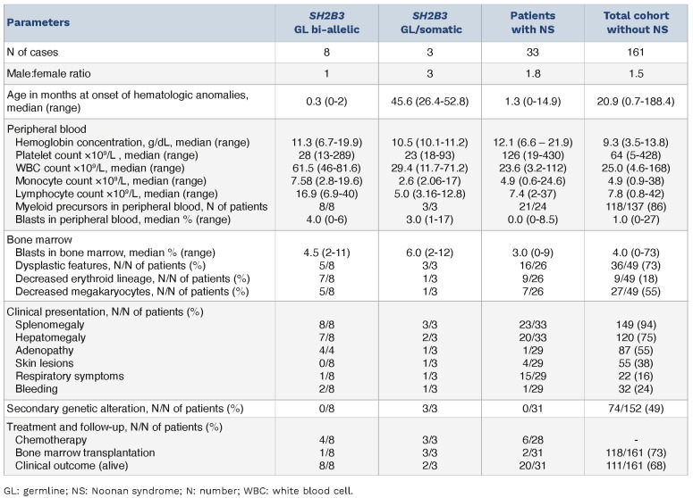
All patients presented with splenomegaly, high monocyte counts and hematologic data meeting the updated World Health Organization’s consensus criteria for the diagnosis of JMML31 (Online Supplementary Table S1). Patients with bi-allelic germline mutations (n=8) had higher counts of white blood cells (P<0.001), monocytes (P=0.045) and lymphocytes (P=0.004) than those with mono-allelic/somatic (n=3) variants or patients from the whole JMML cohort (Table 2). This also held true when these patients were compared to patients with Noonan syndrome-associated JMML (Table 2) or with any of the genetic subgroups of JMML taken separately.9 Notably, all patients with SH2B3 alterations but one displayed marked thrombocytopenia and the median platelet count was significantly lower than in the whole JMML cohort (28×109/L vs. 64×109/L; P=0.02), with frequent lack of megakaryocytes in patients’ BM (Online Supplementary Table S1). BM cellularity was normal or increased, with predominant granulocytic proliferation. Mild dysplastic features were observed in about half of the patients. Blast cells were moderately elevated but lower than 20% in blood and BM.
Table 2.
Clinical and hematologic features of patients with SH2B3-mutated juvenile myelomonocytic leukemia (JMML) in comparison with patients with Noonan syndrome-JMML47 (Strullu et al., updated) and with the whole French JMML cohort (excluding patients with Noonan syndrome).
In patients with bi-allelic germline mutations, JMML onset was neonatal. Most patients (5/6) were not transplanted and had spontaneous resolution.
In striking contrast, the median age of onset of JMML with mono-allelic germline or somatic SH2B3 variants was 3.8 years, which is later than patients with bi-allelic germline SH2B3 variants. The former had a markedly more severe course compared to the latter, and have all undergone transplantation.
Longitudinal follow-up of patients with bi-allelic SH2B3 germline variants shows that after an initial period of thrombocytopenia associated with profound impairment of the megakaryocytic lineage, the children’s platelet counts increased rapidly, reaching above-normal levels in several of them, with even thrombocytosis in patient #79.2 (Online Supplementary Figure S4).
Genetic makeup of SH2B3 mutated JMML
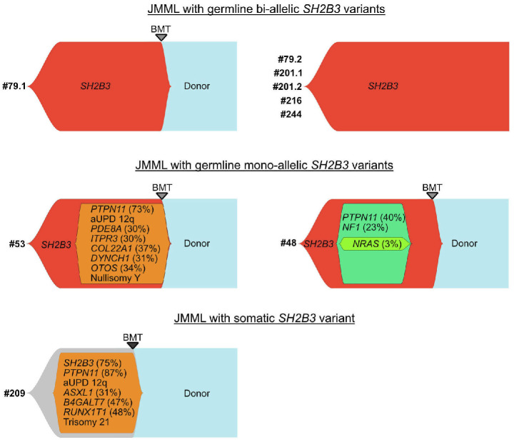
To determine the mutational makeup of JMML associated with SH2B3 variants, genome-wide DNA array analysis and high-depth sequencing of recurrently mutated genes in JMML were carried out. Whole-genome sequencing was also performed on patients with available germline samples (Online Supplementary Figure S1). All patients with bi-allelic SH2B3 variants had a normal karyotype and none had deleterious variants in known JMML drivers. Extensive genetic analysis did not evidence any other germline or somatic pathogenic or likely pathogenic variant or copy number alteration (Figure 3).
In contrast, patients with a mono-allelic germline SH2B3 variant acquired several additional somatic variants. Regardless of their germline or somatic status, mono-allelic SH2B3 mutations were systematically associated with a PTPN11 mutation (Figure 3).
Figure 3.
Clonal architecture of SH2B3 mutated juvenile myelomonocytic leukemia. Patients with a bi-allelic germline mutation of SH2B3 (red clones) (#79, #201, #216, #244) all have a monomorphic profile with no additional genetic alterations. Only patient #79.1 received a bone marrow transplant (upper panel). Patients with a germline mono-allelic SH2B3 mutation (#53, #48) and/or somatic SH2B3 mutation (orange clones) acquired several additional somatic mutations. Patient #53 had a somatic PTPN11 variant and patient #48 had somatic variants of PTPN11 and NF1, whose order of appearance could not be inferred from the allelic frequencies. Both patients underwent bone marrow transplantation (middle panel). In patient #209, the allelic frequency of the somatic SH2B3 variant was consistent with its presence in the full leukemia clone. Multiple other somatic alterations were acquired, including a driver somatic mutation in PTPN11. Here again, all potentially driver mutations were present at variant allele frequencies consistent with early co-occurrence during the course of the disease and order or appearance could not be determined (lower panel). JMML: juvenile myelomonocytic leukemia; BMT: bone marrow transplant; aUPD: acquired uniparental isodisomy.
Notably, two patients (#53; #209) with a mono-allelic SH2B3 truncating variant and a PTPN11 mutation subsequently acquired chromosome 12q uniparental disomy (12q21.1-q24.31 and 12q13.2-q24.31, respectively), resulting in copy-neutral loss of heterozygosity of the SH2B3 variants, but also of PTPN11, which is located on chromosome 12q24.13, close to SH2B3 (12q24.12). Such a pattern of events is rarely found in patients with PTPN11 mutations, which have a gain-of-function behavior, but is a typical mechanism for the complete inactivation of tumor suppressor genes. In our patients, it leads to the bi-allelic loss of function of LNK. Taken together, this supports the view that the loss of function of SH2B3 is a driving event in JMML in these cases as well. It is likely that the proximity to PTPN11 is the reason why a copy-neutral loss of heterozygosity is privileged over a simple deletion of SH2B3 in these cases.
DNA methylation profiling
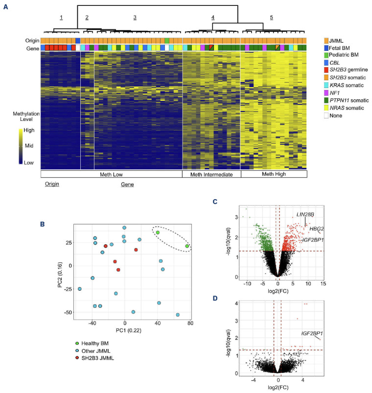
DNA methylation was also analyzed. A reference group of 54 cases of JMML of known genetic groups and two samples (one postnatal BM, one prenatal BM) from healthy subjects were used for comparison. As expected, hierarchical clustering of reference samples delineated three main groups, corresponding to methylation-low, intermediate and high JMML subtypes. The patients with a mono-allelic germline (#53) or somatic (#209) SH2B3 mutation clustered within the intermediate and hypermethylated groups, respectively. In contrast, samples with bi-allelic germline SH2B3 variants defined a distinct cluster (cluster 1) with the strongest hypomethylation pattern (Figure 4A). Interestingly, the prenatal BM clustered with the SH2B3 cluster whereas the postnatal BM clustered in cluster 3 (Figure 4A).
Altogether, methylation profiling is consistent with the idea that JMML with bi-allelic germline SH2B3 mutations represents a specific entity that may be linked to prenatal features, in line with the neonatal presentation.
RNA sequencing of JMML mononucleated cells
Total RNA sequencing was performed on mononuclear cells obtained at diagnosis from four patients with bi-allelic germline SH2B3 mutations. The gene expression profile was compared to that of 17 reference JMML samples and two postnatal BM samples from healthy subjects. Principal component analysis showed that the four SH2B3 cases clustered within the group of JMML samples (Figure 4B). Analysis of differential gene expression between SH2B3 JMML and postnatal BM samples evidenced 447 genes overexpressed, compared with 717 genes underexpressed, in SH2B3 JMML cases (Figure 4C). Interestingly, the oncofetal transcript LIN28B and its two targets HBG2 and IGF2BP1 were among the most overexpressed genes in SH2B3 JMML samples. When compared with other JMML cases, the number of overexpressed genes fell to only 13, with the oncofetal transcript IGF2BP1 ranking first (Figure 4D).
Discussion
SH2B3 encodes the lymphocyte adaptor protein LNK, a member of the SH2B adaptor family of proteins. LNK is predominantly expressed in hematopoietic stem and progenitor cells and is a key negative regulator of numerous cytokine and growth factor receptors.36
We show here that bi-allelic germline loss-of-function SH2B3 mutations represent a new disease predisposing to a JMML-like disorder with autosomal recessive inheritance. In our cohort, it accounted for 50% of JMML cases with unsolved genetics, and all cases experienced spontaneous resolution, in line with a hypomethylated DNA pattern indicative of a favorable prognosis.37
JMML-associated germline SH2B3 mutations were frameshift or missense targeting the SH2 domain. The SH2 domain is required for LNK to bind to the phosphorylated tyrosine (pY813) of JAK2 or JAK3 when activated in response to cytokines. Variants targeting the SH2 domain have been reported to cause severe disruption to LNK function.20 All variants were thus consistent with loss of function of the protein. Deficiency of LNK leads to increased JAK/STAT signaling in a cytokine-independent manner,36 and mice deficient in LNK were demonstrated to have a variety of hematopoietic phenotypes, including expansion of the hematopoietic stem and progenitor cell compartment,19,38 which is consistent with JMML.5,6 It is now known that LNK controls the expansion of hematopoietic stem cells and myeloid progenitors predominantly by modulating the thrombopoietin signaling pathway.20 Indeed, thrompoietin, which was initially identified as the cytokine that stimulates megakaryopoiesis and platelet production, has also been shown to be a major regulator of hematopoietic stem cell self-renewal and quiescence.20
Both germline and somatic LNK loss-of-function mutations have been previously identified in a range of hematologic diseases.24 Heterozygous SH2B3 germline variants, some of which are relatively common in the general population, have been associated with a low penetrance increased risk of myeloproliferative neoplasms25 and idiopathic erythrocytosis.26,27 Homozygous pathogenic variants, by contrast, are absent from reference population databases and have scarcely been described in patients. The first case reported was that of a consanguineous family in which two siblings carrying D231Profs*38 presented with growth retardation, mild developmental delay and autoimmune disorders.28 Although this was not explicitly presented as a JMML, the proband developed a phenotype very similar to that of our patients with high white blood cell count, <20% blasts, thrombocytopenia, and no megakaryocytes in BM at 4 weeks old. Blood cell counts normalized without intervention by the age of 14 months; however, the boy then developed liver cirrhosis alike to one of our patients.
More recently, two unrelated patients with homozygous germline loss-of-function SH2B3 variants R148Profs*40 and V402M with developmental delay, hepatosplenomegaly, myeloproliferation and autoimmune disorders were described.29 Paradoxically, unlike other patients, a major thrombocytosis with megakaryocytic hyperplasia was noted in the BM of these patients. Similarly, even though our patients initially presented with pronounced thrombocytopenia they progressed after spontaneous resolution of JMML towards having thrombocytosis. This could therefore represent two successive phases of the same disease.
Figure 4.
DNA methylation pattern and gene expression profiling suggest persistence of fetal cues in juvenile myelomonocytic leukemia with bi-allelic germline SH2B3 variants. (A) DNA methylation analysis of patients with SH2B3 variants (N=8) and reference juvenile myelomonocytic leukemia (JMML) of known genetic groups (i.e., PTPN11 somatic, NRAS somatic, KRAS somatic, NF1 or CBL) (N=54). Unsupervised hierarchical clustering based on the 1,000 most variant 100 bp tiles identified three main DNA methylation subgroups (low, intermediate, high). JMML with bi-allelic SH2B3 germline mutations defines a subgroup of hypomethylated JMML (cluster 1), whereas JMML with either mono-allelic or somatic SH2B3 mutations cluster in the intermediate (cluster 4) or high (cluster 5) methylation groups. (B-D) Gene expression profile analyses of mononucleated cells of JMML with bi-allelic SH2B3 germline alteration (N=4) in comparison with other types of JMML (N=17) and bone marrow samples from healthy subjects. Principal component analysis (B) and volcano plots showing differential gene expression between JMML with bi-allelic SH2B3 germline alteration and healthy bone marrow (C) or other types of JMML (D). A qval threshold of ≤0.05 and a minimum fold-change of 1.5 were used to define differentially expressed genes. JMML: juvenile myelomonocytic leukemia; BM: bone marrow; Meth: methylation; PC: principal component; FC: fold change.
How LNK-deficient patients progress from a major defect in platelet production to excess production is unclear. The effect of LNK loss on megakaryopoiesis may be age-dependent.39,40 Indeed, the role of thrombopoietin appears to be different in fetal hematopoietic stem cells, which have a high capacity for proliferation and self-renewal for up to 6 months after birth, and in adult hematopoietic stem cells, which are maintained quiescent.41 In this respect, it is interesting to note that all JMML in patients with bi-allelic germline SH2B3 mutations had a neonatal onset and retained fetal characteristics, such as a DNA methylation pattern resembling that of fetal BM and expression of the oncofetal transcripts LIN28B and IGF2BP1.42 The progressive loss of this fetal context may then explain the spontaneous regression of JMML and the switch to thrombocytosis. RASopathies may be associated with immune disease,43 and the combination of spontaneously resolving forms of JMML associated with a propensity to subsequently develop immune-related disorders is particularly reminiscent of what is observed in patients with CBL syndrome.16 Interestingly, LNK recruits the CBL E3 ubiquitin ligase via its tyrosine residue at position 572 and thus inactivates JAK2 by degradation via the proteasome.44 This functional link has recently been extended to RAS activation. Indeed, JAK2 stabilization induced by loss of function of CBL dramatically enhances palmitoylation of NRAS, thereby activating the RAS-MAPK signaling pathway and promoting myeloid leukemogenesis.45 Such interplay between the JAK-STAT and RAS-MAPK signaling pathways highlights how the loss of LNK could induce JMML whose pathogenesis is considered to be strictly linked to activation of the RAS pathway.46 Besides immune-hematologic defects, several of our patients, as well as those reported in the literature,28,29 had multiple extra-hematologic disorders suggestive of a syndromic presentation. However, these signs were inconstant and differed widely from one patient to another, even within a family. It is therefore difficult to incriminate loss of SH2B3 function in these associated features, which could simply result from the increased risk of autosomal recessive disorders in a context of consanguinity.
Our analysis also identified JMML patients with mono-allelic SH2B3 variants. Regardless of their germline or somatic status, these patients had a strikingly different presentation than that of patients with bi-allelic germline variants, with a later age of onset and a much more severe clinical course requiring BM transplantation. In line with the clinical presentation, the mutational make-up and DNA methylation profile of patients with SH2B3 mutations differed greatly depending on the allelic pattern. Unlike patients with bi-allelic germline SH2B3 mutation, for whom this alteration seems to represent the only initiating driver of the JMML-like disorder, patients with mono-allelic germline or somatic SH2B3 variants presented with multiple somatic alterations, systematically including a somatic variant in PTPN11, although we could not determine which lesion was the initiating driver.
In conclusion, our findings show that SH2B3 can act as a driver in JMML in two ways, highlighting the importance of deregulation of the JAK-STAT pathway in this disease. On the one hand, mono-allelic SH2B3 alterations, either germline or somatic, drive a severe ‘classical’ JMML associated with frequent loss of heterozygosity and additional somatic mutations. On the other hand, bi-allelic germline variants of SH2B3 define a singular recessive clinical entity associated with a predisposition to develop a neonatal JMML-like disorder. This new entity is estimated to account for half of the cases of JMML that remained unresolved on the genetic level. Its biology and clinical course distinguish it clinically from cases of JMML associated with somatic or constitutional mono-allelic pathogenic SH2B3 variants. The absence of secondary somatic changes is reminiscent of the JMML-like disorder seen in some patients with Noonan syndrome. However, apart from the neonatal presentation, none of the unique hematologic features of patients with germline SH2B3 bi-allelic mutations is shared with Noonan syndrome-JMML.47 Furthermore, all of our cases had a favorable outcome, which is less common in Noonan syndrome-JMML.47 This, together with the immunological landscape, makes them clinically more similar to patients with CBL syndrome. Indeed, strikingly, all previously reported patients were also described to develop autoimmune disorders (thyroiditis, hepatitis) at a later age.28,29 Similarly, although their follow-up is still short, one of the present patients developed autoimmune thyroiditis and another presented with a severe liver disorder requiring transplantation. Thus, in addition to the JMML-like disorder, this disease seems to cause serious immune disorders, which should be monitored to favor prompt management and effective care.
Supplementary Material
Acknowledgments
We are profoundly grateful to the patients and families for their participation in the research. We thank Stacey Foulane and Aldjia Assous for help with analyses and clinical data collection, respectively. We also thank Dr Bruno Cassinat (Laboratoire de Biologie Cellulaire, Hôpital Saint-Louis) for in vitro culture of myeloid progenitors. Finally, we thank the CRB-K of the Robert Debré Hospital (CRB-K, BB-0033-00076).
Funding Statement
Funding: This work was supported, in part, by grants from AIRC (IG21614 and IG-28768), EJP-RD (NSEuroNet), and the Italian Ministry of Health (RF-2021-12374963).
Data-sharing statement
Whole-genome sequencing, RNA-sequencing and DNA methylation data are deposited in the Sequence Read Archive (SRA) (https://www.ncbi.nlm.nih.gov/sra) with access number SUB13849483 .
References
- 1.Chang TY, Dvorak CC, Loh ML. Bedside to bench in juvenile myelomonocytic leukemia: insights into leukemogenesis from a rare pediatric leukemia. Blood. 2014;124(16):2487-2497. [DOI] [PubMed] [Google Scholar]
- 2.Locatelli F, Niemeyer CM. How I treat juvenile myelomonocytic leukemia. Blood. 2015;125(7):1083-1090. [DOI] [PubMed] [Google Scholar]
- 3.Locatelli F, Nöllke P, Zecca M, et al. Hematopoietic stem cell transplantation (HSCT) in children with juvenile myelomonocytic leukemia (JMML): results of the EWOG-MDS/ EBMT trial. Blood. 2005;105(1):410-419. [DOI] [PubMed] [Google Scholar]
- 4.Lapidot T, Grunberger T, Vormoor J, et al. Identification of human juvenile chronic myelogenous leukemia stem cells capable of initiating the disease in primary and secondary SCID mice. Blood. 1996;88(7):2655-2664. [PubMed] [Google Scholar]
- 5.Caye A, Rouault-Pierre K, Strullu M, et al. Despite mutation acquisition in hematopoietic stem cells, JMML-propagating cells are not always restricted to this compartment. Leukemia. 2020;34(6):1658-1668. [DOI] [PMC free article] [PubMed] [Google Scholar]
- 6.Louka E, Povinelli B, Rodriguez-Meira A, et al. Heterogeneous disease-propagating stem cells in juvenile myelomonocytic leukemia. J Exp Med. 2021;218(2):e20180853. [DOI] [PMC free article] [PubMed] [Google Scholar]
- 7.Emanuel PD, Bates LJ, Castleberry RP, Gualtieri RJ, Zuckerman KS. Selective hypersensitivity to granulocyte-macrophage colony-stimulating factor by juvenile chronic myeloid leukemia hematopoietic progenitors. Blood. 1991;77(5):925-929. [PubMed] [Google Scholar]
- 8.Lasho T, Patnaik MM. Juvenile myelomonocytic leukemia - a bona fide RASopathy syndrome. Best Pract Res Clin Haematol. 2020;33(2):101171. [DOI] [PubMed] [Google Scholar]
- 9.Niemeyer CM. JMML genomics and decisions. Hematology. 2018;2018(1):307-312. [DOI] [PMC free article] [PubMed] [Google Scholar]
- 10.Flex E, Jaiswal M, Pantaleoni F, et al. Activating mutations in RRAS underlie a phenotype within the RASopathy spectrum and contribute to leukaemogenesis. Hum Mol Genet. 2014;23(16):4315-4327. [DOI] [PMC free article] [PubMed] [Google Scholar]
- 11.Niemeyer CM, Flotho C. Juvenile myelomonocytic leukemia: who’s the driver at the wheel? Blood. 2019;133(10):1060-1070. [DOI] [PubMed] [Google Scholar]
- 12.Caye A, Strullu M, Guidez F, et al. Juvenile myelomonocytic leukemia displays mutations in components of the RAS pathway and the PRC2 network. Nat Genet. 2015;47(11):1334-1340. [DOI] [PubMed] [Google Scholar]
- 13.Kratz CP, Niemeyer CM, Castleberry RP, et al. The mutational spectrum of PTPN11 in juvenile myelomonocytic leukemia and Noonan syndrome/myeloproliferative disease. Blood. 2005;106(6):2183-2185. [DOI] [PMC free article] [PubMed] [Google Scholar]
- 14.Tartaglia M, Niemeyer CM, Fragale A, et al. Somatic mutations in PTPN11 in juvenile myelomonocytic leukemia, myelodysplastic syndromes and acute myeloid leukemia. Nat Genet. 2003;34(2):148-150. [DOI] [PubMed] [Google Scholar]
- 15.Pérez B, Mechinaud F, Galambrun C, et al. Germline mutations of the CBL gene define a new genetic syndrome with predisposition to juvenile myelomonocytic leukaemia. J Med Genet. 2010;47(10):686-691. [DOI] [PubMed] [Google Scholar]
- 16.Niemeyer CM. RAS diseases in children. Haematologica. 2014;99(11):1653-1662. [DOI] [PMC free article] [PubMed] [Google Scholar]
- 17.Stieglitz E, Taylor-Weiner AN, Chang TY, et al. The genomic landscape of juvenile myelomonocytic leukemia. Nat Genet. 2015;47(11):1326-1333. [DOI] [PMC free article] [PubMed] [Google Scholar]
- 18.Oh ST, Simonds EF, Jones C, et al. Novel mutations in the inhibitory adaptor protein LNK drive JAK-STAT signaling in patients with myeloproliferative neoplasms. Blood. 2010;116(6):988-992. [DOI] [PMC free article] [PubMed] [Google Scholar]
- 19.Takaki S, Morita H, Tezuka Y, Takatsu K. Enhanced hematopoiesis by hematopoietic progenitor cells lacking intracellular adaptor protein, Lnk. J Exp Med. 2002;195(2):151-160. [DOI] [PMC free article] [PubMed] [Google Scholar]
- 20.Bersenev A, Wu C, Balcerek J, Tong W. Lnk controls mouse hematopoietic stem cell self-renewal and quiescence through direct interactions with JAK2. J Clin Invest. 2008;118(8):2832-2844. [DOI] [PMC free article] [PubMed] [Google Scholar]
- 21.Morales CE, Stieglitz E, Kogan SC, Loh ML, Braun BS. Nf1 and Sh2b3 mutations cooperate in vivo in a mouse model of juvenile myelomonocytic leukemia. Blood Adv. 2021;5(18):3587-3591. [DOI] [PMC free article] [PubMed] [Google Scholar]
- 22.Bersenev A, Wu C, Balcerek J, et al. Lnk constrains myeloproliferative diseases in mice. J Clin Invest. 2010;120(6):2058-2069. [DOI] [PMC free article] [PubMed] [Google Scholar]
- 23.Oh ST. When the brakes are lost: LNK dysfunction in mice, men, and myeloproliferative neoplasms. Ther Adv Hematol. 2011;2(1):11-19. [DOI] [PMC free article] [PubMed] [Google Scholar]
- 24.Maslah N, Cassinat B, Verger E, Kiladjian J-J, Velazquez L. The role of LNK/SH2B3 genetic alterations in myeloproliferative neoplasms and other hematological disorders. Leukemia. 2017;31(8):1661-1670. [DOI] [PubMed] [Google Scholar]
- 25.Coltro G, Lasho TL, Finke CM, et al. Germline SH2B3 pathogenic variant associated with myelodysplastic syndrome/ myeloproliferative neoplasm with ring sideroblasts and thrombocytosis. Am J Hematol. 2019;94(9):E231-E234. [DOI] [PubMed] [Google Scholar]
- 26.McMullin MF, Wu C, Percy MJ, Tong W. A nonsynonymous LNK polymorphism associated with idiopathic erythrocytosis. Am J Hematol. 2011;86(11):962-964. [DOI] [PMC free article] [PubMed] [Google Scholar]
- 27.Spolverini A, Pieri L, Guglielmelli P, et al. Infrequent occurrence of mutations in the PH domain of LNK in patients with JAK2 mutation-negative “idiopathic” erythrocytosis. Haematologica. 2013;98(9):e101-102. [DOI] [PMC free article] [PubMed] [Google Scholar]
- 28.Perez-Garcia A, Ambesi-Impiombato A, Hadler M, et al. Genetic loss of SH2B3 in acute lymphoblastic leukemia. Blood. 2013;122(14):2425-2432. [DOI] [PMC free article] [PubMed] [Google Scholar]
- 29.Blombery P, Pazhakh V, Albuquerque AS, et al. Biallelic deleterious germline SH2B3 variants cause a novel syndrome of myeloproliferation and multi-organ autoimmunity. EJHaem. 2023;4(2):463-469. [DOI] [PMC free article] [PubMed] [Google Scholar]
- 30.Arber DA, Orazi A, Hasserjian R, et al. The 2016 revision to the World Health Organization classification of myeloid neoplasms and acute leukemia. Blood. 2016;127(20):2391-2405. [DOI] [PubMed] [Google Scholar]
- 31.Khoury JD, Solary E, Abla O, et al. The 5th edition of the World Health Organization classification of haematolymphoid tumours: myeloid and histiocytic/dendritic neoplasms. Leukemia. 2022;36(7):1703-1719. [DOI] [PMC free article] [PubMed] [Google Scholar]
- 32.Elghetany MT, Cavé H, De Vito R, Patnaik MM, Solary E, Khoury JD. Juvenile myelomonocytic leukemia; moving forward. Leukemia. 2023;37(3):720-722. [DOI] [PubMed] [Google Scholar]
- 33.Motta M, Pannone L, Pantaleoni F, et al. Enhanced MAPK1 function causes a neurodevelopmental disorder within the RASopathy clinical spectrum. Am J Hum Genet. 2020;107(3):499-513. [DOI] [PMC free article] [PubMed] [Google Scholar]
- 34.Rentzsch P, Schubach M, Shendure J, Kircher M. CADD-spliceimproving genome-wide variant effect prediction using deep learning-derived splice scores. Genome Med. 2021;13(1):31. [DOI] [PMC free article] [PubMed] [Google Scholar]
- 35.Ioannidis NM, Rothstein JH, Pejaver V, et al. REVEL: an Ensemble method for predicting the pathogenicity of rare missense variants. Am J Hum Genet. 2016;99(4):877-885. [DOI] [PMC free article] [PubMed] [Google Scholar]
- 36.Morris R, Butler L, Perkins A, Kershaw NJ, Babon JJ. The role of LNK (SH2B3) in the regulation of JAK-STAT signalling in haematopoiesis. Pharmaceuticals (Basel). 2021;15(1):24. [DOI] [PMC free article] [PubMed] [Google Scholar]
- 37.Schönung M, Meyer J, Nöllke P, et al. International consensus definition of DNA methylation subgroups in juvenile myelomonocytic leukemia. Clin Cancer Res. 2021;27(1):158-168. [DOI] [PMC free article] [PubMed] [Google Scholar]
- 38.Velazquez L, Cheng AM, Fleming HE, et al. Cytokine signaling and hematopoietic homeostasis are disrupted in Lnk-deficient mice. J Exp Med. 2002;195(12):1599-1611. [DOI] [PMC free article] [PubMed] [Google Scholar]
- 39.Nakamura-Ishizu A, Suda T. Multifaceted roles of thrombopoietin in hematopoietic stem cell regulation. Ann N Y Acad Sci. 2020;1466(1):51-58. [DOI] [PubMed] [Google Scholar]
- 40.Buza-Vidas N, Antonchuk J, Qian H, et al. Cytokines regulate postnatal hematopoietic stem cell expansion: opposing roles of thrombopoietin and LNK. Genes Dev. 2006;20(15):2018-2023. [DOI] [PMC free article] [PubMed] [Google Scholar]
- 41.Pietras EM, Passegué E. Linking HSCs to their youth. Nat Cell Biol. 2013;15(8):885-887. [DOI] [PubMed] [Google Scholar]
- 42.Helsmoortel HH, Bresolin S, Lammens T, et al. LIN28B overexpression defines a novel fetal-like subgroup of juvenile myelomonocytic leukemia. Blood. 2016;127(9):1163-1172. [DOI] [PubMed] [Google Scholar]
- 43.Bader-Meunier B, Cavé H, Jeremiah N, et al. Are RASopathies new monogenic predisposing conditions to the development of systemic lupus erythematosus? Case report and systematic review of the literature. Semin Arthritis Rheum. 2013;43(2):217-219. [DOI] [PubMed] [Google Scholar]
- 44.Lv K, Jiang J, Donaghy R, et al. CBL family E3 ubiquitin ligases control JAK2 ubiquitination and stability in hematopoietic stem cells and myeloid malignancies. Genes Dev. 2017;31(10):1007-1023. [DOI] [PMC free article] [PubMed] [Google Scholar]
- 45.Ren J-G, Xing B, Lv K, et al. RAB27B controls palmitoylation-dependent NRAS trafficking and signaling in myeloid leukemia. J Clin Invest. 2023;133(12):e165510. [DOI] [PMC free article] [PubMed] [Google Scholar]
- 46.Kotecha N, Flores NJ, Irish JM, et al. Single-cell profiling identifies aberrant STAT5 activation in myeloid malignancies with specific clinical and biologic correlates. Cancer Cell. 2008;14(4):335-343. [DOI] [PMC free article] [PubMed] [Google Scholar]
- 47.Strullu M, Caye A, Lachenaud J, et al. Juvenile myelomonocytic leukaemia and Noonan syndrome. J Med Genet. 2014;51(10):689-697. [DOI] [PubMed] [Google Scholar]
Associated Data
This section collects any data citations, data availability statements, or supplementary materials included in this article.
Supplementary Materials
Data Availability Statement
Whole-genome sequencing, RNA-sequencing and DNA methylation data are deposited in the Sequence Read Archive (SRA) (https://www.ncbi.nlm.nih.gov/sra) with access number SUB13849483 .


