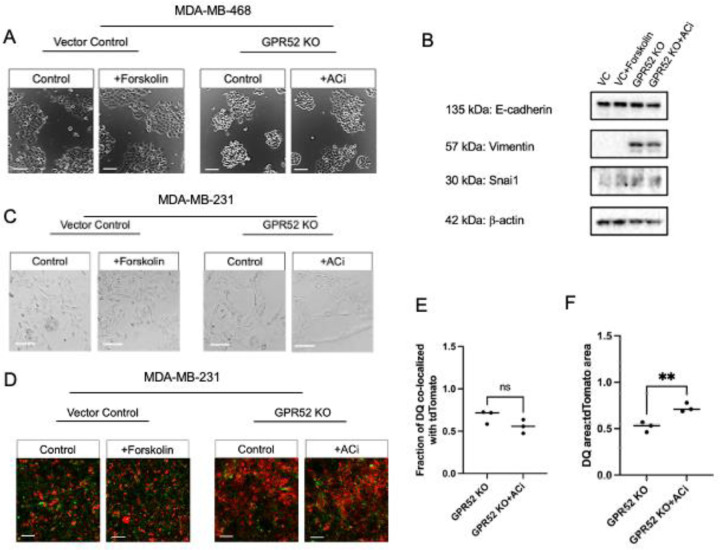
Figure 6. cAMP partially mediates phenotypes associated with GPR52 loss.
A) Vector control (VC) and GPR52 KO (sgRNA2) MDA-MB-468 cells were cultured in monolayer and treated with 1 μM FSK, 1.4 μM ACi, or the vehicle control. Treatment was replaced every 72 hours over 6–8 days. Scalebar=100 μm. B) Western blotting of MDA-MB-468 cells that were treated as in (D) for 24 hours. C) VC and GPR52 KO (sgRNA2) MDA-MB-231 cells were cultured as in (D). Scalebar=100 μm. D) MDA-MB-231 cells were plated to confluency in monolayer and treated the next day with 1 μM FSK, 1.4 μM ACi, and the vehicle control treatments. Invasion assays were conducted as in Fig. 4G. Scalebar=100 μm. The fraction of DQ co-localized with tdTomato (E) and area of DQ normalized to the area of tdTomato (F) at z=10 μm at t=24 hours. n=3, Student’s t-test, P-value<0.05; *P <0.05, **P <0.005, ns=not significant. Line=median.

